Arq. Bras. Oftalmol. 2015;78 (5 )
:278-282
| DOI: 10.5935/0004-2749.20150074
Abstract
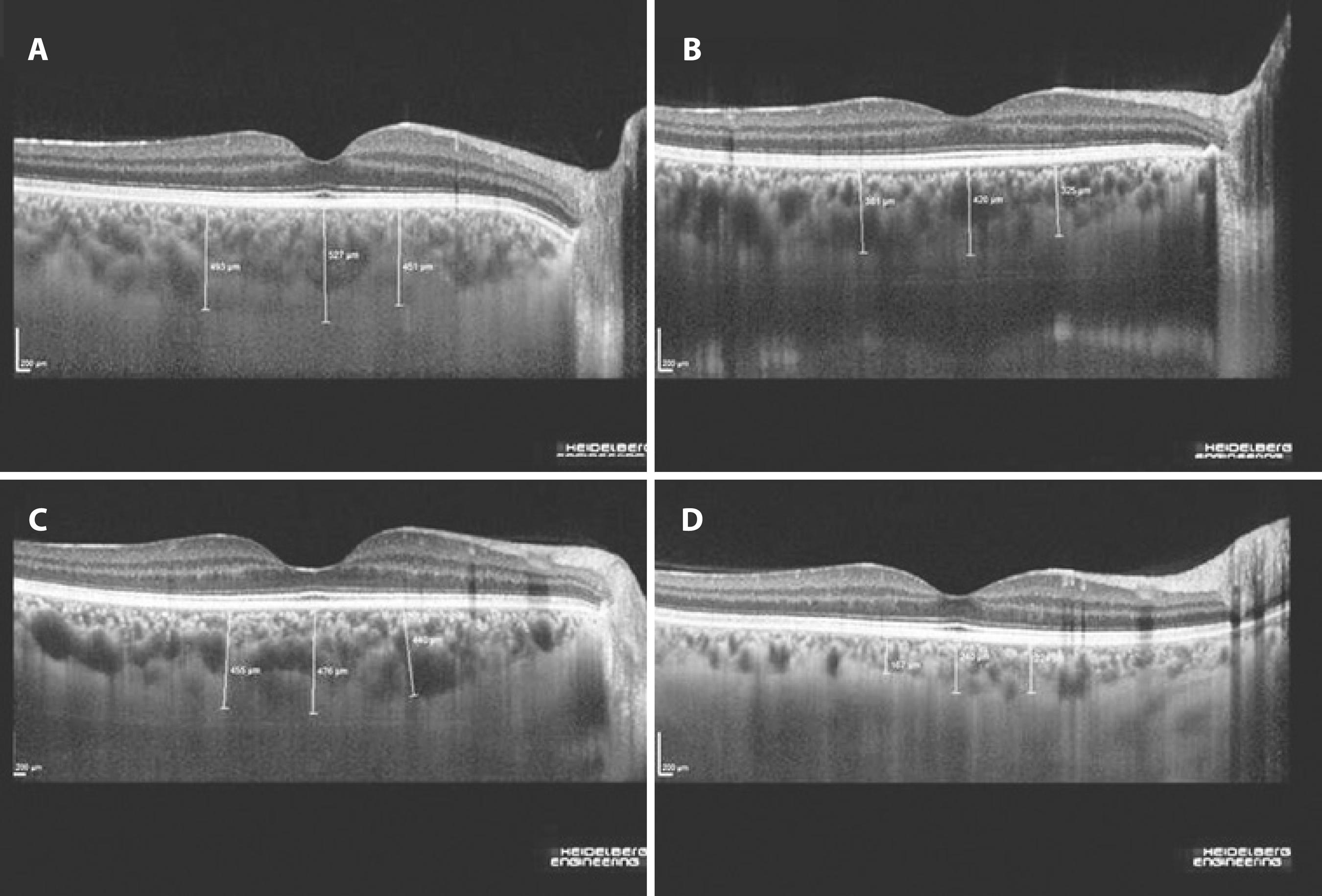
RESUMOObjetivos:Inflamação ocular é uma manifestação extra-intestinal comum de doença inflamatória do intestino (IBD) e pode ser paralela a atividade da doença. Neste estudo, investigamos se a espessura da coroideia pode ser útil para avaliar a atividade da IBD.Método:Um total de 62 olhos de 31 pacientes com IBD [10 com doença de Crohn (CD) e 21 colite ulcerosa (UC)] além de 104 olhos de 52 doadores de sangue saudáveis foram incluídos neste estudo. A espessura da coróide foi medida utilizando-se imagens de tomografia de coerência óptica com profundidade aprimorada. O índice de atividade da doença Crohn (CDAI) e o índice de Truelove Witts modificado foram usados para avaliar atividade da doença em CD e UC, respectivamente.Resultados:Não houve diferença significativa entre os pacientes com IBD e controles saudáveis em termos de medições da espessura da coróide subfoveal média em região 3000 μm nasal e 3000 μm temporal (p>0,05). Com base na análise univariada; idade, tabagismo, local do envolvimento em CD (ileal ou íleo-cecal), CDAI, atividade CD e índice de atividade endoscópica da UC foram significativamente correlacionados com a espessura da coróide (p<0,05). No entanto, fumar (p<0,05) e o local de envolvimento em CD (p<0,01) foram os únicos parâmetros independentes associados com um aumento na espessura da coroideia em todos os pontos de medida.Conclusões:A espessura da coroide não é um marcador útil para refletir a atividade da doença em pacientes com IBD, mas pode ser um indicador de envolvimento ileal em pacientes com CD.
Keywords: Coroide/patologia; Doenças de Crohn; Doenças inflamatórias intestinais/ complicações; Tomografia de coerência óptica; Colite ulcerativa; Uveíte
Arq. Bras. Oftalmol. 2024;87 (3 )
:1-6
| DOI: 10.5935/0004-2749.2022-0369
Abstract
PURPOSE: To evaluate the choroidal vascular alterations and effect of surgical treatment in the setting of idiopathic epiretinal membranes.
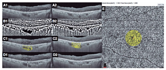
METHODS: The structure of the choroid was studied in 33 patients with unilateral idiopathic epiretinal membrane using optical coherence tomography with enhanced depth imaging and optical coherence tomography angiography. Eyes with epiretinal membrane underwent 25-gauge vitrectomy with epiretinal membrane and internal limiting membrane peeling. The choroidal vascularity index, Haller layer/choroidal thickness ratio, and choriocapillaris flow density were used to evaluate changes in choroidal structure after surgery and compare with the healthy fellow eyes.
RESULTS: The choroidal vascularity index and Haller layer/choroidal thickness ratio of the eyes with epiretinal membrane were higher than those of the fellow eyes at baseline (p=0.009 and p=0.04, respectively) and decreased postoperatively compared with preoperative values (p=0.009 and p=0.001, respectively). The choriocapillaris flow of eyes with epiretinal membrane was lower than that of the fellow eyes at baseline (p=0.001) and increased after surgery compared with the preoperative value (p=0.04). The choroidal vascularity index, Haller layer/choroidal thickness ratio, and choriocapillaris flow values of the healthy fellow eyes were comparable at baseline and final visit. In eyes with epiretinal membrane, the final choroidal vascularity index correlated with the final choriocapillaris flow (r=-0.749, p=0.008) in the multivariate analysis.
CONCLUSION: Idiopathic epiretinal membrane appears to affect the choroidal structure with increased choroidal vascularity index and Haller layer/choroidal thickness ratio and decreased choriocapillaris flow. These macrovascular (choroidal vascularity index and Haller layer/choroidal thickness) and microvascular (choriocapillaris flow) alterations appear to be relieved by surgical treatment of the epiretinal membranes.
Keywords: Epiretinal membrane/surgery; Vitrectomy; Choroid/pathology; Choroid/blood supply; Tomography, optical coherence/methods; Optical coherence tomography angiography; Humans


 ABO is licensed under a Creative Commons Attribution-NonComercial 4.0 Internacional.