Arq. Bras. Oftalmol. 2026;89 (2 )
:1-6
| DOI: 10.5935/0004-2749.2025-0244
Abstract
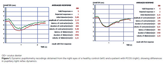
PURPOSE: Polycystic ovary syndrome is frequently associated with autonomic nervous system dysfunction, even in the absence of obesity or overt metabolic abnormalities. Alterations in pupillary responses may reflect early autonomic involvement and serve as a potential tool for early diagnosis, risk stratification, and disease monitoring. This study aimed to investigate pupillary reflex parameters using dynamic pupillometry in newly diagnosed non-obese women with polycystic ovary syndrome and to compare the findings with those of healthy controls. Methods: This prospective cross-sectional study included 48 newly diagnosed women with polycystic ovary syndrome and 44 age- and sex-matched healthy controls. Pupillary function parameters were measured using dynamic pupillometry (MonPackOne; Metrovision, France). Results: The mean age did not differ significantly between the groups (p=0.870). Initial pupil diameter, pupil contraction amplitude, and contraction velocity were significantly lower in the PCOS group than in the control group, whereas pupillary dilation duration was significantly longer (p<0.001, p<0.001, p=0.007, and p=0.032, respectively). No significant differences were observed between the groups regarding contraction latency, contraction duration, dilation latency, or dilation velocity (p=0.749, p=0.925, p=0.653, and p=0.310, respectively). Conclusion: Newly diagnosed non-obese women with polycystic ovary syndrome exhibit significant alterations in pupillary dynamics, suggesting a generalized reduction in both sympathetic and parasympathetic activity. Dynamic pupillometry may represent a practical, noninvasive tool for detecting early autonomic hypoactivity and identifying patients at risk for future metabolic or cardiovascular complications.
Keywords: Autonomic nervous system; IPupil/physiology; Reflex, Pupillary/physiology; Polycystic ovary syndrome/diagnosis; Menstruation disturbances; Ideal body weight
Arq. Bras. Oftalmol. 2026;89 (2 )
:1-8
| DOI: 10.5935/0004-2749.2025-0257
Abstract
PURPOSE: This study aimed to analyze ocular surface parameters and evaluate meibomian gland dysfunction using meibography in newly diagnosed patients with atopic dermatitis compared with healthy controls.
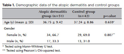
METHODS: This cross-sectional clinical study included 51 newly diagnosed patients with atopic dermatitis and 45 age- and sex-matched healthy controls. Disease severity was assessed using the Eczema Area Severity Index. The Ocular Surface Disease Index questionnaire, Schirmer test, tear meniscus height, noninvasive tear break-up time, conjunctival redness grading, and meibography staging were performed. Meibomian gland dropout was graded for each eyelid from 0 (no loss) to 3 (loss of >2/3 of the total gland area).
RESULTS: Schirmer test values, tear meniscus height, and noninvasive tear break-up time were significantly lower in the atopic dermatitis group than in the control group (p<0.001, p<0.001, and p<0.001, respectively). In contrast, Ocular Surface Disease Index scores, conjunctival redness grades, and total meiboscores were significantly higher in the atopic dermatitis group than in the control group (p<0.001, p<0.001, and p<0.001, respectively). Moreover, in the atopic dermatitis group, a significant positive correlation was observed between the Eczema Area Severity Index score and total meiboscore (rₛ=0.390, p=0.005), while a significant negative correlation was found between the Eczema Area Severity Index score and Schirmer test results (rₛ=−0.301, p=0.032). Conclusions: Newly diagnosed patients with atopic dermatitis exhibit significant alterations in tear film parameters and meibomian gland morphology compared with healthy individuals. These patients should be monitored for early development of dry eye disease and meibomian gland dysfunction to prevent associated complications.
Keywords: Dermatitis, atopic; Ocular surface; Meibomian gland dysfunction; Meibography; Dry eye syndromes
 ABO is licensed under a Creative Commons Attribution-NonComercial 4.0 Internacional.