Arq. Bras. Oftalmol. 2020;83 (1 )
:39-42
| DOI: 10.5935/0004-2749.20200005
Abstract
Objetivo: Revisar os resultados de cirurgias de suspensão ao músculo frontal com o uso de fio de politetrafluoretileno em pacientes com blefaroptose.
Métodos: Em um estudo observacional retrospectivo, foram analisados os resultados das cirurgias de músculo frontal de uma instituição, realizadas entre 2003 e 2018. Todos os procedimentos foram realizados com incisão fechada e técnica de pentágono. Os desfechos foram classificados como satisfatórios ou insatisfatórios com definição satisfatória definida como distância margem-reflexo >3mm e <1mm entre as pálpebras e insatisfatória como hipocorreção, complicações cirúrgicas e assimetria.
Resultados: Incluímos um total de 76 pálpebras de 52 pacientes em nosso estudo. Com um tempo médio de seguimento pós-operatório de 16,8 ± 18,5 meses (intervalo 3-95), 59 (77,6%) pálpebras apresentaram desfecho satisfatório e 17 (22,4%) insatisfatórios (8 casos de assimetria, 3 granulomas, 3 extrusões de sutura, 2 abscessos e 1 caso de celulite). Nove pálpebras do grupo insatisfatório necessitaram de reoperação. Entre os pacientes com seguimento≥12 meses (38 cirurgias), resultados duradouros foram observados na maioria dos casos, exceto por 2 extrusões de sutura de surgimento tardio.
Conclusão: O uso de politetrafluoretileno na cirurgia de músculo frontal mostrou ser previsível, seguro e duradouro, Nossos achados corroboram com estudos prévios que mostraram resultados funcionais adequados e baixos índices de complicação.
Keywords: Pálpebras/cirurgia; Blefaroptose/cirurgia; Blefaroplastia/métodos Politetrafluoretileno/utilização; Sutura; Período pós-operatório
Arq. Bras. Oftalmol. 2025;88 (4 )
:1-6
| DOI: 10.5935/0004-2749.2024-0278
Abstract
PURPOSE: This study aimed to evaluate the prevalence of orbital conditions in a tertiary ophthalmic outpatient hospital in Sao Paulo, Brazil, with a focus on the main diagnoses and their distribution.
METHODS: A retrospective chart review was conducted involving patients registered and admitted to the orbital disease unit at the Department of Ophthalmology, University of São Paulo Medical School, from January 2004 to March 2018. A total of 838 medical charts were analyzed, of which 37 were excluded due to incomplete data. The remaining charts were categorized into eight diagnostic groups: Graves’ orbitopathy , inflammatory disorders, tumors, vascular lesions, acquired structural abnormalities, congenital structural abnormalities, infectious diseases, and others.
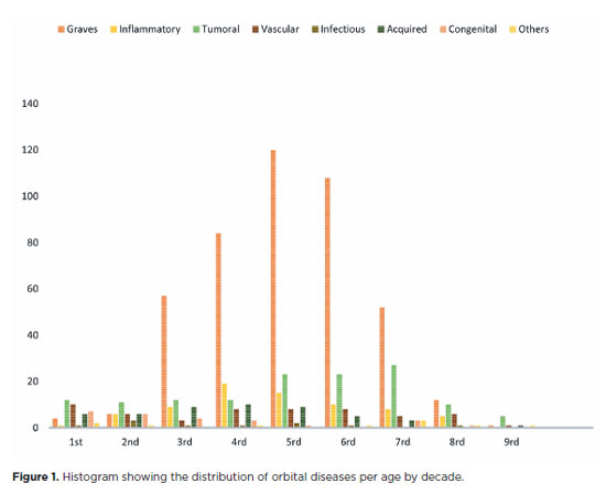
RESULTS: Of the 837,300 ophthalmological appointments, 3,372 (0.4%) were related to orbital diseases. The study included 801 patients, of whom 63.45% were women. The patients’ mean age was 42.86 years. Graves’ orbitopathy was the most common (55%), followed by tumor (17%), inflammatory disorders (9%), vascular lesions (7%), acquired structural abnormalities (5%), congenital structural abnormalities (4%), others (2%), and infectious diseases (1%). The study found significant differences in the incidence and types of orbital diseases, indicating the specialized nature of tertiary care and referral biases.
CONCLUSION: Published data on epidemiological orbital diseases is scarce. Therefore, this study focused on the diverse nature of orbital diseases and their low incidence among ophthalmology appointments. The major trends align with other epidemiological studies, demonstrating a preponderance of Graves’ orbitopathy in middle-aged adults and a bimodal distribution of tumors. These findings are essential in shaping resident training programs and healthcare policies, particularly in tertiary settings. Understanding the epidemiology of orbital diseases can improve diagnostic accuracy, treatment approaches, and patient outcomes as well as support future systemic prospective studies.
Keywords: Orbital diseases; Orbital tumors; Neoplasms; Inflammation; Graves’ ophthalmopathy; Outpatients
Arq. Bras. Oftalmol. 2024;87 (5 )
:1-7
| DOI: 10.5935/0004-2749.2023-0296
Abstract
PURPOSE: To compare inferomedial wall orbital decompression to balanced medial plus lateral wall orbital decompression in patients with Graves’ orbitopathy in the inactive phase with regard to exophthalmos reduction and the effects on quality of life.
METHODS: Forty-two patients with inactive Graves’ orbitopathy were randomly divided into two groups and submitted to one of two orbital decompression techniques: inferomedial wall orbital decompression or medial plus lateral wall orbital decompression. Preoperative and postoperative assessments included Hertel’s exophthalmometry and a validated Graves’ orbitopathy quality of life questionnaire. The results of the two groups were compared.
RESULTS: Compared to preoperative measurement, exophthalmos reduction was statistically significant in both groups (p<0.001) but more so in patients undergoing medial plus lateral wall orbital decompression (p=0.010). Neither orbital decompression techniques increased the visual functioning subscale score on the Graves’ orbitopathy quality of life questionnaire (inferomedial wall orbital decompression p=0.362 and medial plus lateral wall orbital decompression p=0.727), but a statistically significant difference was observed in the score of the appearance subscale in patients submitted to medial plus lateral wall orbital decompression (p=0.006).
CONCLUSIONS: Inferomedial wall orbital decompression is a good alternative for patients who do not require large exophthalmos reduction. However, medial plus lateral wall orbital decompression offers greater exophthalmos reduction and greater improvement in appearance (higher Graves’ orbitopathy quality of life questionnaire scores), making it a suitable option for esthetic-functional rehabilitation.
Keywords: Graves’ ophthalmopathy; Quality of life; Exophthalmos; Strabismus; Diplopia; Decompression, surgical


 ABO is licensed under a Creative Commons Attribution-NonComercial 4.0 Internacional.




09-tab01.jpg)

14-fig01.jpg)