Arq. Bras. Oftalmol. 2022;85 (1 )
:37-45
| DOI: 10.5935/0004-2749.20220006
Abstract
Objetivos: Avaliar a sensibilidade ao contraste em pacientes virgens de tratamento com retinopatia diabética proliferativa de não alto risco, submetidos a panfotocoagulação retiniana com injeções intravítreas de ranibizumabe versus panfotocoagulação isolada.
Métodos: Sessenta olhos de 30 pacientes foram randomizados em dois grupos: um submetido a panfotocoagulação com injeções de ranibizumabe (grupo estudo), e o outro submetimedo a panfotocoagulação isolada (grupo controle). Todos olhos foram tratados em 3 sessões de laser, seguindo recomendação do Early Treatment Diabetic Retinopathy Study (ETDRS). Avaliação da sensibilidade ao contraste foi realizada sob condições fotópicas (85 cd/m2) com tabela Visual Contrast Test Sensitivity 6500, permitindo avaliação de cinco frequências espaciais medidas com redes senoidais: 1.5, 3.0, 6.0, 12.0 e 18.0 ciclos por grau de ângulo visual (cpd). Foram realizadas medidas dos limiares de sensibilidade ao contraste intra e entre grupos na visita inicial, no 1º, 3º, e 6º mês de seguimento.
Resultados: Cinquenta e oito olhos, 28 do grupo estudo e 30 do grupo controle, atingiram o término do estudo. Análise comparativa da SC entre os grupos mostrou diferença estatisticamente significante, nas baixas frequências espaciais, no 1º mês em 1.5 cpd (p=0,001) e 3.0 cpd (p=0,04), no 3º mês em 1.5 cpd (p=0,016) e no 6º mês em 3.0 cpd (p=0,026) a favor do grupo estudo.
Conclusão: O tratamento com panfotocoagulação associada a injeção de ranibizumabe parece causar menos danos a sensibilidade ao contraste quando comparada com panfotocoagulação isolada em olhos com retinopatia diabética proliferativa de não alto risco. Dessa forma, os resultados apresentados podem justificar a associação do ranibizumabe à panfotocoagulação nestes pacientes.
Keywords: Retinopatia diabética; Fotocoagulação; Ranibizumab; Bevacizumab; Sensibilidade de contraste; Fator A de crescimento do endotélio vascular; Injeção intravítrea.
Arq. Bras. Oftalmol. 2025;88 (3 )
:1-11
| DOI: 10.5935/0004-2749.2024-0049
Abstract
PURPOSE: This study aimed to evaluate the total macular thickness as well as the thickness of the inner and outer retinal layers in patients with Parkinson's disease. It also aimed to verify the correlation of these parameters with motor symptoms and cognitive function.
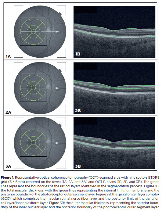
METHODS: A total of 46 eyes of 23 patients with Parkinson's disease and 40 eyes of 20 healthy controls were included in the study. The patients' cognitive, functional, and nonmotor symptoms were evaluated using the Katz Index of Independence and Pfeffer's Activities of Daily Living, Mini-Mental State Examination, Frontal Assessment Battery, Schwab and England Staging Scales, and Movement Disorders Society Nonmotor Symptoms Scale. The macular thickness measurements obtained via total, inner, and outer optical coherence tomography were recorded. Furthermore, the correlation of the parameters of optical coherence tomography with cognitive, functional, and nonmotor symptoms was assessed.
RESULTS: The scores of the Katz Index of Independence and Pfeffer's Activities of Daily Living as well as the Movement Disorders Society Nonmotor Symptoms Scale were significantly lower in patients with Parkinson's disease than in healthy controls. Moreover, the former had greater total macular thickness. The temporal and inferior outer sectors were significantly greater for the ganglion cell complex thickness in patients. A significant correlation was observed between the total macular thickness and the Movement Disorder Society-Unified Parkinson's Disease Rating Scale, Parte III (MDS-UPDRS-III) values. Contrarily, there was a negative correlation between the outer macular thickness and the MDS-UPDRS-III values. Meanwhile, the total macular thickness and ganglion cell complex thickness were significantly correlated with the scores of the Mini-Mental State Examination, Schwab and England Staging Scale, Frontal Assessment Battery, and Katz Index of Independence and Pfeffer's Activities of Daily Living. In addition, the Schwab and England scale was correlated with the outer macular thickness.
CONCLUSION: The total and inner macular thicknesses at the temporal and inferior outer sectors were greater in patients with Parkinson's disease than in the control group. These findings indicate that macular thickness may be greater in those with Parkinson's disease, particularly when associated with mild motor symptoms. In addition, the parameters of the total, inner, and outer optical coherence tomography were significantly associated with motor and nonmotor symptoms as well as cognitive function impairment.
Keywords: Parkinson's disease; Tomography, optical coherence; Neurodegenerative diseases; Cognitive dysfunction; Cognition; Motor perception; Visual acuity; Retina
Arq. Bras. Oftalmol. 2026;89 (3 )
:1-8
| DOI: 10.5935/0004-2749.2025-0330
Abstract
PURPOSE: To assess whether low-concentration brimonidine (0.025%) improves early postoperative signs and symptoms following femtosecond laser-assisted in situ keratomileusis and photorefractive keratectomy without affecting pupil diameter or flap safety.
METHODS: This prospective, randomized, double-masked, contralateral-eye, single-center study was conducted between January and September 2024. In each patient, one eye received 0.025% brimonidine 15–30 min before surgery (mean: 21.3 ± 2.4 min), whereas the fellow eye received 0.15% sodium hyaluronate (control). Primary outcomes on postoperative Day 1 included subconjunctival hemorrhage laser-assisted in situ keratomileusis and patient-reported symptoms (0–10 scale; composite score). Pupil diameter was measured pre-ablation. Statistical analyses included McNemar and paired t tests, with a significant threshold of α=0.05.
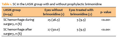
RESULTS: A total of 124 patients were included (54 laser-assisted in situ keratomileusis and 70 photorefractive keratectomy). Pupil diameter did not differ significantly between brimonidine-treated and control eyes (laser-assisted in situ keratomileusis: 2.63 ± 0.47 vs. 2.69 ± 0.42 mm, p=0.273; photorefractive keratectomy: 2.56 ± 0.44 vs. 2.61 ± 0.39 mm, p=0.116). In laser-assisted in situ keratomileusis, subconjunctival hemorrhage occurred less frequently in brimonidine-treated eyes both intraoperatively (9.3% vs. 46.3%, p<0.001) and on postoperative Day 1 (9.3% vs. 50.0%, p<0.001). Composite symptom scores were significantly lower in brimonidine-treated eyes in both laser-assisted in situ keratomileusis and photorefractive keratectomy groups (p=0.001 for both).
CONCLUSIONS: Preoperative administration of low-concentration brimonidine (0.025%) significantly reduced subconjunctival hemorrhage in laser-assisted in situ keratomileusis without comprising flap integrity. It also improved early postoperative symptoms in laser-assisted in situ keratomileusis and photorefractive keratectomy, without affecting pupil diameter. These findings support the use of dilute brimonidine as a safe and effective adjunct to enhance the immediate postoperative experience in refractive surgery.
Keywords: Brimonidine tartrate; Postoperative pain; Subconjunctival hemorrhage; Refractive surgery; Hemorrhage; Keratomileusis, laser in situ; Photorefractive keratectomy
Arq. Bras. Oftalmol. 2024;87 (4 )
:1-6
| DOI: 10.5935/0004-2749.2023-0200
Abstract
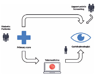
PURPOSE: Timely screening and treatment are essential for preventing diabetic retinopathy blindness. Improving screening workflows can reduce waiting times for specialist evaluation and thus enhance patient outcomes. This study assessed different screening approaches in a Brazilian public healthcare setting.
METHODS: This retrospective study evaluated a telemedicine-based diabetic retinopathy screening implemented during the COVID-19 pandemic and compared it with in-person strategies. The evaluation was conducted from the perspective of a specialized referral center in an urban area of Central-West Brazil. In the telemedicine approach, a trained technician would capture retinal images by using a handheld camera. These images were sent to specialists for remote evaluation. Patient variables, including age, gender, duration of diabetes diagnosis, diabetes treatment, comorbidities, and waiting time, were analyzed and compared.
RESULTS: In total, 437 patients with diabetes mellitus were included in the study (mean age: 62.5 ± 11.0 years, female: 61.7%, mean diabetes duration: 15.3 ± 9.7 years, insulin users: 67.8%). In the in-person assessment group, the average waiting time between primary care referral and specialist evaluation was 292.3 ± 213.9 days, and the referral rate was 73.29%. In the telemedicine group, the average waiting time was 158.8 ± 192.4 days, and the referral rate was 29.38%. The telemedicine approach significantly reduced the waiting time (p<0.001) and significantly lowered the referral rate (p<0.001).
CONCLUSION: The telemedicine approach significantly reduced the waiting time for specialist evaluation in a real-world setting. Employing portable retinal cameras may address the burden of diabetic retinopathy, especially in resource-limited settings.
Keywords: Telemedicine/methods; Diabetic retinopathy; Diagnostic screening programs; Vision screening; Practice patterns, physicians


 ABO is licensed under a Creative Commons Attribution-NonComercial 4.0 Internacional.




10-tab01tb.jpg)
14-fig01.jpg)