Arq. Bras. Oftalmol. 2022;85 (2 )
:166-173
| DOI: 10.5935/0004-2749.20220034
Abstract
Objetivos: Mensurar a perfusão do complexo retina/coróide com ressonância magnética em olhos com fechamento angular primário agudo (FAPA).
Métodos: Três sequências de ressonância magnética, duas anatômicas e uma de perfusão com gadolínio, foram adquiridas em pacientes com fechamento angular primário agudo. Regiões de interesse foram desenhadas na sequência de perfusão e sobrepostas à sequência anatômica. O volume de sangue relativo nos 2 primeiros segundos foi considerado como referência, e sua variação nos 28 segundos subsequentes foi analisada.
Resultados: Cinco olhos de 5 pacientes com fechamento angular primário agudo foram incluídos (3 com crise unilateral e 2 com crise bilateral). Três olhos contralaterais e 2 olhos de 2 pacientes saudáveis, pareados por sexo e idade, foram incluídos no grupo controle. Pacientes com fechamento angular primário agudo incluíam 4 (80%) mulheres, com idade média de 65,8 ± 12,37 anos, pressão intraocular média de 56,2 ± 14,67 mmHg, pressão arterial média de 113,4 ± 8,17 mmHg e pressão de perfusão ocular de 57,2 ± 13,46mmHg. No grupo controle, pressão intraocular média foi de 15,6 ± 2,61 mmHg (p=0,0625), pressão arterial média de 107,4 ± 6,57 mmHg (p=1,00) e pressão de perfusão ocular de 91,8 ± 6,72 mmHg (p=0,0625). O volume de sangue relativo do complexo retina/coróide foi de -0,127 ± 0,048 nos olhos em fechamento angular primário agudo e -0,213 ± 0,116 nos olhos controles (p=0,3125).
Conclusões: A sequência de ressonância magnética com gadolínio não demonstrou diferença na perfusão de retina/coroide em olhos com fechamento angular primário agudo.
Keywords: Glaucoma de ângulo fechado; Imagem por ressonância magnética; Gadolínio; Retina; Perfusão
Arq. Bras. Oftalmol. 2026;89 (1 )
:1-8
| DOI: 10.5935/0004-2749.2025-0025
Abstract
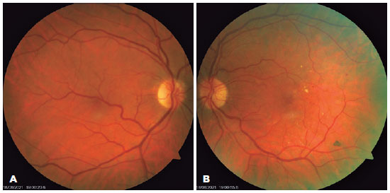
PURPOSE: Diabetic retinopathy screening in low- and middle-income countries is limited by restricted access to specialized care. Portable retinal cameras offer a practical alternative; however, image quality – affected by mydriasis – directly influences the performance of artificial intelligence models. This study evaluated the effect of mydriasis on image gradability and AI-based diabetic retinopathy detection in real-world, resource-limited settings.
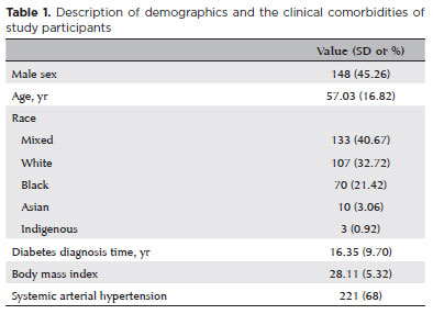
METHODS: The proportions of gradable images were compared between mydriatic and non-mydriatic groups. Generalized estimating equations were used to identify factors associated with image gradability, including age, sex, race, diabetes duration, and systemic hypertension. A ResNet-200d model was trained on the mobile Brazilian Ophthalmological dataset and externally validated on both mydriatic and non-mydriatic images. Model performance was evaluated using accuracy, F1 score, area under the curve, and confusion matrix metrics. Sensitivity differences were assessed using the McNemar test, and area under the curves were compared using DeLong's test. The Youden index was used to determine optimal classification thresholds. Agreement between macula- and disc-centered images was analyzed using Cohen's κ.
RESULTS: The mydriatic group demonstrated a higher proportion of gradable images compared with the non-mydriatic group (82.1% vs. 55.6%; p<0.001). In non-mydriatic images, lower gradability was associated with systemic hypertension, older age, male sex, and longer diabetes duration. The AI model achieved better performance in mydriatic images (accuracy, 85.15%; area under the curve, 0.94) than in non-mydriatic images (accuracy, 79.68%; area under the curve, 0.93). The McNemar test showed a significant difference in sensitivity (p=0.0001), whereas DeLong's test revealed no significant difference in area under the curve (p=0.4666). The Youden index indicated that optimal classification thresholds differed based on mydriasis status. Agreement between image fields was moderate to substantial and improved with mydriasis.
CONCLUSION: Mydriasis significantly improves image gradability and enhances AI performance in diabetic retinopathy screening. Nonetheless, in low- and middle-income countries where pharmacologic dilation may be impractical, optimizing model calibration and thresholding for non-mydriatic images is essential to ensure effective AI implementation in real-world clinical environments.
Keywords: Artificial intelligence; Bias; Diabetic retinopathy; Portable camera; Retina
Arq. Bras. Oftalmol. 2025;88 (3 )
:1-6
| DOI: 10.5935/0004-2749.2024-0112
Abstract
PURPOSE: To describe the ophthalmological findings of dry eye disease and its relation to the quality of life of COVID-19 survivors.
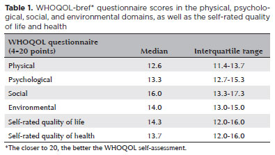
METHODS: COVID-19 survivors who had previously been hospitalized at Hospital das Clínicas de Ribeirão Preto complex underwent an ophthalmological evaluation, which included a dry eye disease questionnaire, break-up time, fluorescein staining, and Schirmer test. We collected the presenting and best-corrected visual acuity, sociodemographic data, personal medical history, and scores from a self-reported quality of life questionnaire (WHOQOL-bref). According to the severity of the acute phase of the disease, the patients were classified into mild-to-moderate, severe, and critical groups.
RESULTS: Ninety-five patients (190 eyes) were evaluated 100 ± 44 days after the onset of COVID-19 symptoms. Of these, 83 patients (87.3%) completed the WHOQOL-bref questionnaire. Ten patients (12.0%) had mild-to-moderate COVID-19, 41 (49.4%) had severe COVID-19, and 32 (38.6%) had critical COVID-19. The median best-corrected visual acuity was logMAR 0 (0-1). Approximately 26.3% patients had a history of dry eye disease or severe dry eye symptoms (frequent or constant ocular dryness and irritation). There was an association between the proportion of patients with dry eye disease and the quality of life (p=0.014) and health (p=0.001). Furthermore, there was a significant trend between the proportion of patients with dry eye disease and how they rated their health and quality of life (p=0.0004 and 0.0027, respectively.
CONCLUSIONS: There is a significant negative correlation between the proportion of patients with dry eye disease and their self-reported quality of life.
Keywords: COVID-19; Coronavirus infections; SARS-CoV-2; Eye diseases; Epidemiology; Ocular surface; Public health


 ABO is licensed under a Creative Commons Attribution-NonComercial 4.0 Internacional.




05-fig01.jpg)