Arq. Bras. Oftalmol. 2022;85 (2 )
:158-165
| DOI: 10.5935/0004-2749.20220071
Abstract
OBJETIVOS: o principal objetivo deste estudo foi descrever pacientes com achados vasculares retinianos temporalmente relacionados à vacinação contra COVID-19. Com maior notificação de possíveis eventos adversos similares, esperamos compreender a real dimensão e relevância do que foi apresentado.
MÉTODOS: Onze pacientes com queixas visuais após vacinação contra COVID-19 foram estudados. Os dados analisados foram: idade, gênero, tipo de vacinação, tempo de aparecimento de sintomas, achados sistêmicos, antecedentes pessoais, acuidade visual com melhor correção, biomicroscopia e imagem retiniana multimodal (retinografia colorida, red-free, SD-OCT, OCTA e angiofluoresceinografia). Os critérios de inclusão foram a presença de alterações oftalmológicas ocorridas dentro de 30 dias após a primeira ou segunda dose de qualquer vacina contra COVID-19.
RESULTADOS: Onze pacientes foram incluídos: 5 com oclusão arterial (45,4%), 4 com oclusão venosa (36,4%) e 2 (18,2%) com alterações não específicas vasculares sugestivas de isquemia retiniana como exsudatos algodonosos. A idade média dos pacientes foi de 57 anos (DP=16; com intervalo de 27 a 84 anos). A média de tempo de aparecimento de sintomas após a vacinação foi de 10 dias (DP=5,4; com intervalo de 3 a 16 dias). Nove dos onze pacientes eram do sexo feminino (81,8%). Fatores de risco sistêmicos foram observados em 36,4% dos pacientes. Dois pacientes tiveram sintomas neurológicos e visuais, com oclusão arterial. 36,4% dos pacientes tiveram infecção prévia por COVID-19 no último ano. Sete pacientes (63,6%) receberam a vacina ChAdOx1 nCoV-19 (AZD1222).
CONCLUSÕES: nossos dados sugerem que eventos retinianos temporalmente relacionados à vacinação contra COVID-19 são possíveis, porém raros. A relação entre estes eventos pós-vacinais exigem futura atenção antes de maiores conclusões.
Keywords: COVID-19; Infecções por coronavírus; Vacina; Oclusão arterial; Oclusão venosa; Síndrome de Susac
Arq. Bras. Oftalmol. 2024;87 (3 )
:1-7
| DOI: 10.5935/0004-2749.2022-0374
Abstract
PURPOSE: To describe a 2019 acute toxoplasmosis outbreak in the city of São Paulo, Brazil, and to evaluate the laboratory serological profile for toxoplasmosis for three consecutive years. The ophthalmological manifestations of the patients involved in the outbreak were also studied.
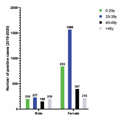
METHODS: A cross-sectional descriptive study of a toxoplasmosis outbreak in São Paulo, Brazil, between February and May 2019. Epidemiological data were described, as were the observed ocular manifestations. As part of this study the number of patients with positive IgM toxoplasmosis serology was obtained from a large laboratory network (DASA) for three consecutive years, including the year of the outbreak (2018, 2019, 2020).
RESULTS: Eighty-three individuals were identified in the outbreak and two clusters were studied. The clinical picture of at least 77% of the patients, the epidemiological analysis, and the short incubation period (5-8 days) suggested contamination by oocysts. Serological laboratory data analysis revealed an increase of positive toxoplasmosis IgM in 2019 of 73% compared to the previous year. Ophthalmological examination revealed that at least 4.8% of the patients developed toxoplasmic retinochoroiditis, none of whom had been treated during the acute systemic disease.
CONCLUSION: Our findings indicate vegetable contamination as the possible source of this outbreak, a high prevalence of toxoplasmosis in São Paulo during the outbreak period, and a drop in the number of tests during the COVID-19 pandemic. Retinochoroiditis was observed in at least 4.8% of the cases. We confirm the need to implement effective means for the prevention, diagnosis, and treatment of the disease. This may involve raising awareness among the population of the importance of vegetable hygiene, and improved quality control of food and water.
Keywords: Toxoplasmosis/etiology; Food parasitology; Water/parasitology; Uveitis, posterior/parasitology; Chorioretinitis/parasitology; Visual acuity; Disease outbreaks; Eye manifestations; Humans.
Arq. Bras. Oftalmol. 2024;87 (4 )
:1-12
| DOI: 10.5935/0004-2749.2023-0042
Abstract
PURPOSE: To assess the quality of life in patients diagnosed as having tuberculous uveitis and its association with sociodemographic, clinical, and psychosocial aspects.
METHOD: By conducting standardized interviews, clinical and demographic data were collected using a measure developed in this study. This measure was applied in addition to other measures, namely SF-12, Hospital Anxiety and Depression Scale, and NEI-VFQ-39, which were used to assess health-related quality of life, anxiety and depression symptoms, and visual functioning.
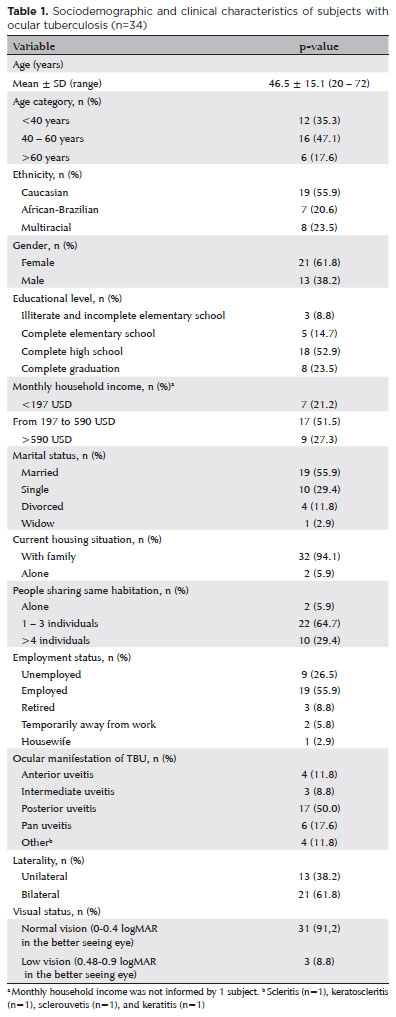
RESULTS: The study included 34 patients [mean age: 46.5 ± 15.1 years, female patients: 21 (61.8%)]. The mean of the VFQ-39 score was 74.5 ± 16.6 and that of SF-12 physical and mental component scores were 45.8 ± 10.1 and 51.6 ± 7.5, respectively, for the health-related quality of life. Anxiety symptoms were the most prevalent compared with depression symptoms and were found in 35.3% of the participants.
CONCLUSION: Tuberculous uveitis affects several scales of quality of life, thereby affecting a population economically active with a social, psychological, and economic burden.
Keywords: Tuberculosis, ocular; Quality of life; Uveitis; Anxiety; Depression; Surveys and questionnaires


 ABO is licensed under a Creative Commons Attribution-NonComercial 4.0 Internacional.


16-fig01tb.jpg)
15-fig01tb.jpg)