Arq. Bras. Oftalmol. 2025;88 (4 )
:1-7
| DOI: 10.5935/0004-2749.2024-0229
Abstract
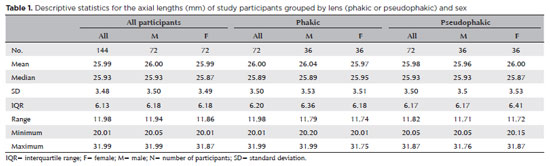
PURPOSE: The volume of the vitreous chamber varies with the size of the eye. The space created in the vitreous cavity by a vitrectomy is called the vitrectomized space. The volume of the vitrectomized space is strongly correlated with the axial length of the eye. This study aims to present guidelines for estimating the using participants stratified by axial length, sex, and history of cataract surgery.
METHODS: This retrospective, observational, cross-sectional study included 144 randomly selected participants who underwent vitrectomies between 2013 and 2023. Before surgery, the axial lengths of participants' eyes were measured using optical biometrics. The axial lengths of the eyes in our sample were between 20-32 mm. In all cases, a complete vitrectomy was performed, followed by complete fluid-air exchange and injection of a balanced saline solution. The volume infused was recorded.
RESULTS: The median (interquartile range; range) volume of the vitrectomized space was 6.1 (3.8; 3.1-11.3) mL in men and 6.1 (3.3; 3.2-11.2) mL in women (p=0.811). The median volume of the vitrectomized space was 5.9 (3.6; 3.1-11.2) mL in patients with phakic lenses and 6.25 (3.6; 3.3-11.3) mL in those with pseudophakic lenses (p=0.533). A positive correlation was found between the axial length and the volume of the vitrectomized space in this sample (r=0.968; p<0.001). In a cubic polynomial regression, the coefficient of determination was 0.948. Similar results were observed in both sexes and in both phakic and pseudophakic patients. The estimated cubic polynomial regression equation for this sample was VVS = 0.000589052857847605 × AL3 - 0.025114926401582700 × AL2 + 0.685961117595624000 × AL - 5.088226672620790000.
CONCLUSION: We developed this axial length estimation of the volume of vitrectomized space as a guideline for the determination of vitrectomized space volume using axial length.
Keywords: Cataract extraction; Retinal perforations/surgery; Epiretinal membrane/surgery; Vitreous body; Axial length, eye; Vitrectomy; Biometry/methods; Diagnostic techniques, ophthalmological; Guidelines as topic.
Arq. Bras. Oftalmol. 2026;89 (6 )
:1-9
| DOI: 10.5935/0004-2749.2025-0294
Abstract
PURPOSE: To compare obstructive sleep apnea screening classifications, anthropometric characteristics, and mechanical behaviors between participants with keratoconus and controls and to explore their associations with disease severity.
METHODS: This prospective cross-sectional study included 74 participants with keratoconus and 49 controls. The paper-based Berlin and STOP-BANG questionnaires were administered face-to-face by a researcher and scored manually. A study-specific questionnaire assessed eye rubbing and sleep position, whereas a separate structured field recorded self-reported ocular compression during sleep. Both groups underwent corneal tomography. The primary logistic regression model included age, sex, body mass index (BMI), eye rubbing, and ocular compression.
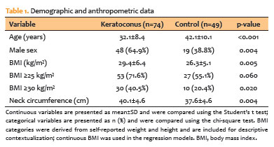
RESULTS: Participants with keratoconus had a higher BMI (29.4±6.4 vs. 26.3±5.1 kg/m²; p=0.005), larger neck circumference (40.1±4.6 vs. 37.6±4.6 cm; p=0.004), more frequent eye rubbing (71.6% vs. 51.0%; p=0.020), and more frequent ocular compression (43.2% vs. 20.4%; p=0.009) than controls. High-risk classifications did not differ significantly between groups according to either the Berlin Questionnaire (p=0.206) or STOP-BANG (p=0.139). In exploratory models adjusted for age and sex, positive associations were observed for the Berlin Questionnaire (OR, 2.58; 95% CI, 0.91-7.28) and STOP-BANG (OR, 3.30; 95% CI, 0.83-13.05). In the primary model, younger age (OR, 0.88; 95% CI, 0.83-0.93), male sex (OR, 2.91; 95% CI, 1.14-7.42), and higher BMI (OR, 1.15; 95% CI, 1.05-1.27) remained associated with keratoconus, whereas the estimates for behavioral factors were attenuated. No association with disease severity was detected.
CONCLUSIONS: Participants with keratoconus had higher anthropometric measurements and more frequent mechanical behaviors; however, the prevalence of high-risk obstructive sleep apnea classifications did not differ significantly between groups. Questionnaire-based and anthropometric findings are not independent because BMI contributes to both screening instruments, whereas neck circumference contributes to the STOP-BANG score. Ocular compression was assessed using a nonvalidated self-report measure rather than objective pressure measurements. Severity analyses were underpowered, and causality cannot be inferred from the study design.
Keywords: Keratoconus; Sleep apnea, obstructive; Risk factors; Body mass index; Sleep; Logistic models; Patient acuity; Tomography; Surveys and questionnaires


 ABO is licensed under a Creative Commons Attribution-NonComercial 4.0 Internacional.