Arq. Bras. Oftalmol. 2007;70 (1 )
:105-108
| DOI: 10.1590/S0004-27492007000100019
Abstract
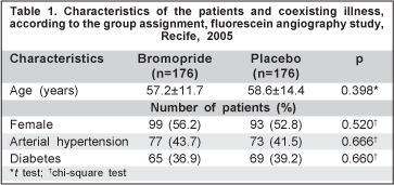
OBJETIVOS: Determinar a eficiência da bromoprida na profilaxia de náuseas na angiofluoresceinografia, quando comparada a um placebo. MÉTODOS: O estudo foi um ensaio clínico aleatório duplo-mascarado, entre dezembro de 2004 e abril de 2005. Os exames foram realizados com fluoresceína sódica a 20% intravenosa em dose única de 2,5 ml. Os pacientes foram divididos em dois grupos: grupo 1, pacientes que receberam 10 mg/ 2 ml de bromoprida via intravenosa e o grupo 2, pacientes que receberam uma dose 2 ml de cloreto de sódio a 0,9% (placebo), ambos 20 minutos antes da injeção do contraste. Foram registrados os casos de náusea durante e após o exame. RESULTADOS: Foram selecionados 352 pacientes, 176 em cada grupo. Foram registrados casos de náusea em 12 (6,8%) pacientes do grupo da bromoprida e 11 (6,3%) pacientes do grupo placebo (p<0,829 - risco relativo=1,09). CONCLUSÃO: Neste estudo a bromoprida não preveniu a ocorrência de náuseas na angiofluoresceinografia, quando comparada a um placebo.
Keywords: Náusea; Metoclopramida; Angiofluoresceinografia; Hipersensibilidade a drogas; Ensaio clínico controlado aleatório
Arq. Bras. Oftalmol. 2007;70 (6 )
:949-952
| DOI: 10.1590/S0004-27492007000600012
Abstract
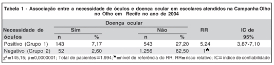
OBJETIVOS: Verificar a relação entre a prescrição de óculos e a presença de afecções oculares encontradas em crianças na idade escolar. MÉTODOS: Crianças na idade escolar que foram examinadas na Campanha " Olho no Olho" em Recife no ano de 2004. Foram seguidas etapas recomendadas pelo Conselho Brasileiro de Oftalmologia para realização desta Campanha, desde a preparação dos professores para triagem das crianças até a consulta com os oftalmologistas. Os pacientes foram divididos em dois grupos de acordo com a necessidade de prescrição de óculos, sendo relacionados com a presença de doença ocular ou não. Trata-se de um estudo descritivo de delineamento transversal que teve seus resultados analisados a partir do programa estatístico Epi Info versão 6.0. RESULTADOS: De uma amostra de 1.994 escolares, 686 deles apresentaram necessidade de óculos (34,4% Grupo1), sendo que 543 (27,2%) não apresentaram qualquer doença ocular, enquanto 143 (7,17%) tinham alguma doença ocular. Em 1.308 crianças (65,5% Grupo 2) não houve necessidade de óculos. Destas, 1.256 (62,5%) não apresentavam doença oftalmológica, enquanto 52 (2,6%) apresentavam algum tipo de afecção ocular. Os grupos 1 e 2 foram comparados entre si verificando que crianças que necessitam de óculos apresentam um risco relativo de possuírem doença ocular de 5,24 (Intervalo de Confiança de 95%: 3,87 a 7,10) vezes maior que as crianças que não precisam dos mesmos, com diferença estatisticamente significativa entre os dois grupos (p= 0.0000001). CONCLUSÃO: Conclui-se que escolares que necessitam de óculos apresentam maior probabilidade de ter doença ocular, sendo necessário um exame oftalmológico completo na infância realizado por oftalmologistas capacitados para a detecção e tratamento das diversas afecções encontradas além da prescrição adequada dos óculos.
Keywords: Saúde escolar; Promoção da saúde; Óculos; Oftalmopatias; Acuidade visual
Arq. Bras. Oftalmol. 2008;71 (1 )
:43-48
| DOI: 10.1590/S0004-27492008000100009
Abstract
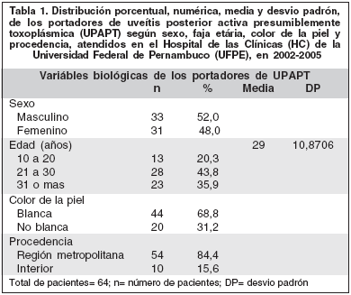
PURPOSE: To describe clinical characteristics of posterior active uveitis presumptively by Toxoplasma gondii (PAUPT) in patients with typical lesion. Tranversal study. METHODS: Sixty-four patients with retinochoroiditis scatter and active satellite lesions examined in Pernambuco, Brazil. All were older than 10 years and immunocompetent. Gender, age, skin color, and residence were recorded. Previous uveitis, visual accuracy, intraocular pressure (IOP), and ocular examination were analyzed. RESULTS: 52% were males, most of them with white skin (68.8%). Mean age 29 years (±10.87). Eighty-four percent of the patients lived in the metropolitan area. 56.2% were having the first episode of uveitis. In the damaged eye, visual accuracy mean was 20/200, IOP mean 14.5 mmHg (±64). Hyperemia of the conjunctiva was observed in 29.7% of the patients and alterations of the cornea in 51.6%. There were cells in the aqueous humor in 62.7%. 6.2% had posterior synechiae. All had vitreous damage and 45.3% retinal vasculitis. In 42.2% of the patients, lesions were located in zone I of Holland and 90.6% had the size of one discus diameter or greater. Neuritis was observed in 28.2%. Uveitis was more frequent in the right eye (54.7%). CONCLUSION: PAUPT affects young people and the main symptom was reduction of visual acuity. IOP mean was normal. Alterations of the vitreous were observed in all cases. Injuries were equal to one discus diameter or greater and located in zone I of Holland.
Keywords: Toxoplasmosis, ocular; Toxoplasma; Uveitis, posterior; Fluorescent antibody technique
 ABO is licensed under a Creative Commons Attribution-NonComercial 4.0 Internacional.