Arq. Bras. Oftalmol. 2003;66 (5 )
:9-15
| DOI: 10.1590/S0004-27492003000600002
Abstract
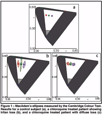
Pacientes que fazem uso de cloroquina ou hidróxi-cloroquina, drogas que são freqüentemente administradas para o tratamento de artrite reumatóide, lúpus eritrematoso ou malária, podem sofrer alterações na visão de cores e na sensibilidade de contraste. O presente estudo avaliou a função visual destes pacientes em um estudo conjunto da Universidade de São Paulo (USP), em São Paulo, e da Universidade Federal do Pará (UFPA), em Belém. Trinta e dois pacientes usuários de cloroquina sem alterações no exame de fundo de olho foram avaliados em São Paulo (n=10, 38 a 71 anos, média=55,8 anos) e em Belém (n=22, 20 a 67 anos, média=40 anos). A dose acumulada de cloroquina prescrita foi de 45 a 430 g (média=213 g; dp=152 g) para o grupo de São Paulo, e de 36 a 540 g (média=174 g; dp=183 g) para o grupo de Belém. Os testes foram realizados monocularmente com o estado refracional corrigido. A discriminação de cor foi avaliada utilizando o Teste de Cor de Cambridge (CCT): o limiar de discriminação de cor foi mensurado primeiro nos eixos protano, deutano e tritano, e em seqüência, três elipses de MacAdam foram determinadas. A visão de cores dos pacientes também foi avaliada com testes de arranjo de cores: o teste de 100 matizes de Farnsworth-Munsell (FM100), o D15 de Farnsworth-Munsell, e o teste Lanthony Dessaturado (D15d). A sensibilidade de contraste foi também medida com grades senoidais preto-e-brancas em 22 pacientes. Os resultados foram comparados com controles sem patologias oftalmológicasou neuro-oftalomológicas. 24 pacientes apresentaram discromatopsia adquirida, com perdas seletivas (11 pessoas) ou difusas (13 pessoas). Embora as perdas estivessem presentes no FM100, não houve correlação entre o escore de erro do FM100 e a área elíptica medida pelo CCT. Além disso, três pacientes que tiveram escores normais no FM100 falharam para alcançar limiares normais no CCT. O teste de Lanthony foi menos sensível do que os outros dois testes, tal que falhou em indicar perda em cerca de metade dos pacientes afetados, e o D15 foi o teste menos sensível, deixando de indicar déficits em 9 de 10 pacientes. A sensibilidade de contraste esteve dentro dos valores normais para pacientes submetidos a este teste. A extensão das perdas na discriminação de cores foi positivamente correlacionada com a dose acumulada. O CCT é recomendado para o acompanhamento destes pacientes, pois forneceu dados quantitativos que podem ser diretamente interpretados no espaço cromático da CIE (Commission Internationelle d'Éclairage).
Keywords: Cloroquina; Retinopatia; Visão de cores; Sensibilidade de contraste; Neurotoxicologia; Bateria de testes neurocomportamentais; Discromatopsia
Arq. Bras. Oftalmol. 2025;88 (4 )
:1-12
| DOI: 10.5935/0004-2749.2023-0263
Abstract
PURPOSE: Amblyopia is a cortical neurological disorder caused by abnormal visual experiences during the critical period for visual development. Recent works have shown that, in addition to the well-known visual alterations, such as changes in visual acuity, several perceptual aspects of vision are affected. This study aims to analyze and compare the effects of different types of amblyopia on visual color processing and determine whether these effects are correlated with visual acuity.
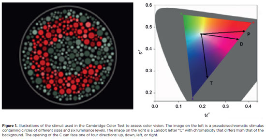
METHODS: Our study sample comprised 42 amblyopic individuals, aged 7-40 years, (strabismus, n=16; anisometropia, n=18; and mixed-cause, n=8) and 33 age-matched controls. Color vision was tested by measuring the chromaticity threshold of each patient on the protan, deutan, and tritan axes using version 02 of the Cambridge Color Test. Spatial stimulation cues were eliminated using spatial noise and luminance.
RESULTS: The color discrimination thresholds on the protan, deutan, and tritan axes were similar between control participants and amblyopic patients (p>0.05). There was no correlation between VA values and color thresholds (p>0.05).
CONCLUSION: Patients with amblyopia have normal color vision in contexts that include luminance and spatial noise. Our results may be indicative of independent neural pathways for spatial and chromatic visual processing.
Keywords: Amblyopia; Anisometropia; Color vision; Strabismus; Vision disorders; Visual acuity
Arq. Bras. Oftalmol. 2024;87 (2 )
:1-6
| DOI: 10.5935/0004-2749.2021-0435
Abstract
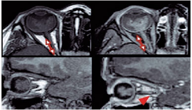
PURPOSE: This study aimed to analyze the association between magnetic resonance imaging apparent diffusion coefficient map value and histopathological differentiation in patients who underwent eye enucleation due to retinoblastomas.
METHODS: An observational chart review study of patients with retinoblastoma that had histopathology of the lesion and orbit magnetic resonance imaging with apparent diffusion coefficient analysis at Hospital de Clínicas de Porto Alegre between November 2013 and November 2016 was performed. The histopathology was reviewed after enucleation. To analyze the difference in apparent diffusion coefficient values between the two major histopathological prognostic groups, Student's t-test was used for the two groups. All statistical analyses were performed using SPSS version 19.0 for Microsoft Windows (SPSS, Inc., Chicago, IL, USA). Our institutional review board approved this retrospective study without obtaining informed consent.
RESULTS: Thirteen children were evaluated, and only eight underwent eye enucleation and were included in the analysis. The others were treated with photocoagulation, embolization, radiotherapy, and chemotherapy and were excluded due to the lack of histopathological results. When compared with histopathology, magnetic resonance imaging demonstrated 100% accuracy in retinoblastoma diagnosis. Optic nerve invasion detection on magnetic resonance imaging showed a 66.6% sensitivity and 80.0% specificity. Positive and negative predictive values were 66.6% and 80.0%, respectively, with an accuracy of 75%. In addition, the mean apparent diffusion coefficient of the eight eyes was 0.615 × 103 mm2/s. The mean apparent diffusion coefficient value of poorly or undifferentiated retinoblastoma and differentiated tumors were 0.520 × 103 mm2/s and 0.774 × 103 mm2/s, respectively.
CONCLUSION: This study revealed that magnetic resonance imaging is useful in the diagnosis of retinoblastoma and detection of optic nerve infiltration, with a sensitivity of 66.6% and specificity of 80%. Our results also showed lower apparent diffusion coefficient values in poorly differentiated retinoblastomas with a mean of 0.520 ×
103 mm2/s, whereas in well and moderately differentiated, the mean was 0.774 × 103 mm2/s.
Keywords: Retinoblastoma; Prognosis; Retinal neoplasms; Orbit; Diffusion magnetic resonance imaging


 ABO is licensed under a Creative Commons Attribution-NonComercial 4.0 Internacional.