Arq. Bras. Oftalmol. 2022;85 (2 )
:158-165
| DOI: 10.5935/0004-2749.20220071
Abstract
OBJETIVOS: o principal objetivo deste estudo foi descrever pacientes com achados vasculares retinianos temporalmente relacionados à vacinação contra COVID-19. Com maior notificação de possíveis eventos adversos similares, esperamos compreender a real dimensão e relevância do que foi apresentado.
MÉTODOS: Onze pacientes com queixas visuais após vacinação contra COVID-19 foram estudados. Os dados analisados foram: idade, gênero, tipo de vacinação, tempo de aparecimento de sintomas, achados sistêmicos, antecedentes pessoais, acuidade visual com melhor correção, biomicroscopia e imagem retiniana multimodal (retinografia colorida, red-free, SD-OCT, OCTA e angiofluoresceinografia). Os critérios de inclusão foram a presença de alterações oftalmológicas ocorridas dentro de 30 dias após a primeira ou segunda dose de qualquer vacina contra COVID-19.
RESULTADOS: Onze pacientes foram incluídos: 5 com oclusão arterial (45,4%), 4 com oclusão venosa (36,4%) e 2 (18,2%) com alterações não específicas vasculares sugestivas de isquemia retiniana como exsudatos algodonosos. A idade média dos pacientes foi de 57 anos (DP=16; com intervalo de 27 a 84 anos). A média de tempo de aparecimento de sintomas após a vacinação foi de 10 dias (DP=5,4; com intervalo de 3 a 16 dias). Nove dos onze pacientes eram do sexo feminino (81,8%). Fatores de risco sistêmicos foram observados em 36,4% dos pacientes. Dois pacientes tiveram sintomas neurológicos e visuais, com oclusão arterial. 36,4% dos pacientes tiveram infecção prévia por COVID-19 no último ano. Sete pacientes (63,6%) receberam a vacina ChAdOx1 nCoV-19 (AZD1222).
CONCLUSÕES: nossos dados sugerem que eventos retinianos temporalmente relacionados à vacinação contra COVID-19 são possíveis, porém raros. A relação entre estes eventos pós-vacinais exigem futura atenção antes de maiores conclusões.
Keywords: COVID-19; Infecções por coronavírus; Vacina; Oclusão arterial; Oclusão venosa; Síndrome de Susac
Arq. Bras. Oftalmol. 2025;88 (6 )
:1-5
| DOI: 10.5935/0004-2749.2025-0085
Abstract
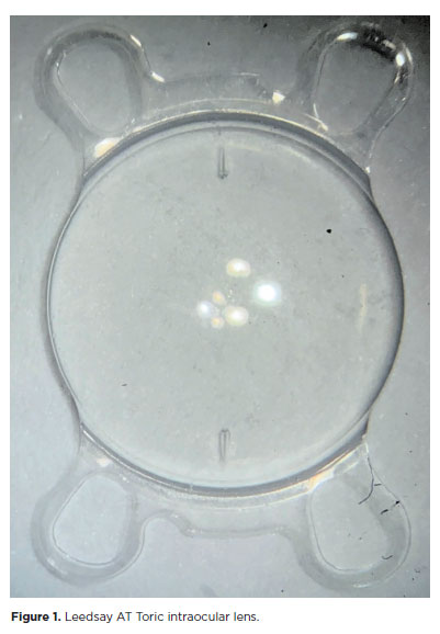
PURPOSE: The purpose of this study was to assess visual outcomes and patient satisfaction following cataract surgery involving the implantation of quad-loop intraocular lenses, including trifocal, bifocal, and toric variants.
METHODS: Information was obtained from both physical and electronic medical records of patients who underwent phacoemulsification cataract surgery with implantation of different intraocular lenses between January 1, 2022, and December 31, 2023. The study included individuals aged over 18 who received bilateral implantation of bifocal, trifocal, or monofocal toric intraocular lenses. Visual acuity was assessed at various postoperative time points using the logMAR scale. Quantitative variables were analyzed using mean and standard deviation.
RESULTS: A total of 92 eyes received premium intraocular lenses: 4 bifocal, 32 trifocal, 52 toric monofocal, and 4 trifocal toric lenses. The average preoperative corrected visual acuity was logMAR 0.478 ± 0.259. On the first postoperative day, the average uncorrected visual acuity was logMAR 0.301 ± 0.207. By day 30, 67.4% of eyes achieved uncorrected distance visual acuity of logMAR 0.2 or better. Patient satisfaction was high, with few reports of glare or halos.
CONCLUSION: Quad-loop intraocular lenses-including trifocal, bifocal, and toric models-demonstrated effective improvement in visual acuity and high levels of patient satisfaction. These lenses represent a suitable option for enhancing visual outcomes after cataract surgery. Additional studies with larger cohorts are recommended to confirm these results.
Keywords: Cataract extraction; Aberrometry/methods; Lenses, intraocular; Lens implantation, intraocular; Prosthesis design
Arq. Bras. Oftalmol. 2025;88 (6 )
:1-8
| DOI: 10.5935/0004-2749.2025-0053
Abstract
PURPOSE: This pilot study evaluated the diagnostic accuracy of a deep learning model for detecting pterygium in anterior segment photographs taken using smartphones in the Brazilian Amazon. The model’s performance was benchmarked against assessments made by experienced ophthalmologists, considered the clinical gold standard.
METHODS: In this cross-sectional study, 38 participants (76 eyes) from Barcelos, Brazil, were enrolled. Trained nonmedical health workers captured high-resolution anterior segment images using smartphones. These images were analyzed using a deep learning model based on the MobileNet-V2 convolutional neural network. Diagnostic metrics–including sensitivity, specificity, accuracy, positive predictive value, negative predictive value, and area under the receiver operating characteristic curve–were calculated and compared with the ophthalmologists’ evaluations.
RESULTS: The deep learning model achieved a sensitivity of 91.43%, specificity of 90.24%, positive predictive value of 88.46%, negative predictive value of 92.79%, and an area under the curve of 0.91. Logistic regression revealed no statistically significant association between pterygium and demographic variables such as age or gender.
CONCLUSIONS: The deep learning model demonstrated high diagnostic performance in identifying pterygium in a remote Amazonian population. These preliminary findings support the potential use of artificial intelligence–based tools to facilitate early detection and screening in underserved regions, thereby enhancing access to ophthalmic care.
Keywords: Pterygium/diagnostic imaging; Smartphone; Diagnostic techniques, ophthalmological; Deep learning; Telemedicine; Artificial intelligence; Cross-sectional studies; Brazil/epidemiology
Arq. Bras. Oftalmol. 2024;87 (3 )
:1-7
| DOI: 10.5935/0004-2749.2022-0374
Abstract
PURPOSE: To describe a 2019 acute toxoplasmosis outbreak in the city of São Paulo, Brazil, and to evaluate the laboratory serological profile for toxoplasmosis for three consecutive years. The ophthalmological manifestations of the patients involved in the outbreak were also studied.
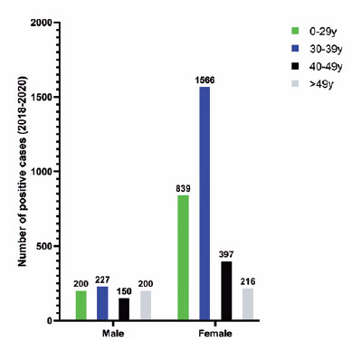
METHODS: A cross-sectional descriptive study of a toxoplasmosis outbreak in São Paulo, Brazil, between February and May 2019. Epidemiological data were described, as were the observed ocular manifestations. As part of this study the number of patients with positive IgM toxoplasmosis serology was obtained from a large laboratory network (DASA) for three consecutive years, including the year of the outbreak (2018, 2019, 2020).
RESULTS: Eighty-three individuals were identified in the outbreak and two clusters were studied. The clinical picture of at least 77% of the patients, the epidemiological analysis, and the short incubation period (5-8 days) suggested contamination by oocysts. Serological laboratory data analysis revealed an increase of positive toxoplasmosis IgM in 2019 of 73% compared to the previous year. Ophthalmological examination revealed that at least 4.8% of the patients developed toxoplasmic retinochoroiditis, none of whom had been treated during the acute systemic disease.
CONCLUSION: Our findings indicate vegetable contamination as the possible source of this outbreak, a high prevalence of toxoplasmosis in São Paulo during the outbreak period, and a drop in the number of tests during the COVID-19 pandemic. Retinochoroiditis was observed in at least 4.8% of the cases. We confirm the need to implement effective means for the prevention, diagnosis, and treatment of the disease. This may involve raising awareness among the population of the importance of vegetable hygiene, and improved quality control of food and water.
Keywords: Toxoplasmosis/etiology; Food parasitology; Water/parasitology; Uveitis, posterior/parasitology; Chorioretinitis/parasitology; Visual acuity; Disease outbreaks; Eye manifestations; Humans.
 ABO is licensed under a Creative Commons Attribution-NonComercial 4.0 Internacional.
16-fig01tb.jpg)