Arq. Bras. Oftalmol. 2023;86 (4 )
:353-358
| DOI: 10.5935/0004-2749.20230055
Abstract
Objetivo: Examinar a eficácia da ceratectomia fototerapêutica para o tratamento de patologias variáveis que apresentarem opacidades anteriores da córnea, e avaliar a distribuição das indicações de ceratectomia fototerapêutica nos últimos 10 anos.
Métodos: Foram revisados retrospectivamente os prontuários de 276 pacientes, com 334 olhos tratados com ceratectomia fototerapêutica entre março de 2010 e o ano de 2020. As etiologias dos pacientes submetidos à ceratectomia fototerapêutica foram anotadas e suas alterações foram examinadas. Os resultados refrativos e de acuidade visual antes e após a operação foram registrados e analisados de acordo com a etiologia.
Resultados: A idade média dos pacientes foi de 40,7 ± 16,2 anos (faixa: 19-84). O tempo médio de acompanhamento foi de 25,5 ± 19,1 meses (faixa: 3-96). A ceratectomia fototerapêutica foi aplicada com mais frequência para distrofias estromais corneanas (44%, 151 olhos de 111 pacientes); entre as distrofias corneanas como um todo, a distrofia granular foi a indicação terapêutica mais comum desse procedimento. Ao contrário de outras indicações, nos últimos 10 anos houve um aumento na aplicação de ceratectomia fototerapêutica em casos de opacidade subepitelial persistente causada por conjuntivite adenoviral. Houve um aumento significativo na acuidade visual em todos os grupos, exceto para o grupo com defeito epitelial recorrente (p<0,05). A maior melhora na acuidade visual foi detectada em casos de distrofia estromal, no subgrupo das distrofias granulares.
Conclusão: Apesar das mudanças nas tendências de indicação, a ceratectomia fototerapêutica continua sendo uma abordagem terapêutica eficaz e confiável para tratar lesões da córnea anterior.
Keywords: Ceratectomia fotorrefrativa; Opacidade da córnea; Lesões da córnea; Distrofias da córnea; Fototerapia.
Arq. Bras. Oftalmol. 2025;88 (2 )
:1-6
| DOI: 10.5935/0004-2749.2023-0229
Abstract
PURPOSE: To compare the outcomes of intravitreal dexamethasone implant used as either an adjuvant or a switching therapy for diabetic macular edema in patients with poor anatomic response after three consecutive monthly injections of ranibizumab.
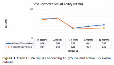
METHODS: This retrospective study included patients with diabetic macular edema who received three consecutive doses of ranibizumab as initial therapy and demonstrated poor response. A single dose of intravitreal dexamethasone implant was administered to these patients. The patients were divided into two groups according to the treatment modalities: the adjuvant therapy group, consisting of patients who continued treatment with ranibizumab injection after receiving intravitreal dexamethasone implant, and the switch therapy group, consisting of patients who were switched from ranibizumab treatment to intravitreal dexamethasone implant as needed. The main outcome measurements were best corrected visual acuity and central retinal thickness at baseline and at 3, 6, 9, and 12 months of follow-up.
RESULTS: In this study that included 64 eyes of 64 patients, the best corrected visual acuity and central retinal thickness values did not significantly differ between the groups at baseline and at 6 months of follow-up (p>0.05). However, at 12 months, the best corrected visual acuity values in the adjuvant and switch therapy groups were 0.46 and 0.35 LogMAR, respectively (p=0.012), and the central retinal thickness values were 344.8 and 270.9, respectively (p=0.007).
CONCLUSIONS: In a real-world setting, it seems more reasonable to use intravitreal dexamethasone implant as a switch therapy rather than an adjuvant therapy for diabetic macula edema refractory to ranibizumab despite three consecutive monthly injections of ranibizumab. Patients switched to intravitreal dexamethasone implant were found to have better anatomic and visual outcomes at 12 months than those who continued ranibizumab therapy despite their less-than-optimal responses.
Keywords: Diabetic retinopathy; Macular edema/drug therapy; Dexamethasone/administration & dosage; Drug implants; Intravitreal injections; Ranibizumab/administration & dosage; Tomography, optical coherence; Endothelial growth factors
 ABO is licensed under a Creative Commons Attribution-NonComercial 4.0 Internacional.
06-tab01.jpg)