Arq. Bras. Oftalmol. 2025;88 (3 )
:1-8
| DOI: 10.5935/0004-2749.2023-0115
Abstract
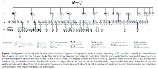
PURPOSE: To evaluate the presence of congenital hypertrophy of the retinal pigment epithelium in a large family affected by familial adenomatous polyposis and identify the causative mutation in the adenomatous polyposis coli gene. Thus, we aimed to determine the significance of congenital hypertrophy of the retinal pigment epithelium as a phenotypic marker of the disease.
METHODS: A family consisting of 95 individuals was evaluated. Among these, 45 individuals were randomly selected by convenience sampling method to undergo ophthalmological evaluation. A funduscopic exam, including slit lamp and indirect ophthalmoscopy, were performed in the selected patients. In those with retinal lesions, a retinography was obtained. The adenomatous polyposis coli gene was sequenced in one affected family member to identify the pathogenic mutation. Once the variant was identified, six undiagnosed family members were tested for the mutation via capillary electrophoresis sequencing.
RESULTS: Congenital hypertrophy of the retinal pigment epithelium was observed in 13 (28.9%) of the 45 individuals evaluated. Of these, nine patients were confirmed to have familial adenomatous polyposis (via colonoscopy or molecular testing). However, four patients had not been investigated. Of the 32 (71.1%) family members without the lesion, 14 did not have familial adenomatous polyposis and 18 were yet to be evaluated. The lesions were bilaterally present and exhibited a peculiar fish-tail shape in all the evaluated individuals. Adenomatous polyposis coli gene sequencing revealed a pathogenic variant c.4031del. (Ser1344*), in heterozygosity (49.27%), in exon 16.
CONCLUSIONS: The study findings confirmed the significance of congenital hypertrophy of the retinal pigment epithelium as a phenotypic marker for familial adenomatous polyposis. Furthermore, it is an effective first-line screening method for at risk family members of such patients. The novel mutation identified in our study participants, which is yet to be described in the literature, causes an aggressive form of the disease.
Keywords: Retinal diseases/congenital; Retinal pigment epithelium; Hypertrophy/congenital; Adenomatous polyposis coli / genetics; Phenotype; Optical coherence tomography
Arq. Bras. Oftalmol. 2025;88 (3 )
:1-3
| DOI: 10.5935/0004-2749.2023-0290
Abstract
PURPOSE: The microbiology pattern of neonatal conjunctivitis has changed over time, and the incidence of gonococcal conjunctivitis is almost nil. This study aimed to determine the etiology of neonatal conjunctivitis cases referred to a tertiary health center in Brazil.
METHODS: From 2017 to 2020, conjunctival swabs were taken from neonates with clinical signs of conjunctivitis and tested with bacterial culture and polymerase chain reaction for Neisseria gonorrhoeae and Chlamydia trachomatis.
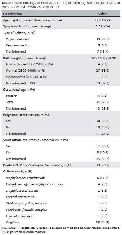
RESULTS: A total of 51 neonates were included in the 3-year study. Chlamydial conjunctivitis was diagnosed in 39 (76.5%) patients, and microbial growth was detected in 13 (25.5%) patients. The most isolated bacterium was Staphylococcus epidermidis (n=6, 11.8%), followed by other coagulase-negative Staphylococcus species (n=4, 7.8%) and S. aureus (n=2, 3.9%). One S. aureus isolate was resistant to oxacillin. There were no cases of gonococcal conjunctivitis. Ten (19.6%) patients showed polymerase chain reaction-negative C. trachomatis and negative bacterial culture test results.
CONCLUSION: Findings show that C. trachomatis is the most common pathogen causing neonatal conjunctivitis. The high prevalence of C. trachomatis infection highlights the importance of screening and treating pregnant woman.
Keywords: Conjunctivitis; Infant, newborn, diseases; Ophthalmia neonatorum; Chlamydia infections; Sexually transmitted diseases


 ABO is licensed under a Creative Commons Attribution-NonComercial 4.0 Internacional.