Arq. Bras. Oftalmol. 2026;89 (1 )
:1-10
| DOI: 10.5935/0004-2749.2024-0344
Abstract
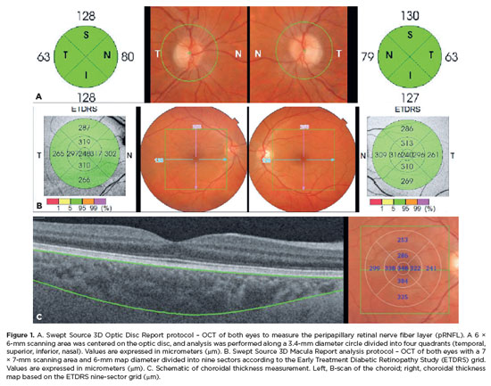
PURPOSE: To evaluate the relationships between retinal changes identified by optical coherence tomography in patients with migraine with aura or migraine without aura and the duration and severity of the disease, as measured by the questionnaire.
METHODS: In this cross-sectional study, 16 patients with migraine with aura and 15 patients with migraine without aura were compared with 16 age- and sex-matched controls without migraine. Retinal parameters were assessed using optical coherence tomography, and disease severity was evaluated using scores.
RESULTS: Foveal choroidal thickness was significantly reduced in migraine patients to controls (p=0.040). Negative correlations were observed between disease duration and total macular thickness (p=0.037), ganglion cell complex thickness (p=0.017), and choroidal thickness (p=0.039) in the migraine without aura Group. Additionally, scores showed an inverse correlation with peripapillary retinal nerve fiber layer thickness (p=0.027) in the migraine without aura Group.
CONCLUSION: Individuals with migraine exhibited significant choroidal thinning compared with controls. Longer disease duration was associated with reduced total macular, ganglion cell complex, and choroidal thickness, while greater migraine-related disability correlated with thinner peripapillary retinal nerve fiber layers.
Keywords: Migraine with aura; Migraine without aura; Nerve fibers; Retina; Macula; Optic disc; Choroid; Tomography, optical coherence; Surveys and questionnaires.
Arq. Bras. Oftalmol. 2025;88 (3 )
:1-11
| DOI: 10.5935/0004-2749.2024-0049
Abstract
PURPOSE: This study aimed to evaluate the total macular thickness as well as the thickness of the inner and outer retinal layers in patients with Parkinson's disease. It also aimed to verify the correlation of these parameters with motor symptoms and cognitive function.
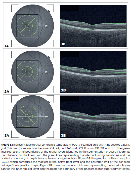
METHODS: A total of 46 eyes of 23 patients with Parkinson's disease and 40 eyes of 20 healthy controls were included in the study. The patients' cognitive, functional, and nonmotor symptoms were evaluated using the Katz Index of Independence and Pfeffer's Activities of Daily Living, Mini-Mental State Examination, Frontal Assessment Battery, Schwab and England Staging Scales, and Movement Disorders Society Nonmotor Symptoms Scale. The macular thickness measurements obtained via total, inner, and outer optical coherence tomography were recorded. Furthermore, the correlation of the parameters of optical coherence tomography with cognitive, functional, and nonmotor symptoms was assessed.
RESULTS: The scores of the Katz Index of Independence and Pfeffer's Activities of Daily Living as well as the Movement Disorders Society Nonmotor Symptoms Scale were significantly lower in patients with Parkinson's disease than in healthy controls. Moreover, the former had greater total macular thickness. The temporal and inferior outer sectors were significantly greater for the ganglion cell complex thickness in patients. A significant correlation was observed between the total macular thickness and the Movement Disorder Society-Unified Parkinson's Disease Rating Scale, Parte III (MDS-UPDRS-III) values. Contrarily, there was a negative correlation between the outer macular thickness and the MDS-UPDRS-III values. Meanwhile, the total macular thickness and ganglion cell complex thickness were significantly correlated with the scores of the Mini-Mental State Examination, Schwab and England Staging Scale, Frontal Assessment Battery, and Katz Index of Independence and Pfeffer's Activities of Daily Living. In addition, the Schwab and England scale was correlated with the outer macular thickness.
CONCLUSION: The total and inner macular thicknesses at the temporal and inferior outer sectors were greater in patients with Parkinson's disease than in the control group. These findings indicate that macular thickness may be greater in those with Parkinson's disease, particularly when associated with mild motor symptoms. In addition, the parameters of the total, inner, and outer optical coherence tomography were significantly associated with motor and nonmotor symptoms as well as cognitive function impairment.
Keywords: Parkinson's disease; Tomography, optical coherence; Neurodegenerative diseases; Cognitive dysfunction; Cognition; Motor perception; Visual acuity; Retina


 ABO is licensed under a Creative Commons Attribution-NonComercial 4.0 Internacional.