

Arq. Bras. Oftalmol. 2026; 89 (4): 10.5935/0004-2749.2025-0373
Total: 890
Gülistan Oyur; Muhammed Mustafa Mazlum; Fatma Savur
DOI: 10.5935/0004-2749.2025-0373
ABSTRACT
PURPOSE: The purpose of this study was to assess the clinical factors associated with dacryocystitis and the need for surgical intervention in infants with congenital dacryocystocele.
METHODS: This retrospective study included 26 infants diagnosed with congenital dacryocystocele and divided them into two groups: complicated-congenital dacryocystocele (with dacryocystitis or preseptal cellulitis) and uncomplicated-congenital dacryocystocele (without infection). Demographic and perinatal characteristics such as age at diagnosis, gender, birth weight, gestational age, delivery method, and laterality were compared between the groups. For an uncomplicated-congenital dacryocystocele, treatment included conservative management and intravenous antibiotics, followed by probing with intraoperative nasal endoscopy and endonasal marsupialization for a complicated-congenital dacryocystocele.
RESULTS: Of the 26 infants, 14 (53.8%) had complicated-congenital dacryocystocele, while 12 (46.2%) had uncomplicated-congenital dacryocystocele. There were no significant differences between the groups in terms of demographic or perinatal characteristics (p>0.05). Surgical intervention was necessary for all complicated-congenital dacryocystocele cases (100%) and two uncomplicated-congenital dacryocystocele cases (16.7%; p<0.001). During a 6-month median follow-up period, all patients demonstrated complete clinical recovery with no intraoperative complications.
CONCLUSION: In conclusion, approximately half of infants with congenital dacryocystocele developed infection-related complications. While perinatal factors were similar across groups, infectious presentation was linked to the need for surgical intervention. These findings suggest that early detection, prompt conservative management, and close follow-up can help reduce the risk of dacryocystitis and the need for surgery.
Keywords: Dacryocystocele; Dacryocystitis; Lacrimal duct obstruction; Marsupialization; Postoperative complication; Gestational age
INTRODUCTION
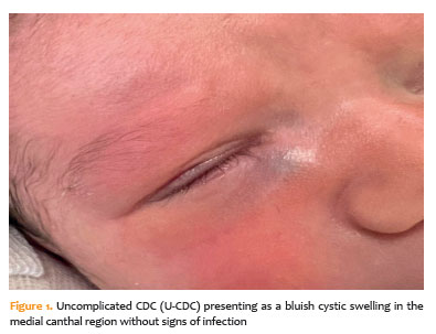
Congenital dacryocystocele (CDC) is a rare congenital anomaly, accounting for only 0.1%-0.3% of all cases of congenital nasolacrimal duct obstruction. It is distinguished by cystic dilation of the lacrimal sac caused by both proximal (Rosenmüller valve) and distal (Hasner valve) obstructions of the tear drainage pathway(1). It typically appears within the first 12 weeks of life as a bluish, cystic swelling in the medial canthal region. It is frequently accompanied by epiphora and mucopurulent discharge during the neonatal period(2,3). The primary complications reported in CDC are acute nasal extension of the cyst, dacryocystitis, and periorbital cellulitis(4,5). However, despite a well-established understanding of its clinical presentation and complications, the CDC has limited data on the factors associated with dacryocystitis and the need for surgical intervention. The purpose of this study was to assess the clinical factors linked to the development of dacryocystitis and the subsequent need for surgical intervention in infants with CDC.
METHODS
This retrospective study examined infants diagnosed with CDC between January 2023 and June 2025 at the Ophthalmology Departments of Başakşehir Çam and Sakura City Hospital in Istanbul, Turkey. Patients were divided into two groups based on their clinical presentation: U-CDC describes CDC cases with no signs of infection (Figure 1), and C-CDC describes complicated cases with dacryocystitis and/or preseptal cellulitis (Figure 2). To assess factors associated with C-CDC and the need for surgical intervention, demographic and perinatal variables such as age at diagnosis, gender, gestational age, birth weight, mode of delivery, laterality, and associated anomalies were compared between the groups.
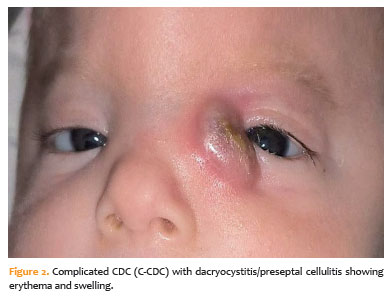
Infants with U-CDC were initially treated conservatively, with regular lacrimal sac massage and topical and/or systemic antibiotic therapy. If the cyst did not resolve or symptoms persisted, a surgical intervention involving probing and marsupialization was performed under general anesthesia.
Infants with C-CDC were given intravenous antibiotics when they were diagnosed. Following clinical improvement and control of the acute infection, surgical intervention (probing with intraoperative nasal endoscopy and endonasal marsupialization) was performed within 48 h. Two oculoplastic surgeons performed all of the surgical procedures.
The study was conducted in accordance with the Declaration of Helsinki and approved by the Institutional Ethics Committee of Basaksehir Çam and Sakura City Hospital (Approval No. 299). The parents or legal guardians of all infants in the study provided written informed consent for participation and the use of medical records.
Statistical analysis
Descriptive statistics for the data included mean, standard deviation, median, minimum, maximum, frequency, and percentage values. The distribution of variables was evaluated using the Kolmogorov-Smirnov and Shapiro-Wilk tests. For quantitative independent variables with a normal distribution, an independent sample t-test was used. The Mann-Whitney U test was applied to quantitative independent variables that did not follow a normal distribution. The chi-square test was used for categorical independent variables, while Fisher’s exact test was used when the chi-square test assumptions were not met. All analyses were carried out using SPSS software (version 28.0).
Procedure
All patients were examined and treated by two oculoplastic surgeons. For infected infants, the family was instructed on the Crigler technique and advised to perform lacrimal sac massage four times daily. Topical moxifloxacin 0.5% eye drops (Vigamox, Alcon, USA) were used for one week, and mucopurulent discharge was treated with oral amoxicillin-clavulanate suspension (Augmentin, GlaxoSmithKline, UK). If the cyst did not heal within 2 weeks, probing and marsupialization were carried out under general anesthesia.
Following diagnosis of dacryocystitis, infants received intravenous ampicillin-sulbactam (Ampisid, Pfizer, Turkey). After clinical improvement and resolution of acute inflammation, probing and marsupialization were performed within 48 h.
All surgically treated cases underwent intraoperative nasal endoscopy to evaluate the inferior meatus for an intranasal cyst, followed by endonasal marsupialization of the intranasal cystic component in the same session. During the procedure, a No. 0 or No. 1 stainless steel Bowman probe (Storz, Germany) was carefully inserted into the nasolacrimal duct via the superior or inferior punctum. Saline irrigation with free flow into the nasal cavity was used under endoscopic visualization to confirm the nasolacrimal system’s patency. The intranasal cystic component of the dacryocystocele was visualized endoscopically under direct illumination to aid in marsupialization. The cyst’s medial wall was incised longitudinally, and a portion was removed to allow for wide and permanent communication with the nasal cavity, adequate drainage, and prevention of recurrence.
Antibiotics were administered systemically and locally for 10 days after surgery. All patients were followed up at 1 day, 1 week, 1 month, 3 months, and 6 months after surgery to assess symptom resolution and monitor for recurrence or other complications.
RESULTS
This study included 26 infants diagnosed with CDC. The average age at diagnosis was 70.3 ± 123.7 days (median 29; range 1-470 days). Of these, 17 (65.4%) were women, and nine (34.6%) were men. The mean gestational age was 38.4 ± 1.2 weeks, and the average birth weight was 3,096 ± 312. Fifteen infants (57.7%) were delivered via cesarean section, while 11 (42.3%) were delivered vaginally. The lesion was unilateral in 25 (96.2%) of the patients and bilateral in one patient (3.8%; Table 1).
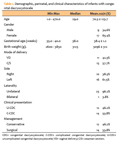
At presentation, 12 infants (46.2%) had U-CDC, while 14 (53.8%) had C-CDC, which included dacryocystitis or preseptal cellulitis. There were no significant differences between the two groups in terms of age at diagnosis, gender distribution, gestational age, mode of delivery, or laterality (p>0.05 for all; see Table 2).
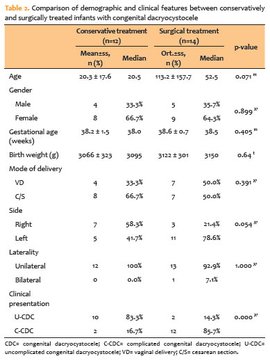
All 14 (100%) infants with C-CDC required surgical intervention, while only 2 of the 12 (16.7%) infants in the U-CDC group did (p<0.001; Table 2). Intraoperative nasal endoscopy revealed an intranasal cystic component in all 14 surgically treated C-CDC infants. Following treatment, all patients made full clinical recoveries. Anatomical success was defined as patent irrigation with free flow into the nasal cavity, while functional success was defined as complete resolution of swelling/discharge with no recurrence at follow-up. There were no intraoperative or postoperative complications, such as bleeding, mucosal injury, or recurrence, during a 6-month average follow-up period.
DISCUSSION
CDC is a rare type of congenital nasolacrimal duct obstruction that can cause a variety of clinical symptoms, ranging from simple asymptomatic bluish swelling in the medial canthal region to secondary dacryocystitis, preseptal cellulitis, and even respiratory distress in neonates due to intranasal cyst extension(3). This condition is caused by the simultaneous obstruction of both the proximal (Rosenmüller valve) and distal (Hasner valve) portions of the nasolacrimal drainage system, resulting in cystic dilation of the lacrimal sac and the accumulation of mucus or amniotic fluid(6).
Female predominance was observed in our study, with 17 out of 26 infants (65.4%) being female. Similar to previous reports indicating a higher incidence of CDC in females, our study found a significant female predominance. This has been attributed to anatomical differences, such as a narrower nasolacrimal duct and a more acute angle between the canal and nasal floor, which may increase tear drainage resistance and increase the risk of obstruction(7,8).
The optimal treatment for CDC remains controversial, as management strategies vary according to the presence of infection, intranasal extension, or respiratory failure(8,9). While some cases resolve spontaneously or with conservative measures such as lacrimal sac massage and topical antibiotics, if complications such as infection and respiratory distress develop, prompt surgical intervention, typically involving catheterization with marsupialization, may be required(6,8). Congenital dacryocystocele is caused by an obstruction of the lacrimal drainage pathway, resulting in tear stasis and the accumulation of mucus or amniotic fluid within the sac. This stagnant environment promotes bacterial proliferation, which can result in dacryocystitis. Once infected, local inflammation can quickly progress to preseptal cellulitis or abscess formation, emphasizing the importance of early diagnosis and prompt treatment to avoid complications(9-12). In this study, 53.8% of infants had dacryocystitis associated with CDC whereas previous studies found infection rates ranging from 18% to 75%(3-5). The relatively high proportion of dacryocystitis cases in our series could be explained by our institution’s role as a tertiary referral center, where patients are frequently admitted after infection has developed. Notably, in our cohort, most infants developed dacryocystitis at admission rather than during follow-up, implying that many congenital dacryocystocele cases go undetected until infection occurs. This finding emphasizes the importance of early clinical recognition and management to avoid infectious complications.
In our study, patients with dacryocystitis required surgical treatment. Following adequate infection control, probing and endonasal marsupialization were performed, resulting in complete clinical recovery in all cases, with no intraoperative or postoperative complications. Early surgical management after acute inflammation resolution was found to be safe and effective in restoring lacrimal drainage patency, relieving symptoms quickly, and preventing recurrence(13-15).
Infants who presented with only a dacryocystocele received regular lacrimal sac massage and antibiotic treatment. In the majority of these cases, the cyst resolved within 2 weeks without the development of dacryocystitis, and surgery was postponed if symptoms persisted or did not recur during follow-up(3,4,6). Previous research has shown that early lacrimal sac massage, when combined with topical or systemic antibiotic therapy, promotes the nasolacrimal system by relieving distal obstruction and reducing tear stasis. This conservative approach promotes spontaneous recovery while lowering the risk of secondary infection(12,14,16). In our study, most infants with U-CDC responded well to conservative treatment, demonstrating the efficacy of early massage and antibiotic therapy. Despite regular massage, two infants (16.7%) developed dacryocystitis, highlighting the risk of conservative treatment failing even when appropriate, as well as the importance of close follow-up. Our findings are consistent with those of these studies, which show that delayed or inadequate conservative treatment can result in secondary bacterial infections that necessitate surgical intervention(3,8,13,15,17). This delayed presentation indicates a lack of early diagnosis and medical evaluation. These findings further support previous reports that infants who received timely lacrimal sac massage and antibiotic therapy did not develop dacryocystitis, suggesting that early conservative management is effective(14,16). As a result, timely recognition of dacryocystoceles by pediatricians and caregivers is critical for initiating early treatment, preventing progression to acute infection, and reducing the need for emergency surgery.
Our findings are consistent with previous literature regarding the incidence and treatment outcomes of CDC and provide additional clinical insights. In contrast to most prior studies, which predominantly included infants diagnosed with U-CDC(3,4,10), our cohort mainly consisted of patients presenting after the onset of infection, reflecting a more advanced disease stage. This distinction emphasizes the clinical importance of early diagnosis and referral, as delayed presentation and/or delayed initiation of conservative treatment may result in secondary infection and an increased likelihood of surgical intervention. These findings suggest that starting conservative treatment before infection develops may help reduce the risk of complications and the need for urgent surgery. In addition to infection-related morbidity, functional nasal obstruction should be considered in certain infants. Because newborns breathe primarily through their noses, bilateral CDC and/or a prominent intranasal cystic component may increase the risk of nasal obstruction(3-5). As a result, an early evaluation, including a nasal examination and multidisciplinary assessment if clinically indicated, may be critical for timely treatment and follow-up.
However, intranasal findings were only available for surgically treated infants because nasal endoscopy was performed intraoperatively in this subgroup; thus, we were unable to assess nasal cyst presence as an independent risk factor for infection across the entire cohort. Furthermore, because endoscopic images were not routinely archived in our retrospective dataset, representative intraoperative endoscopic photographs could not be included.
CDC’ s are typically isolated anomalies not associated with syndromic conditions. Rare conditions reported in the literature include choanal atresia (in three cases), cleft palate, hydrocele, gastroschisis, and sphenoidal meningocele(5,10,18,19). There were no associated congenital anomalies in any of the infants studied.
According to the literature, the lack of significant differences between the groups in terms of gestational age, birth weight, delivery mode, sex, laterality, and prenatal diagnosis indicates that perinatal and demographic factors are unlikely to influence infection development(17,19,20). This finding implies that dacryocystitis is more likely to be influenced by postnatal factors, particularly the absence or delay of conservative management as well as other factors that are not yet fully understood than by inherent perinatal characteristics.
Overall, our findings indicate that infectious presentation is strongly associated with the need for surgical intervention in congenital dacryocystocele. Given the retrospective design, causal relationships cannot be inferred; however, early detection by pediatricians and primary care providers, prompt initiation of conservative treatment, and close follow-up may help reduce the risk of infection and related complications. When infection is present or symptoms persist, our series shows that probing with intraoperative nasal endoscopy and endonasal marsupialization after infection control is both safe and effective. Larger prospective multicenter studies are required to confirm these findings and improve management strategies.
Finally, in this retrospective series, initial conservative management was effective in most uncomplicated cases, whereas complicated or persistent cases required surgical intervention after infection control. All patients ultimately achieved complete clinical recovery without complications. These findings support early clinical evaluation and structured follow-up to guide treatment escalation as needed. Prospective multicenter studies are needed to improve management algorithms and identify predictors of infection and surgical need.
AUTHORS’ CONTRIBUTIONS:
Significant contribution to conception and design: Gülistan Oyur. Data Acquisition: Muhammed Mustafa Mazlum. Data Analysis and Interpretation: Gülistan Oyur. Manuscript Drafting: Gülistan Oyur. Significant Intellectual Content Revision of the Manuscript: Gülistan Oyur, Fatma Savur. Final Approval of the Submitted Manuscript: Gülistan Oyur, Fatma Savur, Muhammed Mustafa Mazlum. Statistical Analysis: Gülistan Oyur. Obtaining Funding: not applicable. Supervision of Administrative, Technical, or Material Support: not applicable. Research Group Leadership: Gülistan Oyur.
REFERENCES
1. MacEwen CJ, Young JD. Epiphora during the first year of life. Eye (Lond). 1991;5(Pt 5):596-600.
2. Cavazza S, Laffi GL, Lodi L, Tassinari G, Dall’Olio D. Congenital dacryocystocele: diagnosis and treatment. Acta Otorhinolaryngol Ital. 2008;28(6):298-301.
3. Davies R, Watkins WJ, Kotecha S, et al. Presentation, clinical features, complications, and treatment of congenital dacryocystocele. Eye. 2018;32:522-526.
4. Wong RK, VanderVeen DK. Presentation and management of congenital dacryocystocele. Pediatrics. 2008;122(5):e1108-12.
5. Paysse EA, Coats DK, Bernstein JM, Go C, de Jong AL. Management and complications of congenital dacryocele with concurrent intranasal mucocele. J AAPOS. 2000;4(1):46-53.
6. Ahmad TR, Lindquist KJ, Oatts JT, de Alba Campomanes AG, Kersten RC, Chan DK, et al. Probing versus primary nasal endoscopy for the treatment of congenital dacryocystoceles. J AAPOS. 2024;28(2):103865.
7. Shigeta K, Takegoshi H, Kikuchi S. Sex and age differences in the bony nasolacrimal canal: an anatomical study. Arch Ophthalmol. 2007;125(12):1677-81.
8. Singh S, Ali MJ. Congenital dacryocystocele: A major review. Ophthalmic Plast Reconstr Surg. 2019;35(4):309-317.
9. Hulka GF, Kulwin DR, Weeks SM, Cotton RT. Congenital lacrimal sac mucoceles with intranasal extension. Otolaryngol Head Neck Surg. 1995;113(5):651-5.
10. Mansour AM, Cheng KP, Mumma JV, Stager DR, Harris GJ, Patrinely JR, et al. Congenital dacryocele. A collaborative review. Ophthalmology. 1991;98(11):1744-51.
11. Schnall BM, Christian CJ. Conservative treatment of congenital dacryocele. J Pediatr Ophthalmol Strabismus. 1996;33(5):219-22.
12. Pur DR, Husein M, Makar I. Management of congenital dacryocystocele: a case series and literature review. J Pediatr Ophthalmol Strabismus. 2023; 60(3):e31-4.
13. Dogan E, Yüksel NG, Ecevit MC, Yaman A, Berk AT, Sütay S. Microdebrider assisted endoscopic marsupialization of congenital intranasal nasolacrimal duct cysts. Int J Pediatr Otorhinolaryngol. 2012;76(4):488-91.
14. Levy NS. Conservative management of congenital amniotocele of the nasolacriminal sac. J Pediatr Ophthalmol Strabismus. 1979;16(4):254-6.
15. Ali MJ, Singh S, Naik MN. Long-term outcomes of cruciate marsupialization of intra-nasal cysts in patients with congenital dacryocele. Int J Pediatr Otorhinolaryngol. 2016;86:34-6.
16. Mimura M, Ueki M, Oku H, Sato B, Ikeda T. Process of spontaneous resolution in the conservative management of congenital dacryocystocele. Clin Ophthalmol. 2014;8:465-9.
17. Ali MJ, Baig F, Lakshman M, et al. Simple versus complex congenital nasolacrimal duct obstructions: etiology, management, and outcomes. Int Forum Allergy Rhinol. 2015;5(6):598-601.
18. Kim YH, Lee YJ, Song MJ, Han BH, Lee YH, Lee KS. Dacryocystocele on prenatal ultrasonography: diagnosis and postnatal outcomes. Ultrasonography. 2015;34(1):51-7.
19. Sullivan TJ, Clarke MP, Morin JD, Pashby RC. Management of congenital dacryocystocoele. Aust N Z J Ophthalmol. 1992;20(2):105-8.
20. Dagi LR, Bhargava A, Melvin P, Prabhu SP. Associated signs, demographic characteristics, and management of dacryocystocele in 64 infants. J AAPOS. 2012;16(3):255-60.
Submitted for publication:
December 10, 2025.
Accepted for publication:
April 8, 2026.
Approved by the following research ethics committee: Başakşehir Çam and Sakura City Hospital (Approval No. 299, 24.09.2025).
Research Data Availability: The datasets generated and/or analyzed during the current study are included in manuscript.
Edited by
Editor-in-Chief: Newton Kara-Júnior
Associate Editor: Dácio C. Costa
Funding: This study received no specific financial support.
Disclosure of potential conflicts of interest: The authors declare no potential conflicts of interest.

How to cite this article:
 ABO is licensed under a Creative Commons Attribution-NonComercial 4.0 Internacional.

