Arq. Bras. Oftalmol. 2022;85 (5 )
:465-471
| DOI: 10.5935/0004-2749.20220067
Abstract
Objetivos: Relatar a distribuição dos motivos de encaminhamento de crianças para ambulatório de glaucoma infantil em um serviço oftalmológico terciário.
Métodos: Dados médicos de pacientes menores que 18 anos encaminhados para ambulatório de glaucoma infantil na cidade de São Paulo, Brasil, de 2012 a 2018 foram retrospectivamente analisados. Os dados incluíram os motivos de encaminhamento, a idade, a origem e quem detectou a alteração ocular. Para definição diagnóstica, a classificação do Childhood Glaucoma Research Network foi usada.
Resultados: 563 olhos de 328 pacientes foram incluídos no estudo. O diagnóstico de glaucoma foi confirmado em 162 crianças (49%). 83 (25%) pacientes tiveram diagnóstico de glaucoma descartado, e 83 (25%) continuaram em acompanhamento como glaucoma suspeito. Os principais motivos de encaminhamento foram relação escavação-disco >0,5 ou assimetria ≥0,2 (24%), pressão intraocular >21 mmHg (21%) e opacidade corneana (15%). No período neonatal, os motivos de encaminhamento foram opacidade corneana, buftalmo, lacrimejamento e fotofobia. Após o período neonatal, além desses sinais externos, outros sinais foram também motivos de encaminhamento, como escavação-disco >0,5 ou assimetria ≥0,2, pressão intraocular >21 mmHg e miopização. Os encaminhamentos ocorreram por profissionais de saúde em 69% e preocupação dos pais em 30%. Os pais foram os primeiros a identificar as alterações e procurar cuidado médico em 97% dos casos de glaucoma congênito primário.
Conclusões: Os motivos de encaminhamento de crianças para um serviço de glaucoma de glaucoma terciário foram determinados e diferem em diferentes faixas etárias e grupos. Os autores reforçam a necessidade de alertar diferentes grupos para os sinais mais comuns, a fim de evitar, não só o adiamento do diagnóstico, o que impacta no prognóstico, mas também despender recursos importantes direcionados ao enfrentamento dessas doenças, com encaminhamentos imprecisos.
Keywords: Glaucoma/congênito; Glaucoma/fisiopatologia; Opacidade da córnea; Criança; Acuidade visual; Encaminhamento e consulta; Serviços de saúde ocular
Arq. Bras. Oftalmol. 2021;84 (5 )
:454-461
| DOI: 10.5935/0004-2749.20210071
Abstract
Objetivo: Comparar a estrutura da córnea e as alterações morfológicas endoteliais após cirurgia de catarata por facoemulsificação sem intercorrências entre pacientes com diabetes mellitus tipo 2 e não diabéticos; e determinar quais fatores pré e intra-operatórios relacionados com a maior redução da densidade celular endotelial.
Métodos: Quarenta e cinco diabéticos (45 olhos) e 43 (43 olhos) controlos com catarata relacionada à idade foram incluídos neste estudo observacional prospectivo. Os parâmetros da córnea (espessura e volume) e do segmento anterior foram medidos pela tomografia Scheimpflug; a densidade e morfologia celular endotelial (coeficiente de variação do tamanho das células, células hexagonais) foram registrados usando microscopia especular não contato. Os pacientes foram avaliados no pré-operatório, 1 e 6 meses após a cirurgia. Foi realizada uma análise de regressão linear uni e multivariada para avaliar a relação entre os parâmetros demográficos, clínicos, oculares e intra-operatórios com a redução da densidade celular endotelial aos 6 meses.
Resultados: Nos dois grupos houve uma perda significativa de células endoteliais ao 1º mês pós-operatório (p<0,001), que permaneceu estável até ao 6º mês; sem diferenças estatisticas entre os grupos diabetes mellitus e não diabetes mellitus em qualquer avaliação. A espessura média da córnea no pós-operatório central aos 1 e 6 meses não mudou significativamente em relação ao valor médio pré-operatório nos dois grupos (p>0.05). A análise de regressão multivariada linear mostrou que a idade avançada (p=0.042) e os graus mais elevados de catarata (p=0.001) foram significativamente associados à maior redução densidade celular endotelial aos 6 meses de seguimento.
Conclusão: Este estudo mostrou que a idade avançada e as cataratas mais densas podem predispor a uma maior redução densidade celular endotelial após a cirurgia de catarata. Outros fatores, como diabetes mellitus e parâmetros pré-operatórios do segmento anterior, não influenciaram significativamente as alterações pós-operatórias da densidade celular endotelial.
Keywords: Catarata; Facoemulsificação; Diabetes mellitus tipo 2; Retinopatia diabética; Epitélio posterior; Paquimetria corneana; Perda de células endoteliais da córnea
Arq. Bras. Oftalmol. 2023;86 (4 )
:353-358
| DOI: 10.5935/0004-2749.20230055
Abstract
Objetivo: Examinar a eficácia da ceratectomia fototerapêutica para o tratamento de patologias variáveis que apresentarem opacidades anteriores da córnea, e avaliar a distribuição das indicações de ceratectomia fototerapêutica nos últimos 10 anos.
Métodos: Foram revisados retrospectivamente os prontuários de 276 pacientes, com 334 olhos tratados com ceratectomia fototerapêutica entre março de 2010 e o ano de 2020. As etiologias dos pacientes submetidos à ceratectomia fototerapêutica foram anotadas e suas alterações foram examinadas. Os resultados refrativos e de acuidade visual antes e após a operação foram registrados e analisados de acordo com a etiologia.
Resultados: A idade média dos pacientes foi de 40,7 ± 16,2 anos (faixa: 19-84). O tempo médio de acompanhamento foi de 25,5 ± 19,1 meses (faixa: 3-96). A ceratectomia fototerapêutica foi aplicada com mais frequência para distrofias estromais corneanas (44%, 151 olhos de 111 pacientes); entre as distrofias corneanas como um todo, a distrofia granular foi a indicação terapêutica mais comum desse procedimento. Ao contrário de outras indicações, nos últimos 10 anos houve um aumento na aplicação de ceratectomia fototerapêutica em casos de opacidade subepitelial persistente causada por conjuntivite adenoviral. Houve um aumento significativo na acuidade visual em todos os grupos, exceto para o grupo com defeito epitelial recorrente (p<0,05). A maior melhora na acuidade visual foi detectada em casos de distrofia estromal, no subgrupo das distrofias granulares.
Conclusão: Apesar das mudanças nas tendências de indicação, a ceratectomia fototerapêutica continua sendo uma abordagem terapêutica eficaz e confiável para tratar lesões da córnea anterior.
Keywords: Ceratectomia fotorrefrativa; Opacidade da córnea; Lesões da córnea; Distrofias da córnea; Fototerapia.
Arq. Bras. Oftalmol. 2026;89 (2 )
:1-5
| DOI: 10.5935/0004-2749.2025-0208
Abstract
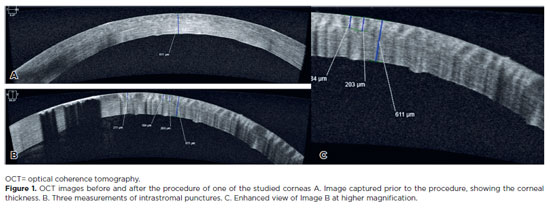
PURPOSE: To assess the reliability and penetration depth of an automated micropuncture system using a tattoo machine.
METHODS: Twenty human corneas were obtained and subjected to intrastromal micropuncture using a tattoo machine. Each cornea was divided into two halves: one received pigment, while the other received saline solution as a control. The Cheyenne tattoo machine was operated at 60 Hz, with standardized needle exposure (six passes per application). The machine used cartridges containing five microneedles. The study was registered with Agência Nacional de Vigilância Sanitária ANVISA (numbers 80281110015, 80281110016, and 80281110019). The pigment used was Electric Ink black ink, with a density of 1,271,460 μg/mL. Puncture depth was measured before and after the procedure using both anterior segment optical coherence tomography and histopathological analysis. Puncture depth measurements were analyzed using ImageJ software. Each cornea was measured thrice, and the results were subsequently compared.
RESULTS: No corneal perforations were observed with the use of the tattoo machine, and puncture depth measurements ranged from 107 to 486 µm.
CONCLUSIONS: The use of a tattoo machine represents a viable and accessible approach for keratopigmentation, with potential for both cosmetic and therapeutic applications. Its adaptation for controlled intrastromal drug delivery may enable the targeted treatment of deep infectious keratitis, corneal neovascularization, and stromal inflammatory disorders, representing a promising approach for corneal stromal diseases. Further research is needed to optimize techniques and evaluate long-term safety and efficacy, particularly for the delivery of antimicrobial, anti-inflammatory, and anti-vascular endothelial growth factor agents.
Keywords: Eye banks; Cadaver; Cornea; Corneal stroma; Drug delivery systems; Tissue donors; Tattooing/instrumentation; Punctures
Arq. Bras. Oftalmol. 2025;88 (6 )
:1-7
| DOI: 10.5935/0004-2749.2025-0120
Abstract
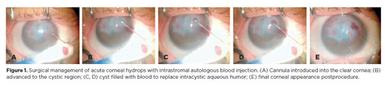
PURPOSE: To describe the technique and outcomes of intrastromal autologous blood injection in patients with severe corneal hydrops.
METHODS: Nineteen patients with corneal hydrops underwent intrastromal autologous blood injection. Postoperative assessments included best-corrected visual acuity and time to resolution of corneal edema
RESULTS: Corneal edema resolved within 1 week in 5 patients, within 1 month in 11, and within 3 months in 3. The mean duration of edema persistence was 37.94 ± 33.05 days (range, 6–124). Corneal thickness decreased from 2.06 ± 0.71-mm preoperatively to 1.34 ± 0.65-mm at day 7, 0.85 ± 0.56-mm at day 30, and 0.57 ± 0.13-mm at day 90 (p<0.001). Descemet’s membrane (DM) detachment decreased from 1.01 ± 0.75-mm to 0.44 ± 0.57-mm, 0.24 ± 0.36-mm, and 0.08 ± 0.11-mm on postoperative days 7, 30, and 90, respectively (p<0.001). DM break size decreased from 1.12 ± 1.19-mm to 0.62 ± 0.84-mm at 3 months (p<0.005). Three patients developed hematocornea; no other major complications were observed. At 3 months, mean best-corrected visual acuity improved from 2.37 ± 0.66 to 0.41 ± 0.17 logMAR with hard contact lenses (p<0.001).
CONCLUSIONS: Intrastromal autologous blood injection is an effective treatment for severe corneal hydrops, promoting faster edema resolution and visual improvement with minimal complications.
Keywords: Corneal edema; Corneal diseases; Edema; Visual acuity; keratoconus.
Arq. Bras. Oftalmol. 2025;88 (5 )
:1-7
| DOI: 10.5935/0004-2749.2024-0217
Abstract
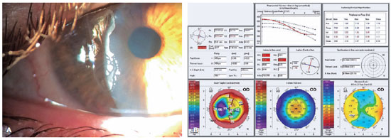
PURPOSE: This study aimed to evaluate the influence of intrastromal corneal ring segment implants on the intraocular pressure measurements using Goldmann applanation tonometry, rebound tonometry, and noncontact tonometry in keratoconic corneas and analyze the intertonometer agreement.
METHODS: We enrolled 74 eyes in this observational and prospective study. Each participant had a complete eye examination, corneal analysis with Scheimpflug Tomography (Pentacam®), and intraocular pressure evaluation with Goldmann applanation tonometry, rebound tonometry, and noncontact tonometry, before and after intrastromal corneal ring segment implantation (on postoperative days 1, 7, 45, and 90). Intertonometer agreement was assessed using Bland-Altman analysis.
RESULTS: The mean age was 29.9 ± 10.2 years, and 47 (63.5%) eyes had keratoconus grade II. Intraocular pressures were higher for noncontact tonometry preoperatively and on 90 postoperative day (mean ± SD: 12.4 ± and 12.1 ± 2.2 mmHg, respectively), followed by Goldmann applanation tonometry (11.1 ± 3.0 and 11.2 ± 2.7 mmHg, respectively), and were lower for rebound tonometry (9.7 ± and 9.4 ± 3.2 mmHg, respectively). The variation from the Goldmann tonometry on 7 postoperative day to the baseline (p=0.022) and that of noncontact tonometry on 90 postoperative day to the baseline (p=0.021) were statistically significant. The rebound tonometry underestimated intraocular pressure when compared with the Goldmann applanation tonometry by a mean of 1.47 ± 5.19 mmHg. Noncontact tonometry, when compared with Goldmann applanation tonometry, overesti-mated intraocular pressure by a mean of 1.23 ± 4.15 mmHg.
CONCLUSION: Despite statistically significant differences between some postoperative periods, the intraocular pressure measurement differences may not be clinically relevant.
Keywords: Keratoconus; Intraocular pressure; Cornea; Corneal stroma; Postoperative period; Tonometry ocular; Prostheses and implants
Arq. Bras. Oftalmol. 2024;87 (4 )
:1-6
| DOI: 10.5935/0004-2749.2022-0128
Abstract
Objetivo: Relatar um experimento projetado para determinar alterações anatômicas em córneas porcinas após a colocação de um novo implante depolímero na córnea.
Métodos: Foi utilizado olho de porco ex vivo. Um novo agente modelador biocompatível, de colágeno tipo 1, com 6mm de diâmetro foi moldado com excimer laser em sua face posterior, para criar três formatos planocôncavos. Os implantes foram inseridos dentro de um bolsão, dissecado manualmente, a 200 micrômetros (µm). Foram definidos três grupos de tratamento: grupo A (n=3), teve a profundidade máxima de ablação de70 µm; o grupo B (n=3), profundidade máxima de ablação de 64 µm; e o grupo C (n=3), profundidade máxima de ablação de 104 µm, com buraco central. O grupo controle, D (n=3), foi incluído, com a criação do bolsão estromal, porém sem inserir o material. A avaliação desses olhos foi realizada por tomografia de coerência óptica (OCT) e por tomografia corneana.
Resultados: A tomografia corneana mostrou uma tendência para diminuição da ceratometria média em todos os 4 grupos. A tomografia de coerência óptica mostrou córneas com implantes localizados no estroma anterior e aplanamento visível, enquanto as córneas não mudaram qualitativamente o formato no grupo controle.
Conclusões: O novo implante de biomaterial planocôncavo descrito aqui foi capaz de remodelar a córnea em modelo de animal ex vivo, resultando no aplanamento corneano. Novos estudos são necessários usando modelos animais in vivo para confirmar tais achados.
Keywords: Córnea; Cirurgia da córnea a laser; Substância própria; Proteses e implantes; Lasers de excimer; Materiais biocompatíveis; Animais; Suínos
Arq. Bras. Oftalmol. 2024;87 (3 )
:1-7
| DOI: 10.5935/0004-2749.2023-0049
Abstract
PURPOSE: To investigate the association of pre-photorefractive keratectomy Schirmer-1 test value with post-photorefractive keratectomy central corneal epithelial thickness, ocular surface disease index score, and uncorrected distance visual acuity.
METHODS: Patients were categorized according to preoperative Schirmer-1 value: the normal Schirmer Group (n=54; Schirmer-1 test value, >10 mm) and the low Schirmer Group (n=52; Schirmer-1 test value, between 6 and 10 mm). We analyzed ablation depth, visual acuity, result of Schirmer-1 test (with anesthesia), tear film break-up time, ocular surface disease index score, central corneal epithelial thickness, and spherical equivalent refraction.
RESULTS: We found significant differences between the groups in Schirmer-1 test value, tear film break-up time, and ocular surface disease index score, both preoperatively and postoperatively (p<0.001). The preoperative central corneal epithelial thicknesses of the two groups were similar (p>0.05). After photorefractive keratectomy, the Schirmer-1 test value and spherical equivalent refraction decreased in both groups (p<0.05), and ocular surface disease index scores and central corneal epithelial thickness values increased in the low Schirmer Group (p<0.001) but not in the normal Schirmer Group (p>0.05). The postoperative central corneal epithelial thicknesses of the low Schirmer Group were significantly higher than those of the normal Schirmer Group (p<0.001). Postoperative uncorrected distance visual acuity did not differ significantly between the two groups (p>0.05).
CONCLUSIONS: In patients with low Schirmer-1 test values before photorefractive keratectomy, the corneal epithelium thickened and ocular surface complaints increased during the postoperative period. However, changes in the corneal epithelium did not affect the postoperative uncorrected distance visual acuity. To reduce postoperative problems on the ocular surface in these patients, we recommend that dry eye be treated before photorefractive keratectomy.
Keywords: Epithelium, corneal; Cornea; Photorefractive keratectomy; Schirmer test; Visual acuity
Arq. Bras. Oftalmol. 2024;87 (3 )
:1-8
| DOI: 10.5935/0004-2749.2023-0109
Abstract
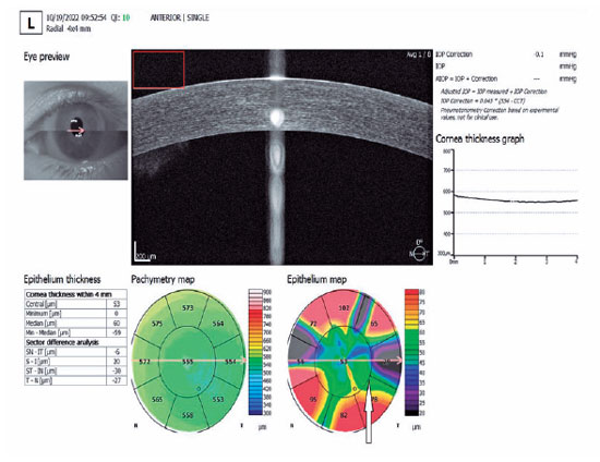
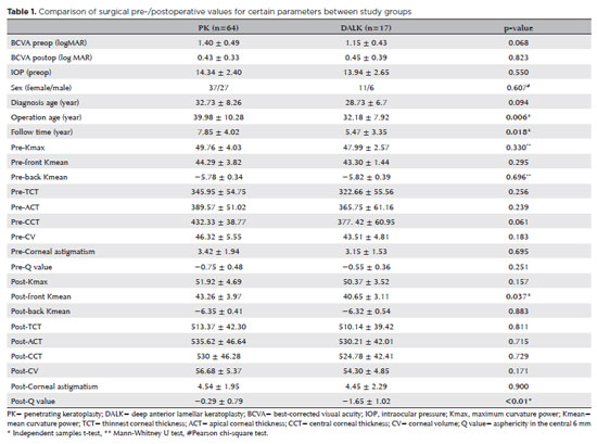
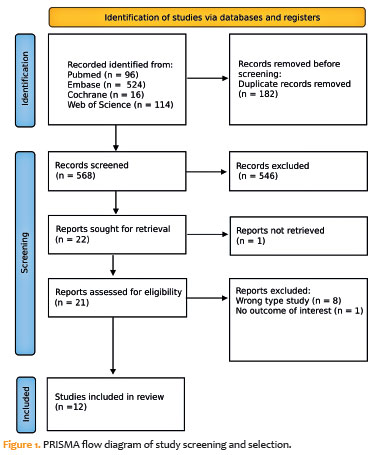
PURPOSES: This study aims to assess and compare the postoperative visual and topographic outcomes, complications, and graft survival rates following deep anterior lamellar keratoplasty and penetrating keratoplasty in patients with macular corneal dystrophy.
METHODS: In this study we enrolled 59 patients (23 male; and 36 female) with macular corneal dystrophy comprising 81 eyes. Out of these, 64 eyes underwent penetrating keratoplasty, while 17 eyes underwent deep anterior lamellar keratoplasty. The two groups were analyzed and compared based on best-corrected visual acuity, corneal tomography parameters, pachymetry, complication rates, and graft survival rates.
RESULTS: After 12 months, 70.6% of the patients who underwent deep anterior lamellar keratoplasty (DALK) and 75% of those who had penetrating keratoplasty (PK) achieved a best-corrected visual acuity of 20/40 or better (p=0.712). Following surgery, DALK group showed lower front Kmean (p=0.037), and Q values (p<0.01) compared to the PK group. Postoperative interface opacity was observed in seven eyes (41.2%) in the DALK group. Other topography values and other complications (graft rejection, graft failure, cataract, glaucoma, microbial keratitis, optic atrophy) did not show significant differences between the two groups. The need for regrafting was 9.4% and 11.8% in the PK and DALK groups, respectively (p=0.769). Graft survival rates were 87.5% and 88.2% for PK and DALK; respectively (p=0.88 by Log-rank test).
CONCLUSION: Both PK and DALK are equally effective in treating macular corneal dystrophy, showing similar visual, topographic, and survival outcomes. Although interface opacity occurs more frequently after DALK the visual results were comparable in both groups. Therefore, DALK emerges as a viable surgical choice for patients with macular corneal dystrophy without Descemet membrane involvement is absent.
Keywords: Macular corneal dystrophy; Corneal dystrophies; Hereditary; Keratoplasty; Penetrating; Corneal transplantation
 ABO is licensed under a Creative Commons Attribution-NonComercial 4.0 Internacional.
14-fig01tb.jpg)
06-tab01tb.jpg)
06-tab01.jpg)

09-fig01.jpg)
09-fig01.jpg)