Arq. Bras. Oftalmol. 2022;85 (1 )
:13-18
| DOI: 10.5935/0004-2749.20220003
Abstract
Objetivo: Fornecer informações sobre a ocorrência e a eficácia do aconselhamento sobre o uso de tabaco por oftalmologistas a pacientes com doenças oculares associadas à tireoide.
Métodos: Analisamos os prontuários médicos eletrônicos de uma coorte digital de pacientes atendidos por oftalmologistas no Sistema de Saúde da Universidade da Pensilvânia entre o início de 2012 e o final de 2017 com os códigos da Classificação Internacional de Doenças (CID) para a doença de Graves, exoftalmia tireotóxica ou doença ocular associada à tireoide. Os históricos de uso de tabaco foram registrados na primeira e na última visita ao consultório de Oftalmologia, ou na visita mais próxima no tempo. A quantidade de maços/dia (mpd) e todas as anotações feitas nas visitas ao consultório de Oftalmologia foram analisadas para aconselhamento sobre o uso de tabaco.
Resultados: Um total de 435 indivíduos preencheram os critérios de inclusão, dos quais 72 (16,6%) estavam fumando ativamente no momento do primeiro encontro. Apenas 57 (79,2%) desses indivíduos que fumam ativamente registraram queixas relacionadas ao tabagismo, sendo que 34 (59,6%) deles receberam alguma forma de aconselhamento sobre o uso de tabaco. Ao todo, 9 (26,5%) indivíduos dentre os que receberam aconselhamento sobre tabaco e 1 (4,3%) que não teve aconselhamento registrado pararam de fumar (diferença de risco de 22,1%; IC 95%, [1,7%, 39,1%]; p=0,04). Dentre aqueles que receberam aconselhamento, 17 (50,0%) reduziram seus mpd, além de 7 (30,4%) daqueles que não tiveram aconselhamento (diferença de risco de 19,6%; IC 95% [-6,3%, 41,3%]; p=0,18). No geral, 14 (25,5%) dos 55 oftalmologistas que tiveram um paciente fumante ativo registraram evidências de aconselhamento sobre o uso de tabaco.
Conclusões: Os resultados deste estudo revelam tanto as oportunidades perdidas de aconselhamento sobre o uso do tabaco quanto a eficácia do aconselhamento no contexto de doenças oculares associadas à tireoide.
Keywords: Uso de tabaco; Aconselhamento; Doenças da glândula tireóide; Doença de Graves; Oftalmopatias
Arq. Bras. Oftalmol. 2021;84 (3 )
:220-224
| DOI: 10.5935/0004-2749.20210030
Abstract
OBJETIVO: Avaliar a morfologia da córnea e da câmara anterior em olhos fácicos com inflamação intraocular não infecciosa.
MÉTODOS: Esse estudo incluiu 59 olhos com uveíte ativa, 62 olhos com uveíte inativa e 95 olhos saudáveis. A densidade de células endoteliais da córnea, a proporção de células hexagonais, o coeficiente de variação, o volume e a espessura da córnea, a ceratometria máxima e o volume e profundidade da câmara anterior foram medidos com um microscópio especular e uma Pentacam HR.
RESULTADOS: A duração média da uveíte foi de 24,6 ± 40,5 (0-180) meses. O número médio de crises de uveíte foi de 2,8 ± 3,0 (1-20). O coeficiente de variação foi significativamente maior no grupo com uveíte ativa do que no grupo com uveíte inativa (p=0,017, Tukey post-hoc). Não houve diferença significativa nos demais parâmetros do segmento anterior entre os grupos com uveíte ativa, com uveíte inativa e controle (p>0,05). A análise de regressão linear múltipla demonstrou que o coeficiente de variação foi maior na uveíte ativa do que na uveíte inativa, após ajustes para a duração e tipo de uveíte e a presença ou não de doença reumática e de tratamento imunossupressor (p=0,003). A duração da uveíte e o número de crises não demonstraram correlação significativa com os parâmetros oculares (p>0,05, correlação de Spearman). A diferença nos parâmetros não demonstrou correlação significativa com o tipo de uveíte (p>0,05).
CONCLUSÕES: O coeficiente de variação foi maior nos olhos com uveíte ativa do que naqueles com uveíte inativa, ao passo que a densidade de células endoteliais e a morfologia da câmara anterior não mostraram diferenças significativas entre os grupos com uveíte ativa, com uveíte inativa e controle.
Keywords: Câmara anterior; Inflamação; Epitélio posterior; Contagem de células; Uveites
Arq. Bras. Oftalmol. 2023;86 (1 )
:33-37
| DOI: 10.5935/0004-2749.20230008
Abstract
Objetivo: Este estudo avaliou os níveis de calprotectina fecal em uma série de pacientes com uveíte anterior na tentativa de determinar se pacientes com uveíte associada com espondiloartrites apresentam níveis mais elevados desta proteína do que pacientes com uveíte anterior de outras etiologias. Um terceiro grupo com espondiloartrites sem uveíte também foi incluído na avaliação para entendimento do papel da uveíte anterior no aumento da calprotectina fecal.
Métodos: Estudo transversal de 28 pacientes divididos em três grupos: (a) com espondiloartrites e uveíte (n=9); (b) com espondiloartrites sem uveíte (n=10) e (c) com uveíte sem espondiloartrites (n=9). A dosagem de calprotectina fecal foi avaliada.
Resultados: Pacientes com uveíte anterior associada a espondiloartrites apresentaram valores medianos maiores de calprotectina fecal (101 µg/g) que os valores dos pacientes com uveíte sem espondiloartrites (9 µg/g), pacientes com espondiloartrites sem uveíte que também demonstraram valores maiores (93.0 µg/g) que os dos pacientes com uveíte sem espondiloartrites (p=0,02).
Conclusão: Pacientes com espondiloartrites com e sem uveíte anterior aguda demonstraram níveis significativamente elevados de calprotectina fecal. Este teste pode ser útil na diferenciação entre uveítes associadas com espondiloartrites de uveítes de outras etiologias. Entretanto, não foi possível demonstrar associação entre o aumento dos níveis de calprotectina fecal e a presença da uveíte em espondiloartrites.
Keywords: Calprotectin; Uveitis; Spondyloarthritis; Inflammatory bowel diseases; Biomarkers
Arq. Bras. Oftalmol. 2025;88 (4 )
:1-6
| DOI: 10.5935/0004-2749.2024-0278
Abstract
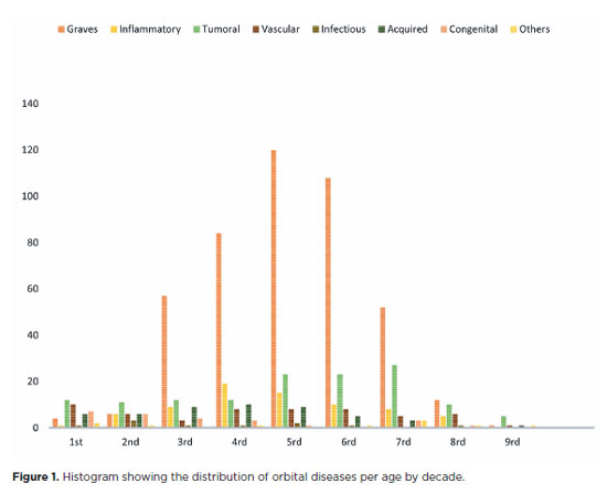
PURPOSE: This study aimed to evaluate the prevalence of orbital conditions in a tertiary ophthalmic outpatient hospital in Sao Paulo, Brazil, with a focus on the main diagnoses and their distribution.
METHODS: A retrospective chart review was conducted involving patients registered and admitted to the orbital disease unit at the Department of Ophthalmology, University of São Paulo Medical School, from January 2004 to March 2018. A total of 838 medical charts were analyzed, of which 37 were excluded due to incomplete data. The remaining charts were categorized into eight diagnostic groups: Graves’ orbitopathy , inflammatory disorders, tumors, vascular lesions, acquired structural abnormalities, congenital structural abnormalities, infectious diseases, and others.
RESULTS: Of the 837,300 ophthalmological appointments, 3,372 (0.4%) were related to orbital diseases. The study included 801 patients, of whom 63.45% were women. The patients’ mean age was 42.86 years. Graves’ orbitopathy was the most common (55%), followed by tumor (17%), inflammatory disorders (9%), vascular lesions (7%), acquired structural abnormalities (5%), congenital structural abnormalities (4%), others (2%), and infectious diseases (1%). The study found significant differences in the incidence and types of orbital diseases, indicating the specialized nature of tertiary care and referral biases.
CONCLUSION: Published data on epidemiological orbital diseases is scarce. Therefore, this study focused on the diverse nature of orbital diseases and their low incidence among ophthalmology appointments. The major trends align with other epidemiological studies, demonstrating a preponderance of Graves’ orbitopathy in middle-aged adults and a bimodal distribution of tumors. These findings are essential in shaping resident training programs and healthcare policies, particularly in tertiary settings. Understanding the epidemiology of orbital diseases can improve diagnostic accuracy, treatment approaches, and patient outcomes as well as support future systemic prospective studies.
Keywords: Orbital diseases; Orbital tumors; Neoplasms; Inflammation; Graves’ ophthalmopathy; Outpatients
Arq. Bras. Oftalmol. 2025;88 (1 )
:1-5
| DOI: 10.5935/0004-2749.2023-0083
Abstract
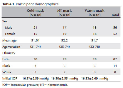
PURPOSE: This study aimed to determine whether early-stage intraocular pressure can be modulated using a thermal face mask.
METHODS:In this prospective clinical study, healthy participants were randomized on a 1:1:1 allocation ratio to three mask groups: hypothermic (G1), normothermic (G2), and hyperthermic (G3). After randomization, 108 eyes from 108 participants were submitted to clinical evaluations, including measurement of initial intraocular pressure (T1). The thermal mask was then applied for 10 minutes, followed by a second evaluation of intraocular pressure (T2) and assessment of any side effects.
RESULTS:The hypothermic group (G1) showed a significant reduction in mean intraocular pressure between T1 (16.97 ± 2.59 mmHg) and T2 (14.97 ± 2.44 mmHg) (p<0.001). G2 showed no significant pressure difference between T1 (16.50 ± 2.55 mmHg) and T2 (17.00 ± 2.29 mmHg) (p=0.054). G3 showed a significant increase in pressure from T1 (16.53 ± 2.69 mmHg) to T2 (18.58 ± 2.95 mmHg) (p<0.001). At T1, there was no difference between the three study groups (p=0.823), but at T2, the mean values of G3 were significantly higher than those of G1 and G2 (p<0.00).
CONCLUSION:Temperature was shown to significantly modify intraocular pressure. Thermal masks allow the application of temperature in a controlled, reproducible manner. Further studies are needed to assess the duration of these effects and whether they are reproducible in patients with pathologies that affect intraocular pressure.
Keywords: Intraocular pressure; Temperature; Masks; Glaucoma; Eye diseases
Arq. Bras. Oftalmol. 2024;87 (5 )
:1-8
| DOI: 10.5935/0004-2749.2022-0172
Abstract
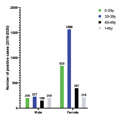
Objetivo: Avaliar a presença de RNA de coronavírus 2 causador de síndrome respiratória aguda grave (SARS-CoV-2) na superfície ocular de indivíduos clinicamente suspeitos com COVID-19 e determinar a precisão de diferentes abordagens de testes moleculares na superfície ocular com base no status de positividade do RT-qPCR de nasofaringe para COVID-19.
Métodos: 152 indivíduos com sintomas suspeitos para a COVID-19 foram submetidos a coleta de reação em cadeia da polimerase de nasofaringe simultaneamente a duas técnicas diferentes de coleta de filme lacrimal para RT-qPCR: aleatoriamente, um olho com a tira filtro do teste de Schirmer e, o olho contralateral, com citologia (swab) conjuntival no fórnice inferior. Todos os indivíduos foram submetidos à biomicroscopia com lâmpada de fenda.
Resultados: Dos 152 pacientes, 86 (56,6%) tiveram a COVID-19 confirmada por PCR de nasofaringe. Ambas as técnicas de coleta detectaram partículas virais: o teste de Schirmer foi positivo em 16,3% (14/86) e a citologia conjuntival em 17,4% (15/86), sem diferenças estatisticamente significativas. Não houve testes oculares positivos entre aqueles com reação em cadeia da polimerase de nasofaringe negativo. A concordância geral dos testes oculares foi de 92,7% e, em combinação, a sensibilidade aumentaria para 23,2%. Os valores médios do limiar de ciclo nos testes de nasofaringe, Schirmer e citologia conjuntival foram 18,2 ± 5,3, 35,6 ± 1,4 e 36,4 ± 3,9, respectivamente.
Conclusão: Os testes de Schirmer (16,3%) e swab conjuntival (17,4%) foram igualmente capazes de detectar RNA de SARS-CoV-2 na superfície ocular por RT-PCR e demonstraram sensibilidade e especificidade indistintas. A coleta simultânea de amostras ao processamento dos testes de RT-PCR de nasofaringe, Schirmer e citologia (swab) conjuntival demonstraram carga viral significativamente menor em ambas as abordagens da superfície ocular em comparação com o teste de nasofaringe. As manifestações oculares detectadas pela biomicroscopia com lâmpada de fenda não foram claramente associadas à positividade do RT-PCR ocular.
Keywords: COVID19; SARS-CoV-2; Túnica conjuntiva; Lágrimas; Reaçao em cadeia da polimerae via transcriptase reversa; RNA; viral.
Arq. Bras. Oftalmol. 2025;88 (5 )
:1-8
| DOI: 10.5935/0004-2749.2024-0312
Abstract
PURPOSE: To evaluate the changes in the rates and indications of eye removal procedures during the recent COVID-19 pandemic.
METHODS: The medical records of all patients who underwent eye removal from 2007 to 2022 were retrospectively reviewed. The patient demographic data and indications for surgery were collected. Data from two groups of patients (prepandemic surgery and postpandemic surgery) were compared. Statistical significance was set at p<0.05.
RESULTS: Fifty-nine patients underwent enucleation (69%), evisceration (27%), or exenteration (3%). The mean (SD) age of the patients was 55.9 (19.4) years, and most (69%) of the patients were males. Most (47%) of the study population were Black. The common indications for eye removal were trauma (41%), painful blind eye (34%), and infection/inflammation (24%). The types of trauma were assault (55%), accidental (39%), and self-inflicted (6%). The mean (SD) monthly rates of eye removal increased from 0.25 (0.50) in the prepandemic period to 0.77 (0.91) during the pandemic (p<0.001). These increases were noted in both males (p=0.003) and females (p=0.001) and were the highest among Black patients [0.42 (0.76); p<0.001]. Among the indications of eye removal, painful blind eyes [0.35 (0.75); p<0.001] and ocular trauma [0.31 (0.47); p=0.051] exhibited the greatest increases following the pandemic.
CONCLUSION: The rate of eye removal procedures increased during the recent pandemic. Although delayed care of chronic eye conditions may have contributed to the increased rates of painful blind eyes, the increased trauma-related eye removals may be attributed to the simultaneous spike in violent assaults in New York City.
Keywords: Eye injuries; Eye enucleation; COVID-19; Pandemics; Ethinicity; Inflammation, Trauma centers
Arq. Bras. Oftalmol. 2024;87 (3 )
:1-7
| DOI: 10.5935/0004-2749.2022-0374
Abstract
PURPOSE: To describe a 2019 acute toxoplasmosis outbreak in the city of São Paulo, Brazil, and to evaluate the laboratory serological profile for toxoplasmosis for three consecutive years. The ophthalmological manifestations of the patients involved in the outbreak were also studied.
METHODS: A cross-sectional descriptive study of a toxoplasmosis outbreak in São Paulo, Brazil, between February and May 2019. Epidemiological data were described, as were the observed ocular manifestations. As part of this study the number of patients with positive IgM toxoplasmosis serology was obtained from a large laboratory network (DASA) for three consecutive years, including the year of the outbreak (2018, 2019, 2020).
RESULTS: Eighty-three individuals were identified in the outbreak and two clusters were studied. The clinical picture of at least 77% of the patients, the epidemiological analysis, and the short incubation period (5-8 days) suggested contamination by oocysts. Serological laboratory data analysis revealed an increase of positive toxoplasmosis IgM in 2019 of 73% compared to the previous year. Ophthalmological examination revealed that at least 4.8% of the patients developed toxoplasmic retinochoroiditis, none of whom had been treated during the acute systemic disease.
CONCLUSION: Our findings indicate vegetable contamination as the possible source of this outbreak, a high prevalence of toxoplasmosis in São Paulo during the outbreak period, and a drop in the number of tests during the COVID-19 pandemic. Retinochoroiditis was observed in at least 4.8% of the cases. We confirm the need to implement effective means for the prevention, diagnosis, and treatment of the disease. This may involve raising awareness among the population of the importance of vegetable hygiene, and improved quality control of food and water.
Keywords: Toxoplasmosis/etiology; Food parasitology; Water/parasitology; Uveitis, posterior/parasitology; Chorioretinitis/parasitology; Visual acuity; Disease outbreaks; Eye manifestations; Humans.
Arq. Bras. Oftalmol. 2024;87 (4 )
:1-7
| DOI: 10.5935/0004-2749.2023-0141
Abstract
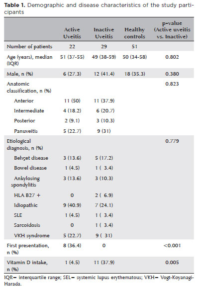
PURPOSE: This study aimed to investigate the correlation between serum vitamin D levels and disease activity in patients with noninfectious uveitis.
METHODS: We conducted a prospective case-control study, assessing 51 patients with noninfectious uveitis, categorized into active (n=22) and inactive (n=29) groups, along with 51 healthy controls. Serum 25-hydroxy vitamin D [25(OH)D] levels were measured. The uveitis group also completed a questionnaire regarding sunlight exposure habits and vitamin D supplementation.
RESULTS: Patients with inflammation-related uveitis exhibited low serum 25(OH)D levels in 68% of cases. The median 25(OH)D level in patients with active uveitis was 17.8 ng/mL (interquartile range [IQR], 15-21 ng/mL), significantly lower compared to the 31.7 ng/mL (IQR, 25-39 ng/mL) in patients with inactive uveitis (p<0.001) and the 27 ng/mL (IQR, 23-31 ng/mL) in the Control Group (p<0.001). Significantly, nearly all patients with uveitis taking vitamin D supplementation were in the Inactive Group (p<0.005). Moreover, reduced sunlight exposure was associated with active uveitis (p<0.003). Furthermore, patients with 25(OH)D levels below 20 ng/mL had ten times higher odds of developing active uveitis (p=0.001).
CONCLUSIONS: This study revealed a prevalent 25(OH)D deficiency among patients with noninfectious uveitis and suggested a link between low 25(OH)D levels and disease activity. To prevent future episodes of intraocular inflammation, vitamin D supplementation and controlled sunlight exposure could be viable options.
Keywords: Vitamin D; 25-hydroxyvitamin D; Uveitis; Vitamin D deficiency; Immunity; Eye/immunology
 ABO is licensed under a Creative Commons Attribution-NonComercial 4.0 Internacional.
06-tab01tb.jpg)
04-tab01.jpg)
06-tab01tb.jpg)
04-tab01tb.jpg)

08-fig01.jpg)
14-fig01tb.jpg)
09-fig01.jpg)
08-tab01.jpg)