Arq. Bras. Oftalmol. 2023;86 (3 )
:1-4
| DOI: 10.5935/0004-2749.20230016
Abstract
A disseminação metastática ocular de tumores sistêmicos é incomum, ocorrendo principalmente na coroide e em pacientes idosos. O câncer de pulmão é considerado o principal tumor metastático ocular em homens, contudo, outras doenças oculares, como as uveítes e lesões retinianas, podem mimetizar os implantes secundários tumorais nos tecidos oculares. O aspecto fundoscópico das neoplasias da coroide pode apresentar similaridade com outros processos infecciosos, especialmente o tuberculoma de coroide. Dessa forma, a investigação clínica detalhada é de grande importância no diagnóstico de pacientes com massas coroideanas, especialmente quando configuram a primeira manifestação de uma doença sistêmica e grave. Relatamos um caso raro de metástase coroideana como primeira manifestação clínica do carcinoma de células renais em um homem jovem, mimetizando um tuberculoma de coroide.
Keywords: Neoplasias renais/complicações; Metástase neoplásica; Carcinoma de células renais; Neoplasias da coroide/etiologia; Humanos; Relatos de casos
Arq. Bras. Oftalmol. 2023;86 (1 )
:79-82
| DOI: 10.5935/0004-2749.20220077
Abstract
Um homem de 53 anos, com história de 3 dias de edema periorbital e perda de visão no olho esquerdo, apresentou trombose séptica do seio cavernoso com envolvimento bilateral das veias orbitais, causando uma orbitopatia congestiva. O paciente foi tratado com uma cantotomia e cantólise de emergência, colírios para redução da pressão intraocular, antibióticos, anticoagulantes e exames seriados. A tomografia de coerência óptica finalmente demonstrou destruição isquêmica difusa de ambas as camadas da retina, sugerindo uma oclusão da artéria oftálmica ou das artérias ciliares posteriores curtas e da artéria retiniana central, com ausência de envolvimento do segmento intracavernoso da artéria carótida interna. O paciente permaneceu sem percepção luminosa no olho esquerdo.
Keywords: Trombose do corpo cavernoso; Doenças orbitárias; Tomografia de coerência óptica; Humanos; Relato de casos
Arq. Bras. Oftalmol. 2024;87 (4 )
:1-5
| DOI: 10.5935/0004-2749.2023-0066
Abstract
Endophthalmitis is a severe form of purulent inflammation caused by the infection of the intraocular tissues or fluids. This infection infrequently occurs through endogenous routes, which are often correlated with major risk factors. Escherichia coli, a gram-negative rod, can cause endophthalmitis through hematogenous spread. We here report a 59-year-old man who presented to our service with acute visual impairment in his left eye, preceded by floaters. He was taking sirolimus and azathioprine for a transplanted kidney, had undergone catheterization for bladder atresia, and had a history of recurrent E. coli urinary tract infections. On evaluation, the left eye exhibited visual acuity of hand motion, anterior chamber reaction (3+/4+), and intense vitritis (4+/4+) with white flake clusters, which prevented appropriate retinal evaluation. Pars plana vitrectomy was performed, and the culture yielded E. coli. The present case highlights the importance of identifying the signs and symptoms of infection early so that diagnosis and treatment of endophthalmitis can be promptly initiated.
Keywords: Endophthalmitis; Escherichia coli; Escherichia coli infections; Eye infections, Bacterial; Sepsis; Vitrectomy; Anti-bacterial agents/therapeutic use; Humans; Case reports
Arq. Bras. Oftalmol. 2024;87 (3 )
:1-4
| DOI: 10.5935/0004-2749.2022-0357
Abstract
We present a rare case of primary caruncle basal cell carcinoma (BCC), a condition with limited occurrences. Our patient, an 80-year-old woman without prior ocular pathological history, presented a 2x2mm pedunculated blackish nodular lesion on the caruncle of her left eye, without local conjunctival or cutaneous involvement. Histological analysis following complete excision confirmed the presence of basal cell carcinoma within the caruncle. Over a span of 30 months, no recurrence has been observed. While scant cases are documented in the literature, we conducted a review of these instances. Despite its infrequent manifestation, this condition should be taken into account when evaluating caruncular tumors, given its tendency to invade the orbit. Complete excision with free surgical margins is the treatment of choice, and adjuvant radiotherapy or chemotherapy might be considered.
Keywords: Conjunctival diseases; Eye neoplasms; Sebaceous gland neoplasms; Conjunctival neoplasms; Carcinoma, basal cell; Diagnosis, differential; Humans; Case reports
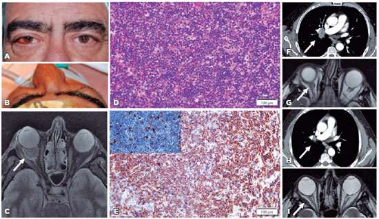
Arq. Bras. Oftalmol. 2024;87 (2 )
:1-3
| DOI: 10.5935/0004-2749.2022-0091
Abstract
Hepatitis C virus infection may be implicated in 12.7% of ocular adnexal marginal zone lymphomas. We present the first case of an orbital-systemic mucosa-associated lymphoid tissue lymphoma that responded to hepatitis C virus medical treatment. A 62-year-old male with a right-sided orbital mass was diagnosed with stage IIA orbital marginal zone lymphoma in addition to hepatitis C virus infection based on clinical, imaging, laboratory, and histological examinations. The systemic and orbital responses were achieved 1 year after undergoing hepatitis C virus treatment with glecaprevir/pibrentasvir. The association between the hepatitis C virus infection and orbital-systemic mucosa-associated lymphoid tissue lymphoma is relevant. Accordingly, patients with orbital mucosa-associated lymphoid tissue lymphoma should be assessed for hepatitis C virus seroreactivity for therapeutic and prognostic purposes.
Keywords: Orbital disease; Orbital neoplasms; Lymphoma, B-cell marginal zone; Hepacivirus; Hepatitis C; Humans; Case reports
 ABO is licensed under a Creative Commons Attribution-NonComercial 4.0 Internacional.
08-fig01.jpg)
11-fig01.jpg)
10-fig01.jpg)
10-fig01.jpg)
14-fig01.jpg)
02-fig01.jpg)
03-fig01.jpg)
02-fig01.jpg)
02-fig01.jpg)
14-fig01.jpg)