Arq. Bras. Oftalmol. 2022;85 (1 )
:13-18
| DOI: 10.5935/0004-2749.20220003
Abstract
Objetivo: Fornecer informações sobre a ocorrência e a eficácia do aconselhamento sobre o uso de tabaco por oftalmologistas a pacientes com doenças oculares associadas à tireoide.
Métodos: Analisamos os prontuários médicos eletrônicos de uma coorte digital de pacientes atendidos por oftalmologistas no Sistema de Saúde da Universidade da Pensilvânia entre o início de 2012 e o final de 2017 com os códigos da Classificação Internacional de Doenças (CID) para a doença de Graves, exoftalmia tireotóxica ou doença ocular associada à tireoide. Os históricos de uso de tabaco foram registrados na primeira e na última visita ao consultório de Oftalmologia, ou na visita mais próxima no tempo. A quantidade de maços/dia (mpd) e todas as anotações feitas nas visitas ao consultório de Oftalmologia foram analisadas para aconselhamento sobre o uso de tabaco.
Resultados: Um total de 435 indivíduos preencheram os critérios de inclusão, dos quais 72 (16,6%) estavam fumando ativamente no momento do primeiro encontro. Apenas 57 (79,2%) desses indivíduos que fumam ativamente registraram queixas relacionadas ao tabagismo, sendo que 34 (59,6%) deles receberam alguma forma de aconselhamento sobre o uso de tabaco. Ao todo, 9 (26,5%) indivíduos dentre os que receberam aconselhamento sobre tabaco e 1 (4,3%) que não teve aconselhamento registrado pararam de fumar (diferença de risco de 22,1%; IC 95%, [1,7%, 39,1%]; p=0,04). Dentre aqueles que receberam aconselhamento, 17 (50,0%) reduziram seus mpd, além de 7 (30,4%) daqueles que não tiveram aconselhamento (diferença de risco de 19,6%; IC 95% [-6,3%, 41,3%]; p=0,18). No geral, 14 (25,5%) dos 55 oftalmologistas que tiveram um paciente fumante ativo registraram evidências de aconselhamento sobre o uso de tabaco.
Conclusões: Os resultados deste estudo revelam tanto as oportunidades perdidas de aconselhamento sobre o uso do tabaco quanto a eficácia do aconselhamento no contexto de doenças oculares associadas à tireoide.
Keywords: Uso de tabaco; Aconselhamento; Doenças da glândula tireóide; Doença de Graves; Oftalmopatias
Arq. Bras. Oftalmol. 2021;84 (5 )
:442-448
| DOI: 10.5935/0004-2749.20210069
Abstract
Objetivo: Verificar se pacientes com dislexia do desenvolvimento (DD) apresentam déficits coerentes com uma disfunção magnocelular visual.
Métodos: Participantes com diagnóstico confirmado de dislexia do desenvolvimento (n=62; faixa etária=8 a 25 anos; Média da idade=13.8 anos, desvio padrão=3.9; 77% homens) foram comparados a um grupo controle com desenvolvimento típico, pareado por idade, sexo, dominância ocular, acuidade visual e compreensão de texto. A perimetria Frequency-Doubling Technology avaliou o limiar de sensibilidade ao contraste do campo visual periférico. O rastreador ocular Visagraph-III registrou os movimentos dos olhos durante leitura de texto.
Resultados: O grupo com dislexia do desenvolvimento apresentou piores limiares de sensibilidade no Frequency-Doubling Technology, com tamanho de efeito forte, do que o grupo controle. O grupo com dislexia do desenvolvimento apresentou mais olhos classificados com déficits na sensibilidade à ilusão de frequência duplicada do que o grupo controle. O grupo com dislexia do desenvolvimento apresentou pior habilidade motora ocular e no desempenho de leitura, revelado pela diferença entre os grupos em relação às fixações oculares, regressões, alcance de reconhecimento, taxa de leitura e eficiência relativa. Foi encontrada correlação significativa entre a sensibilidade ao contraste e as habilidades motoras oculares. Os participantes com boa eficiência relativa apresentaram uma sensibilidade ao contraste significativamente melhor do que os participantes com baixa eficiência relativa.
Conclusões: O grupo com dislexia do desenvolvimento apresentou desempenho inferior nas variáveis visuais relacionadas à função visual magnocelular (i.e., perimetria de frequência duplicada e habilidades motoras oculares), quando comparado ao grupo controle pareado. Os profissionais precisam estar cientes da importância de investigar a visão dos pacientes com dislexia do desenvolvimento além da acuidade visual e incluir nos seus procedimentos diagnósticos instrumentos para avaliar o processamento temporal, com limiar de sensibilidade ao contraste.
Keywords: Dislexia; Leitura; Percepção visual; Transtornos da visão; Músculos oculomotores; Movimentos oculares
Arq. Bras. Oftalmol. 2023;86 (3 )
:1-7
| DOI: 10.5935/0004-2749.20230020
Abstract
Objetivos: Blefaroptose e estrabismo podem ser coexistentes em adultos e ambos afetam a aparência estética e o domínio psicossocial. Ambos também geralmente requerem cirurgia, realizada tradicionalmente em uma abordagem sequencial. O objetivo do presente estudo foi avaliar a eficácia da execução simultânea da ressecção musculoconjuntival de Müller, com ou sem cirurgia de tarsectomia, e da cirurgia de estrabismo em pacientes adultos com ptose e estrabismo coexistentes.
Métodos: Foram retrospectivamente avaliados pacientes com ptose e estrabismo coexistentes submetidos simultaneamente à ressecção musculoconjuntival de Müller, com ou sem tarsectomia, e à cirurgia de estrabismo horizontal. A análise incluiu a mensuração do ângulo de desvio das dioptrias de prisma, a distância do reflexo à margem, a assimetria da altura palpebral e quaisquer complicações após a cirurgia. A ressecção musculoconjuntival de Müller, com ou sem sucesso na tarsectomia, foi considerada bem-sucedida com uma distância reflexo-margem medindo entre 3,5 e 5 mm, e uma diferença entre as duas pálpebras superiores menor que 1 mm. O sucesso da cirurgia de estrabismo foi definido como um alinhamento com ± 10 dioptrias prismáticas de ortotropia.
Resultados: Os pacientes foram 3 mulheres e 5 homens, com média de idade de 37,12 anos (faixa de 22 a 62 anos). A parte de estrabismo da cirurgia foi realizada primeiro em todos os pacientes. Os resultados da simetria palpebral superior foram avaliados como perfeitos (<0,5 mm) em 4 pacientes, bons (≥0,5 mm, <1 mm) em 4 pacientes e regulares (≥1 mm) em nenhum. A ressecção musculoconjuntival de Müller, com ou sem sucesso na tarsectomia, teve sucesso em 6 dos 8 pacientes (75%) e a intervenção para o estrabismo foi bem-sucedida em todos os pacientes. Não foi necessária cirurgia de revisão da pálpebra ou do estrabismo após a cirurgia simultânea em nenhum paciente.
Conclusões: A ressecção musculoconjuntival de Müller, com ou sem tarsectomia, pode ser combinada com a cirurgia de estrabismo em uma abordagem alternativa para pacientes com ptose e estrabismo coexistentes.
Keywords: Blefaroptose/cirurgia; Ambliopia; Estrabismo/cirurgia; Músculos oculomotores/cirurgia; Pálpebras; Procedimentos cirúrgicos oftalmológicos/métodos
Arq. Bras. Oftalmol. 2025;88 (4 )
:1-6
| DOI: 10.5935/0004-2749.2024-0278
Abstract
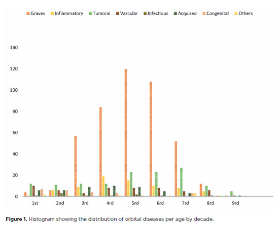
PURPOSE: This study aimed to evaluate the prevalence of orbital conditions in a tertiary ophthalmic outpatient hospital in Sao Paulo, Brazil, with a focus on the main diagnoses and their distribution.
METHODS: A retrospective chart review was conducted involving patients registered and admitted to the orbital disease unit at the Department of Ophthalmology, University of São Paulo Medical School, from January 2004 to March 2018. A total of 838 medical charts were analyzed, of which 37 were excluded due to incomplete data. The remaining charts were categorized into eight diagnostic groups: Graves’ orbitopathy , inflammatory disorders, tumors, vascular lesions, acquired structural abnormalities, congenital structural abnormalities, infectious diseases, and others.
RESULTS: Of the 837,300 ophthalmological appointments, 3,372 (0.4%) were related to orbital diseases. The study included 801 patients, of whom 63.45% were women. The patients’ mean age was 42.86 years. Graves’ orbitopathy was the most common (55%), followed by tumor (17%), inflammatory disorders (9%), vascular lesions (7%), acquired structural abnormalities (5%), congenital structural abnormalities (4%), others (2%), and infectious diseases (1%). The study found significant differences in the incidence and types of orbital diseases, indicating the specialized nature of tertiary care and referral biases.
CONCLUSION: Published data on epidemiological orbital diseases is scarce. Therefore, this study focused on the diverse nature of orbital diseases and their low incidence among ophthalmology appointments. The major trends align with other epidemiological studies, demonstrating a preponderance of Graves’ orbitopathy in middle-aged adults and a bimodal distribution of tumors. These findings are essential in shaping resident training programs and healthcare policies, particularly in tertiary settings. Understanding the epidemiology of orbital diseases can improve diagnostic accuracy, treatment approaches, and patient outcomes as well as support future systemic prospective studies.
Keywords: Orbital diseases; Orbital tumors; Neoplasms; Inflammation; Graves’ ophthalmopathy; Outpatients
Arq. Bras. Oftalmol. 2025;88 (4 )
:1-6
| DOI: 10.5935/0004-2749.2024-0236
Abstract
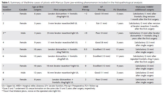
PURPOSE: This study was conducted to report the histopathological and clinical features of the Marcus Gunn phenomenon and other similar conditions of upper eyelid misfiring.
METHODS: This was a retrospective study of patients with congenital ptosis with Marcus Gunn phenomenon who have undergone surgical repair over a period of 12 years and another two patients with upper eyelid misfiring in association with extraocular movements to identify their histopathological findings as subtypes representing ocular congenital cranial dysinnervation disorder.
RESULTS: Among 136 patients with congenital ptosis, 11 (8%) patients with Marcus Gunn phenomenon or misfiring were identified, of whom 9 (6.6%) had typical known Marcus Gunn phenomenon and 2 (1.4%) had eyelid misfiring similar to Marcus Gunn phenomenon. In all patients, the histopathological changes of the excised levator muscle included overall loss and/or atrophy of muscle fibers and irregular-modified Gomori trichrome staining.
CONCLUSION: The Marcus Gunn phenomenon and similar misfiring conditions with synkinetic extraocular muscle movements share findings that are consistent with the neurogenic type of muscle atrophy. This result suggests a common underlying etiology with variable clinical findings, representing the ocular counterpart of congenital cranial dysinnervation disorder, which has been reported as ocular congenital cranial dysinnervation disorder.
Keywords: Eyelid diseases; Ocular motility disorders/surgery; Ophthalmologic surgical procedures
Arq. Bras. Oftalmol. 2026;89 (1 )
:1-6
| DOI: 10.5935/0004-2749.2025-0071
Abstract
PURPOSE: This study aimed to evaluate the outcomes of strabismus surgical correction in patients with Down syndrome.
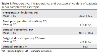
METHODS: We conducted a retrospective chart review of patients with Down syndrome who underwent strabismus surgery between January 1997 and May 2024 at an Ophthalmology Outpatient Clinic in São Paulo, Brazil. The data collected included age, sex, medical and ocular history, surgical details, and follow-up outcomes. The patients were categorized by strabismus type into esotropia, fourth nerve palsy, and mixed groups. Surgical success was defined as final alignment within 10Δ of orthotropia and, where applicable, whether there was resolution of abnormal head posture of ocular origin. Patients with postoperative follow-up <6 months were excluded.
RESULTS: A total of 37 patients (21 females) were included. Of these, 22 (59.5%) were in the esotropia group, 10 (27.0%) in the fourth nerve palsy group, and 5 (13.5%) in the mixed group. The surgical success rate in the esotropia group was 86.4%, with a mean preoperative deviation of 35.2 (± 6.5)Δ, and mean surgical correction of 30.1 (± 10.4)Δ. The success rate in the fourth nerve palsy group was 40.0%, with a mean preoperative deviation of 10.4 (± 4.3)Δ. Overall, success was achieved with a single surgical procedure in 73.0% of the sample. No significant associations were found between surgical success and the clinical and demographic variables, including sex, age at surgery, oblique muscle overaction, pattern strabismus, visual acuity, amblyopia, preoperative deviation, or postoperative follow-up duration (p>0.05).
CONCLUSIONS: When standard surgical tables are applied, strabismus surgery in patients with Down syndrome appears to be safe and effective. We found high success rates, particularly among patients with esotropia. We observed no tendencies toward over- or under-correction. These findings support the use of conventional surgical protocols with this patient population.
Keywords: Down Syndrome/complications; Strabismus/surgery; Esotropia/surgery; Oculomotor nerve diseases/physiopathology; Vision disorders; Humans; Brazil.
Arq. Bras. Oftalmol. 2024;87 (4 )
:1-8
| DOI: 10.5935/0004-2749.2021-0401
Abstract
Objetivos: Avaliar a eficácia do uso de toxina botulínica tipo A no tratamento do estrabismo em pacientes com comprometimento neurológico e avaliar os fatores associados ao sucesso do tratamento.
Métodos: Cinquenta pacientes com estrabismo e comprometimento neurológico foram incluídos no estudo. Em todas as crianças, a toxina botulínica tipo A foi injetada no músculo extraocular apropriado. A relação entre características demográficas, características clínicas e o sucesso do tratamento foram analisadas.
Resultados: No grupo de estudo, 34 pacientes tiveram esotropia e 16 pacientes tiveram exotropia, sendo trinta e seis pacientes com paralisia cerebral e 14 pacientes com hidrocefalia. O tempo médio de acompanhamento foi de 15,3 ± 7,3 meses. O número médio de aplicações foi de 1,4 ± 0,6. O ângulo de desvio médio foi de 42,5 ± 13,2 DP antes do tratamento e diminuiu para 12,8 ± 11,9 DP após o tratamento. Alinhamento motor bem sucedido (ortotropia dentro de 10 DP) foi alcançado em 60% dos pacientes. A análise de regressão logística binária revelou que o desalinhamento esotrópico e uma menor duração do estrabismo foram significativamente associados ao sucesso do tratamento no grupo de estudo. Pacientes esotrópicos com ângulos de desalinhamento menores são mais propensos a serem tratados com uma única aplicação.
Conclusão: O uso da toxina botulínica tipo A para o tratamento de estrabismo em crianças com comprometimento neurológico é uma boa alternativa para a terapia cirúrgica convencional com menor risco de hipercorreção. O resultado do tratamento é melhor em exodesvios e em pacientes com estrabismo de menor duração, implicando em vantagem para o tratamento precoce.
Keywords: Estrabismo; Toxinas botulínicas; Manifestações neurológicas; Doenças do sistema nervoso; Paralisia cerebral; Hodrocefalia; Criança
Arq. Bras. Oftalmol. 2025;88 (2 )
:1-7
| DOI: 10.5935/0004-2749.2024-0029
Abstract
PURPOSE: To evaluate the effect of upper eyelid ptosis repair with Muller muscle-conjunctival resection on meibomian gland function and ocular surface parameters.
METHODS: Thirty-eight patients who underwent ptosis repair with Muller muscle-conjunctival resection were retrospectively reviewed. Meibomian gland loss, Ocular Surface Disease Index OXFORD score, meiboscore, and noninvasive keratograph break-up time were measured preoperatively and at 1st, 3rd, and 6th months postoperatively.
RESULTS: Noninvasive keratograph break-up time values decreased significantly at 1st and 3rd months postoperatively compared to the preoperative level, but were similar to the preoperative level at 6th months postoperatively (p<0.001 and p=0.628, respectively). Ocular surface disease index, OXFORD score, meibomian gland loss, and meiboscore values increased significantly in the 1st and 3rd postoperative months compared to the preoperative period, but these values decreased to preoperative levels in the 6th postoperative month (p<0.001 and p>0.05, respectively).
CONCLUSION: There is a transient deterioration in meibography findings and OSDI score in the early postoperative period after Muller muscle-conjunctival resection. Patients undergoing Muller muscle-conjunctival resection may require topical lubricants, especially in the first 3 postoperative months.
Keywords: Meibomian glands; Blepharoptosis; Preoperative period; Conjunctiva; Muscles; Eyelid diseases; Diagnostic techniques, ophthalmological
Arq. Bras. Oftalmol. 2025;88 (5 )
:1-7
| DOI: 10.5935/0004-2749.2024-0318
Abstract
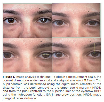
PURPOSE: Ptosis is characterized by drooping of the upper eyelid, often requiring surgical intervention for functional and aesthetic purposes. Müller’s muscle conjunctival resection is a commonly utilized surgical technique to correct mild to moderate ptosis. This retrospective study aimed to evaluate the impact of Hering’s law on the outcomes of unilateral Müller’s muscle conjunctival resection surgery, particularly eyelid and brow symmetry.
METHODS: Thirty patients with unilateral ptosis underwent Müller’s muscle conjunctival resection. Pre- and postoperative assessments included ipsilateral and contralateral side margin-reflex distance and brow position, measured through digital image analysis.
RESULTS: We found significant improvements in postoperative margin-reflex distance measurements in the ipsilateral eyelid but not in the contralateral eyelid, indicating minimal influence of Hering’s law. Brow position showed a statistically significant increase on the contralateral side but not on the ipsilateral side.
CONCLUSION: Müller’s muscle conjunctival resection effectively restores symmetry in eyelid height and maintains brow symmetry. This is the first study to explore bilateral eyelid and brow symmetry after unilateral Müller’s muscle conjunctival resection surgery for mild to moderate ptosis. Further research should be conducted to understand the long-term effects of Müller’s muscle conjunctival resection on facial aesthetics, particularly in relation to brow position.
Keywords: Müller muscle conjunctiva resection; Hering’s law; Eyelids; Blepharoptosis; Reflex; Oculomotor muscles
Arq. Bras. Oftalmol. 2025;88 (2 )
:1-5
| DOI: 10.5935/0004-2749.2024-0113
Abstract
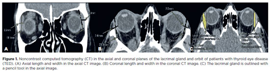
This study aimed to evaluate the morphometric and volumetric dimensions of the lacrimal gland in patients with inactive thyroid eye disease and compare them with the values reported in the literature. This case series evaluated consecutive patients with inactive thyroid eye disease treated at a tertiary eye hospital from 2015 to 2020. The patients' baseline demographics and clinical characteristics were obtained. The axial and coronal length, width, and volume of the lacrimal gland were measured on computed tomography scan images, and the results were statistically analyzed. A total of 21 patients (42 orbits) with inactive thyroid eye disease were evaluated. Their mean age was 49.0 ± 14.6 years, and 12 (57.1%) of them were men. The main complaint was dryness, and the majority of the patients had good vision and mild proptosis. The mean axial length and width of the lacrimal gland were 19.3 ± 3.9 mm and 7.5 ± 2.1 mm, respectively; coronal length and width, 20.4 ± 4.5 mm and 7.5 ± 2.1 mm, respectively; and lacrimal gland volume, 0.825 ± 0.326 mm3. Age, sex, or laterality were not found to be determinants of lacrimal gland enlargement. Patients with thyroid eye disease have enlarged lacrimal gland even in the nonactive phase of the disease multifactorial aspects influence the lacrimal gland in thyroid eye disease, making it difficult to establish a clear correlation with predisposing factors. Further studies are warranted to better understand the association between thyroid eye disease and the lacrimal gland.
Keywords: Graves' ophthalmology; Graves' disease; Lacrimal apparatus; Lacrimal apparatus diseases; X-ray computed tomography
Arq. Bras. Oftalmol. 2024;87 (3 )
:1-7
| DOI: 10.5935/0004-2749.2023-0028
Abstract
PURPOSE: Evaluation of lid contour and marginal peak point changes to compare outcomes of external levator advancement and Müller’s muscle conjunctival resection surgery in unilateral ptosis.
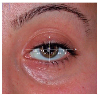
METHODS: We reviewed the charts of unilateral ptosis patients who underwent external levator advancement or Müller’s muscle conjunctival resection. Eyelid contour analysis was conducted on preoperative and 6-month postoperative digital images. This was performed with the multiple margin reflex distances technique, measuring the vertical distance from a line intersecting the center of the pupil to the eyelid margin at 10 positions at 2 mm intervals. The marginal peak point changes were analyzed digitally using the coordinates of the peak point according to the pupil center. Each position’s mean distance was compared preoperatively, postoperatively, and with the fellow eyelid.
RESULTS: Sixteen patients underwent external levator advancement and 16 patients had Müller’s muscle conjunctival resection. The mean margin reflex distance was improved by both techniques (1.46 vs. 2.43 mm and 1.12 vs. 2.25 mm, p=0.008 and p=0.0001 respectively) and approached that of the fellow eyelid (2.43 vs. 2.88 and 2.25 vs. 2.58 mm, p=0.23 and p=0.19, respectively). However, statistically significant lid margin elevation was limited to between the N6 and T6 points in the external levator advancement group. Whereas, significant elevation was achieved along the whole lid margin in the Müller’s muscle conjunctival resection group. The marginal peak point was shifted slightly laterally in the external levator advancement group (p=0.11).
CONCLUSIONS: Both techniques provide effective lid elevation, however, the external levator advancement’s effect lessens toward the canthi while Müller’s muscle conjunctival resection provides more uniform elevation across the lid margin. The margin reflex distance alone is not sufficient to reflect contour changes.
Keywords: Blepharoptosis; Eyelids; Conjunctiva; Oculomotor muscles; Image processing, computer-assisted; Treatment outcome
 ABO is licensed under a Creative Commons Attribution-NonComercial 4.0 Internacional.
06-tab01tb.jpg)
04-tab01tb.jpg)
06-tab01tb.jpg)
12-tab01tb.jpg)

12-fig01.jpg)
02-fig01.jpg)