Arq. Bras. Oftalmol. 2023;86 (5 )
:1-6
| DOI: 10.5935/0004-2749.20230071
Abstract
Objetivo: O melanoma da conjuntiva é um tumor raro e agressivo, com propensão à disseminação metastática regional e distante. Este estudo tem como objetivo analisar os marcadores BRAF e NRAS no melanoma da conjuntiva e sua relação com recidivas tumorais e com o prognóstico do paciente.
Métodos: Este foi um estudo retrospectivo, observacional e unicêntrico de pacientes consecutivos com diagnóstico anatomopatológico de melanoma da conjuntiva feito entre janeiro de 1992 e dezembro de 2019. As mutações BRAF e NRAS foram analisadas com o kit cobas® 4800 (Roche®) em amostras obtidas através de biópsia excisional ou por mapa. Além disso, foi avaliada a presença de lesões pré-cancerosas ou tumorais associadas.
Resultados: Foram incluídos 12 pacientes com amostras histológicas positivas para melanoma da conjuntiva (7 mulheres e 5 homens), com idade média ao diagnóstico de 60 anos e tempo médio de evolução de 6,38 ± 3,4 anos. A mutação BRAF V600E foi encontrada em 3 biópsias (25%), bem como a NRAS Q61X (25%). Ocorreram recidivas em todos os pacientes positivos para mutações de BRAF ou NRAS e 5 desses pacientes desenvolveram disseminação sistêmica (83,33%). Além disso, 4 dos 6 pacientes com BRAF ou NRAS mutante (66,66%) apresentaram achados histopatológicos de lesões tumorais ou pré-cancerosas.
Conclusões: As mutações BRAF e NRAS podem ser fatores de risco para recorrência e menor sobrevida no melanoma da conjuntiva, o que tornaria esses pacientes candidatos a terapias direcionadas e a um acompanhamento mais abrangente e individualizado. Todos esses dados justificam mais estudos prospectivos padronizados.
Keywords: Neoplasias da túnica conjuntiva; Melanoma; Biomarcadores tumorais; Proteínas proto-oncogênicas B-raf; Genes ras.
Arq. Bras. Oftalmol. 2021;84 (3 )
:209-213
| DOI: 10.5935/0004-2749.20210035
Abstract
OBJETIVOS: Identificar vasos linfáticos em espécimes orbitários de cadáveres humanos através de microscopia óptica e análise imunohistoquímica.
MÉTODOS: Um estudo postmortem incluiu dez espécimes orbitários provenientes de dez cadáveres humanos. Todos os espécimes orbitários foram obtidos até 12 horas após a morte com uma técnica cirúrgica de exenteração orbitária e dissecados em glândula lacrimal, nervo óptico, gordura órbitária e músculos extraoculares. Para classificar como um vaso linfático, os critérios histológicos incluíram vasos endoteliais de parede única sem membrana basal bem desenvolvida, irregulares e lúmen sem hemácias, e os critérios imunohistoquímicos incluíram vasos endoteliais de parede única, com formato irregular e lúmen sem hemácias e reagentes a podoplanina D2-40.
RESULTADOS: As lâminas histológicas de glândula lacrimal, nervo óptico, tecido adiposo e músculos extraoculares reagiram positivamente a podoplanina D2-40.
CONCLUSÃO: Este estudo demonstrou vasos linfáticos na órbita humana, mais exatamente, na glândula lacrimal, no nervo óptico, na gordura orbitária e nos músculos extrínsecos extraoculares via microscopia óptica e imunohistoquímica.
Keywords: Vasos linfáticos; Órbita; Nervo óptico; Aparelho lacrimal; Músculos oculomotores; Tecido adiposo; Microscopia
Arq. Bras. Oftalmol. 2023;86 (5 )
:1-5
| DOI: 10.5935/0004-2749.20230063
Abstract
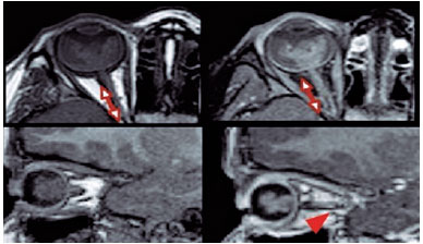
Objetivo: A injeção peribulbar de triancinolona é um tratamento alternativo para doenças oculares da tireoide; no entanto, a segurança desse procedimento continua controversa. O objetivo deste artigo é descrever os efeitos adversos locais e sistêmicos de injeções peribulbares de triancinolona em pacientes com doença ocular da tireoide.
Métodos: Estudo retrospectivo de uma série de casos. Foram analisados os prontuários médicos dos pacientes com doença ocular da tireoide tratados com injeções de triancinolona peribulbar em uma única instituição acadêmica entre 2007 e 2019. Foram documentadas as complicações locais e sistêmicas.
Resultados: Um total de 123 pacientes foram tratados. Apenas 11 (8,9%) pacientes apresentaram complicações locais, sendo a mais frequente a presença de equimoses palpebrais superficiais (7,3%), enquanto 2 (1,6%) pacientes apresentaram complicações sistêmicas (hiperglicemia e inibição da suprarrenal após a interrupção do tratamento). Todas estas complicações foram transitórias e nenhum paciente apresentou sequelas de longo prazo.
Conclusões: As injeções peribulbares de triancinolona nas doenças oculares da tireoide têm uma taxa muito baixa de complicações, tanto locais quanto sistêmicas. São necessários estudos prospectivos para aprofundar este tópico.
Keywords: Órbita/diagnóstico por imagem; Imageamento por ressonância magnética; Oftalmopatia de Graves; Triancinolona/efeitos adversos; Injeções.
Arq. Bras. Oftalmol. 2025;88 (4 )
:1-6
| DOI: 10.5935/0004-2749.2024-0278
Abstract
PURPOSE: This study aimed to evaluate the prevalence of orbital conditions in a tertiary ophthalmic outpatient hospital in Sao Paulo, Brazil, with a focus on the main diagnoses and their distribution.
METHODS: A retrospective chart review was conducted involving patients registered and admitted to the orbital disease unit at the Department of Ophthalmology, University of São Paulo Medical School, from January 2004 to March 2018. A total of 838 medical charts were analyzed, of which 37 were excluded due to incomplete data. The remaining charts were categorized into eight diagnostic groups: Graves’ orbitopathy , inflammatory disorders, tumors, vascular lesions, acquired structural abnormalities, congenital structural abnormalities, infectious diseases, and others.
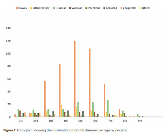
RESULTS: Of the 837,300 ophthalmological appointments, 3,372 (0.4%) were related to orbital diseases. The study included 801 patients, of whom 63.45% were women. The patients’ mean age was 42.86 years. Graves’ orbitopathy was the most common (55%), followed by tumor (17%), inflammatory disorders (9%), vascular lesions (7%), acquired structural abnormalities (5%), congenital structural abnormalities (4%), others (2%), and infectious diseases (1%). The study found significant differences in the incidence and types of orbital diseases, indicating the specialized nature of tertiary care and referral biases.
CONCLUSION: Published data on epidemiological orbital diseases is scarce. Therefore, this study focused on the diverse nature of orbital diseases and their low incidence among ophthalmology appointments. The major trends align with other epidemiological studies, demonstrating a preponderance of Graves’ orbitopathy in middle-aged adults and a bimodal distribution of tumors. These findings are essential in shaping resident training programs and healthcare policies, particularly in tertiary settings. Understanding the epidemiology of orbital diseases can improve diagnostic accuracy, treatment approaches, and patient outcomes as well as support future systemic prospective studies.
Keywords: Orbital diseases; Orbital tumors; Neoplasms; Inflammation; Graves’ ophthalmopathy; Outpatients
Arq. Bras. Oftalmol. 2026;89 (5 )
:1-8
| DOI: 10.5935/0004-2749.2026-0041
Abstract
PURPOSE: To evaluate the efficacy of autologous fat grafting for treating enophthalmos in anophthalmic sockets.
METHODS: In this prospective interventional case series, consecutive patients with anophthalmic sockets and enophthalmos were enrolled between August 2013 and July 2025. Clinical ophthalmologic examination, Hertel exophthalmometry, and orbital computed tomography scans were performed. Enophthalmos was assessed clinically and radiologically before and after orbital lipofilling with autologous fat. Fat was harvested from the inner thigh and injected into the inferolateral orbital compartment. The volume of injected fat was determined according to the degree of enophthalmos measured by Hertel exophthalmometry. Follow-up ranged from 2 to 60 months (median, 7 months).
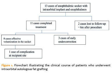
RESULTS: Thirteen patients (13 sockets) underwent orbital autologous fat grafting using Coleman's technique. The injected fat volume ranged from 3.7 to 6 mL per procedure (median, 4.2 mL). Clinically, a reduction in enophthalmos of 1–5 mm was observed in 12 cases (median, 2 mm), which was associated with a decrease in prosthesis thickness. One patient developed a fat cyst due to anterior fat migration, and three patients required repeat grafting because of undercorrection. Computed tomography scans, available for nine patients, demonstrated a 6.8%–10.8% increase in intraorbital volume, particularly in the inferolateral quadrant. Conclusions: Clinical and radiological assessments demonstrated that lipofilling is an effective technique for restoring orbital volume in anophthalmic sockets with enophthalmos.
Keywords: Anophthalmos; Enophthalmos; Orbital volumetry; Fat grafting; Orbital reconstruction
Arq. Bras. Oftalmol. 2024;87 (2 )
:1-6
| DOI: 10.5935/0004-2749.2021-0435
Abstract
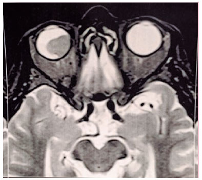
PURPOSE: This study aimed to analyze the association between magnetic resonance imaging apparent diffusion coefficient map value and histopathological differentiation in patients who underwent eye enucleation due to retinoblastomas.
METHODS: An observational chart review study of patients with retinoblastoma that had histopathology of the lesion and orbit magnetic resonance imaging with apparent diffusion coefficient analysis at Hospital de Clínicas de Porto Alegre between November 2013 and November 2016 was performed. The histopathology was reviewed after enucleation. To analyze the difference in apparent diffusion coefficient values between the two major histopathological prognostic groups, Student's t-test was used for the two groups. All statistical analyses were performed using SPSS version 19.0 for Microsoft Windows (SPSS, Inc., Chicago, IL, USA). Our institutional review board approved this retrospective study without obtaining informed consent.
RESULTS: Thirteen children were evaluated, and only eight underwent eye enucleation and were included in the analysis. The others were treated with photocoagulation, embolization, radiotherapy, and chemotherapy and were excluded due to the lack of histopathological results. When compared with histopathology, magnetic resonance imaging demonstrated 100% accuracy in retinoblastoma diagnosis. Optic nerve invasion detection on magnetic resonance imaging showed a 66.6% sensitivity and 80.0% specificity. Positive and negative predictive values were 66.6% and 80.0%, respectively, with an accuracy of 75%. In addition, the mean apparent diffusion coefficient of the eight eyes was 0.615 × 103 mm2/s. The mean apparent diffusion coefficient value of poorly or undifferentiated retinoblastoma and differentiated tumors were 0.520 × 103 mm2/s and 0.774 × 103 mm2/s, respectively.
CONCLUSION: This study revealed that magnetic resonance imaging is useful in the diagnosis of retinoblastoma and detection of optic nerve infiltration, with a sensitivity of 66.6% and specificity of 80%. Our results also showed lower apparent diffusion coefficient values in poorly differentiated retinoblastomas with a mean of 0.520 ×
103 mm2/s, whereas in well and moderately differentiated, the mean was 0.774 × 103 mm2/s.
Keywords: Retinoblastoma; Prognosis; Retinal neoplasms; Orbit; Diffusion magnetic resonance imaging


 ABO is licensed under a Creative Commons Attribution-NonComercial 4.0 Internacional.


13-fig01.jpg)
02-fig01.jpg)
05-tab01.jpg)
01-fig01.jpg)
11-fig01.jpg)
02-fig01.jpg)
03-fig01.jpg)
05-fig01.jpg)