

Showing of 1 until 15 from 136 result(s)
Search for: Trachoma; Personal hygiene; Health infraestructure; Endemic diseases; Brazil; Space-time clustering; Caregivers
05-tab01tb.jpg)
Abstract
Objetivo: Avaliar as razões para não comparecimento à clínica oftalmológica da universidade após triagem oftalmológica realizada usando uma unidade móvel oftalmológica que fornece exame oftalmológico para comunidades não assistidas em uma região do Brasil.
Métodos: Foi realizado um estudo observacional prospectivo no ano de 2017/2018 para avaliar as razões que fizeram com que os indivíduos triados usando uma unidade móvel oftalmológica e referenciados para a clínica oftalmológica da universidade não comparecessem à consulta agendada. A triagem foi feita em 10 municípios da região centro-oeste do estado de São Paulo, Brasil. Todos os 1.928 participantes fizeram o exame oftalmológico sem custo e 37,1% deles necessitaram de encaminhamento para a clínica oftalmológica da universidade para exames especializados ou cirurgias. O estudo usou duas ferramentas: (1) análise comparativa entre os dados dos indivíduos encaminhados que compareceram ao agendamento com os que não compareceram; (2) busca ativa dos indivíduos que não compareceram à consulta agendada, aplicando-se um questionário para avaliar os motivos para o não comparecimento.
Resultados: Fatores como idade, sexo, distância entre a cidade de origem e o hospital universitário, número de oftalmologistas na cidade de procedência, renda familiar média e acuidade visual não influenciaram no comparecimento ao encaminhamento. Catarata foi a maior causa para o encaminhamento (350 casos). O não comparecimento foi maior nos portadores de glaucoma/glaucoma suspeitos (54,1%), estrabismo (45%) e afecções do segmento anterior (33,6%). Muitos indivíduos que não compareceram ao serviço de referência procuraram por outro local para o atendimento oftalmológico.
Conclusão: O não comparecimento para tratamento oftalmológico sem custo depende de fatores relacionados ao paciente ou à falta de conhecimento das próprias condições oftalmológicas. Campanhas educativas nas comunidades assistidas devem ser feitas para alcançar maior comparecimento às consultas e melhor prevenir a cegueira evitável.
Keywords: Serviços de saúde ocular; Unidades móveis de saúde; Acesso aos serviços de saúde; Pacientes desistentes do tratamento; Promoção da saúde
01-tab01.jpg)
Abstract
Objetivo: Determinar a incidência de emergências oculares em um centro oftalmológico de referência no Brasil.
Métodos: O banco de dados de prontuários da Fundação Altino Ventura, Recife, Brasil, foi analisado retrospectivamente e incluiu pacientes atendidos, entre janeiro de 2017 e janeiro de 2018, na sala de emergência oftalmológica. Foram excluídos os prontuários com dados incompletos e com quadros ambulatoriais. Apenas o primeiro atendimento na emergência foi considerado para análise.
Resultados: Em um ano, 134.788 pacientes (idade média de 38,7 ± 22 anos [0-99 anos]) foram admitidos na emergência da Fundação Altino Ventura. Os diagnósticos mais frequentes foram conjuntivite (52.732 casos [37,3%]), blefarite (7.213 casos [5,1%]) e corpo estranho na córnea/conjuntiva (6.925 casos [4,9%]). Corpo estranho na córnea/conjuntiva e trauma ocular foram cerca de 8 vezes e 2 vezes mais incidente em indivíduos do sexo masculino, respectivamente (p<0,001 em ambos). Triquíase e blefarite afetaram ~2 vezes mais pacientes do sexo feminino, respectivamente (p<0,001 em ambos). Corpo estranho na córnea/conjuntiva e trauma ocular afetaram mais pacientes em idade produtiva (>15 anos), enquanto úlcera, blefarite e triquíase da córnea afetaram mais pacientes idosos. Todos os grupamentos de diagnóstico (doenças infecciosas, trauma ocular, corpos estranhos, retinopatias, doenças das pálpebras, doenças da córnea, crise glaucomatosa e doenças neurooftalmológicas) foram mais incidentes na primavera (valor de p<0,001).
Conclusão: As emergências oftalmológicas mais comuns no presente estudo foram as doenças infecciosas e o corpo estranho. Porém, a incidência das emergências oculares são fluências pela faixa etária e gênero do paciente, além da época do ano.
Keywords: Emergência; Oftalmopatia; Transtorno da visão; Conjuntivite; Corpo estranho; Traumatismo oculare; Estações do ano; Brasil
Abstract
PURPOSE: This study evaluated macular thickness using spectral-domain optical coherence tomography in healthy participants from a population-based eye survey.
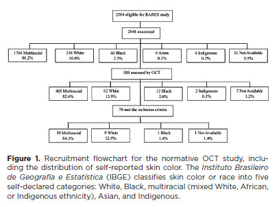
METHODS: The Brazilian Amazon Region Eye Survey was a population-based study assessing the prevalence and causes of visual impairment, blindness, and ocular diseases in adults aged ≥45 years from urban and rural areas of Parintins. A subgroup was selected based on inclusion criteria for both eyes: best-corrected visual acuity ≥20/32, normal eye examination results, and no prior ocular surgery. Scans were performed using the iVue optical coherence tomography device. Measurements were taken from the nine subfields defined by the Early Treatment Diabetic Retinopathy Study, examining the full retina as well as the inner and outer retinal layers. Associations of retinal thickness with age and sex were also analyzed. Statistical significance was set at p≤0.05.
RESULTS: In total, 70 healthy participants (25 males), aged 45–65 years (mean=52 ± 5), were included. Mean central foveal thickness was 248.71 ± 18.73 μm. A significant age-related reduction in macular thickness was observed, particularly in the inner superior parafovea (p=0.036), nasal perifovea (p=0.001), superior perifovea (p=0.028), outer layer of inferior parafovea (p=0.049), and the inferior perifovea of the full retina (p=0.029). Males showed significantly greater thickness in the outer layer, especially in the outer parafovea (p=0.004) and perifovea (p<0.0001).
CONCLUSIONS: This study established normative macular thickness values for healthy older adults in the Brazilian Amazon region using spectral-domain optical coherence tomography. Age and sex were found to significantly influence macular thickness and should be considered when interpreting measurements. These data will support future studies of retinal diseases in this population.
Keywords: Retinal diseases/diagnosis; Macula lutea/pathology; Macular degeneration/diagnosis; Diabetic retinopathy/diagnosis; Vision, low; Vision tests; Tomography, optical coherence/methods; Young adult; Cross-sectional studies; Brazil/epidemiology
13-tab01.jpg)
Abstract
Objetivo: Avaliar o perfil clínico e epidemiológico dos transplantes de córnea realizados em um centro de referência oftalmológica de Recife no estado de Pernambuco, localizado no nordeste do Brasil.
Métodos: Esse estudo transversal coletou através de prontuários médicos dados clínicos e epidemiológicos de pacientes submetidos a ceratoplastia na Fundação Altino Ventura, de janeiro a dezembro de 2017.
Resultados: Um total de 356 procedimentos foram realizados em 327 pacientes dos quais 165 (50.5%) eram mulheres. A média de idade na cirurgia foi de 50.9 ± 22.6 anos (variação, 10 - 89 anos). A maioria dos pacientes (n=152 [46.5%]) era da capital e região metropolitana. A média de tempo de espera na fila para o transplante de córnea foi de 52.4 ± 58.9 dias (variação, 0 - 460 dias). As principais indicações de transplante foram ceratite infecciosa (n=88 [24.7%]), ceratocone (n=80 [22.5%]) e falência de transplante prévio (n=75 [21.1%]). Transplante penetrante foi a técnica mais realizada (n=213 [59.9%]) e foi mais comum em homens (n=132 [76.7%]), enquanto os transplantes lamelares posteriores (n=143 [41.1%]) foram mais realizados nas mulheres (p<0.001).
Conclusão: Ceratites infecciosas foram a causa mais comum de transplante, com prevalência similar em adultos economicamente ativos de ambos os sexos. Transplante penetrantes foram os prevalentes em homens e os transplantes lamelares em mulheres.
Keywords: Doença da córnea/epidemiologia; Transplante de córnea; Ceratoplastia penetrante; Brasil/epidemiologia
Abstract
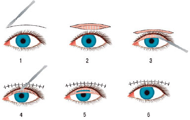
Objetivo: Compartilhar os resultados dos pacientes submetidos à rotação de retalho tarsal anterior, combinados com a reposição lamelar anterior devido à entrópio cicatricial da pálpebra superior e determinar a eficácia e a confiabilidade desta técnica cirúrgica.
Métodos: Foram incluídos neste estudo quinze olhos de 11 pacientes em quem realizamos cirurgia de rotação de retalho tarsal anterior combinada com reposição lamelar anterior devido ao entrópio cicatricial. Os registros médicos dos pacientes foram analisados retrospectivamente e as causas da entrópio cicatricial, bem como os achados do exame oftalmológico pré-operatório e pós-operatório foram registrados. A integridade anatômica e funcional da pálpebra foi considerada como sucesso cirúrgico.
Resultados: A idade média foi de 59,81 ± 18 anos. O período médio de seguimento foi de 21,72 ± 14 meses (intervalo 5-43 meses). As causas da entrópio cicatricial foram o desenvolvimento de cicatrizes pós-operatórias devido a eletrólises múltiplas para triquíase e/ou distiquiase em 7 olhos, tracoma em 6 olhos e trauma em 2 olhos. Todos os pacientes foram tiveram irritação e lacrimejamento pré-operatório, enquanto que 10 pacientes apresentavam opacidade e erosão da córnea e 1 paciente apresentava apenas erosão epitelial. O sucesso total anatômico e funcional foi alcançado em todos os casos.
Conclusão: A rotação do retalho tarsal anterior combinada com a reposição lamelar anterior no reparo da entrópio cicatricial é um procedimento cirúrgico alternativo efetivo e confiável.
Keywords: Tracoma/complicações; Pálpebras/cirurgia; Entrópio/cirurgia; Cicatriz; Retalhos cirúrgicos; Procedimentos cirúrgicos oftalmológicos/métodos
Abstract
PURPOSE: This study aimed to evaluate the outcomes of strabismus surgical correction in patients with Down syndrome.
METHODS: We conducted a retrospective chart review of patients with Down syndrome who underwent strabismus surgery between January 1997 and May 2024 at an Ophthalmology Outpatient Clinic in São Paulo, Brazil. The data collected included age, sex, medical and ocular history, surgical details, and follow-up outcomes. The patients were categorized by strabismus type into esotropia, fourth nerve palsy, and mixed groups. Surgical success was defined as final alignment within 10Δ of orthotropia and, where applicable, whether there was resolution of abnormal head posture of ocular origin. Patients with postoperative follow-up <6 months were excluded.
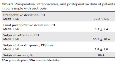
RESULTS: A total of 37 patients (21 females) were included. Of these, 22 (59.5%) were in the esotropia group, 10 (27.0%) in the fourth nerve palsy group, and 5 (13.5%) in the mixed group. The surgical success rate in the esotropia group was 86.4%, with a mean preoperative deviation of 35.2 (± 6.5)Δ, and mean surgical correction of 30.1 (± 10.4)Δ. The success rate in the fourth nerve palsy group was 40.0%, with a mean preoperative deviation of 10.4 (± 4.3)Δ. Overall, success was achieved with a single surgical procedure in 73.0% of the sample. No significant associations were found between surgical success and the clinical and demographic variables, including sex, age at surgery, oblique muscle overaction, pattern strabismus, visual acuity, amblyopia, preoperative deviation, or postoperative follow-up duration (p>0.05).
CONCLUSIONS: When standard surgical tables are applied, strabismus surgery in patients with Down syndrome appears to be safe and effective. We found high success rates, particularly among patients with esotropia. We observed no tendencies toward over- or under-correction. These findings support the use of conventional surgical protocols with this patient population.
Keywords: Down Syndrome/complications; Strabismus/surgery; Esotropia/surgery; Oculomotor nerve diseases/physiopathology; Vision disorders; Humans; Brazil.
Abstract
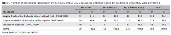
PURPOSE: Trachoma is the major infectious cause of preventable blindness in the world, and its sequelae include the presence of cicatricial entropion and trachomatous trichiasis. Trachoma can be corrected by surgical treatment of the eyelids and, if left untreated, may result in corneal opacification, low vision, and blindness. There are limited data on trachomatous trichiasis in Brazil. This study was conducted to estimate the frequency of entropion and trichiasis surgeries of trachomatous origin based on the records of procedures performed in specialized hospitals that served the Unified Health System (SUS) in the years 2016 and 2017.
METHODS: This was a retrospective study conducted in the oculoplastic sectors of the ophthalmology services of the following three hospitals in the state of São Paulo: Hospital das Clínicas da Faculdade de Medicina de Botucatu (HC Botucatu), Hospital das Clínicas da Faculdade de Medicina de Ribeirão Preto da Universidade de São Paulo (HC Ribeirão Preto), and Hospital Estadual de Bauru (HE Bauru). Medical records corresponding to the codes of interest were evaluated.
RESULTS: In total, 462 medical records were evaluated, including 170 (36.8%) at HC Botucatu, 61 (13.2%) at HE Bauru, and 231 (50.0%) at HC Ribeirão Preto. There were 39 (8.4%) cases of trachomatous trichiasis, ranging from 9 (14.8%) at HE Bauru to 15 (6.5%) at HC Ribeirão Preto.
CONCLUSIONS: The frequency of surgery due to trachoma was low in these oculoplastic services. The state of São Paulo might have reached the goal for trachoma elimination in the surgical component. The questionnaire used for data collection was successfully tested despite some difficulties in collecting data from the medical records. Studies with the same methodology are recommended in other services in the areas of endemic trachoma in the past to understand the frequency of eye lid surgeries performed for treating trachomatous sequelae.
Keywords: Trachoma; Trichiasis; Medical records; Epidemiology; Neglected diseases; Unified Health System; Brazil
Abstract
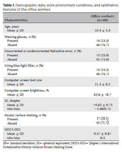
PURPOSE: To examine how ophthalmological features, screen exposure duration, and break habits among office employees affect ocular surface parameters.
METHODS: This single-center cross-sectional study involved two assessments on the same day: one before and one after a visual display terminal task. During the initial assessment, information on screen use was gathered, and refractive error, anterior segment examination, tear breakup time, and Schirmer test measurements were conducted. Participants tracked their screen usage and break durations throughout the day. At the end of the workday, tear breakup time and Schirmer I tests were repeated. Baseline and follow-up results were compared, and regression analysis was performed to identify factors linked to tear breakup time reduction.
RESULTS: The study enrolled 60 female office employees. Their mean screen time was 269.26 ± 70.21 min, with an average break duration of 151.93 ± 46.24 min. Tear breakup time at the second assessment (6.38 ± 2.70) was significantly lower than at baseline (8.62 ± 2.73) (p<0.001), whereas Schirmer test scores showed no significant change (p>0.05). Tear breakup time reduction was noted in 54 participants (90.0%), with a significant association between tear breakup time decrease percentage and screen exposure (p=0.001, r=0.463). Regression analysis showed that uncorrected or undercorrected refractive error was an independent risk factor for a ≥30% tear breakup time reduction, while taking more frequent short breaks (<15 min) acted as a protective factor.
CONCLUSIONS: Taking more frequent short breaks (<15 min) and correcting refractive errors help prevent intra-day tear breakup time decline during visual display terminal use. Structuring breaks to support tear film stability is advisable for occupations that require regular visual display terminal tasks.
Keywords: Tear film; Screen time; Tear breakup time; Office workers; Protective factors; Lacerations; Refractive errors; Risk factors.
Abstract
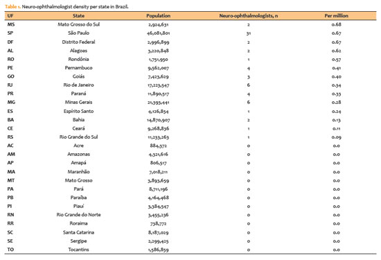
PURPOSE: Neuro-ophthalmology is a highly specialized subspecialty at the interface of neurology and ophthalmology. Globally, workforce shortages and geographic maldistribution limit access to neuro-ophthalmic care. Although Brazil has a large ophthalmology workforce, regional disparities may restrict access to subspecialty services. The national distribution of neuro-ophthalmologists has not previously been quantified. This study aimed to characterize the geographic distribution of neuro-ophthalmologists in Brazil and identify potential gaps in access to neuro-ophthalmic care.
METHODS: This cross-sectional, population-based study used data from the registry of the Brazilian Neuro-Ophthalmology Association (Associação Brasileira de Neuro-Oftalmologia [ABNO]) to identify practicing neuro-ophthalmologists (data extracted on January 1, 2026). Municipal- and state-level population estimates were obtained from the Brazilian Institute of Geography and Statistics. Specialist density was calculated as the number of neuro-ophthalmologists per million inhabitants. States were classified according to the presence or absence of at least one specialist and were further grouped into Brazil's five macroregions for comparative analyses. Descriptive statistical analyses were performed.
RESULTS: A total of 66 neuro-ophthalmologists were identified across 14 of Brazil's 27 states. Thirteen states had no registered neuro-ophthalmologists, representing approximately 49.4 million people (23.2% of the national population). Five states—São Paulo (n=31), Rio de Janeiro (n=6), Minas Gerais (n=6), Pernambuco (n=4), and Paraná (n=4)—accounted for more than two-thirds of all specialists. Specialist density ranged from 0 to more than 0.7 per million inhabitants, with higher concentrations in the Southeast and no specialists in several states in the North and Northeast. The distribution was highly clustered in metropolitan areas.
CONCLUSION: Brazil exhibits marked geographic maldistribution of neuro-ophthalmologists, with nearly one-quarter of the population lacking local access to specialized care. The concentration of specialists in urban and economically developed regions mirrors international trends and may exacerbate inequities within the public health system. Expanding the workforce and integrating teleophthalmology into neuro-ophthalmic services may help reduce regional disparities and improve access to specialized care.
Keywords: Ophthalmologists; Public health; Health policy; Workforce; Professional practice location; Telemedicine; Teleophthalmology; Brazil
Abstract
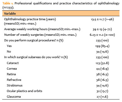
PURPOSE: To assess musculoskeletal symptoms, identify the most affected body areas, and investigate factors associated with the development of musculoskeletal disorders among ophthalmologists in Brazil.
METHODS: A survey was conducted using an online questionnaire and snowball sampling. Statistical analyses were performed using Jamovi version 2.3.28, and graphs were generated using RStudio version 2023.06.2 + 561.
RESULTS: A total of 233 participants (42 ophthalmology residents and 191 ophthalmologists) were included, with a mean age of 40.4 years (standard deviation 11.3; range 25–73 years). Musculoskeletal symptoms were reported by 83% of participants. The cervical region (57.1%), upper back (54.5%), and lumbar region (53.6%) were the most frequently reported sites of pain. A high body mass index was identified in 54.9% of the sample, and 50.2% of participants reported using painkillers in the previous year for musculoskeletal symptoms. The mean duration of professional activity in ophthalmology was 13.5 years, and the mean weekly workload was 39 hours. A significant association was observed between weekly workload and the presence of musculoskeletal disorders (p=0.045).
CONCLUSION: This study demonstrated a high prevalence of musculoskeletal disorders among ophthalmologists in Brazil, particularly involving the cervical, lumbar, and upper back regions, consistent with findings reported in international studies. Important contributing factors include long working hours, a high patient volume, and repetitive or awkward postures during examinations and procedures. Preventive strategies and improvements in working conditions are needed to protect the health and well-being of ophthalmologists.
Keywords: Musculoskeletal Diseases/epidemiology; Back pain; Lumbar Vertebrae; Occupational diseases/epidemiology; Ergonomics; Ophthalmic practice; Ophthalmologists/statistics & numerical data; Brazil/epidemiology
Abstract
PURPOSE: To describe the ophthalmological findings of dry eye disease and its relation to the quality of life of COVID-19 survivors.
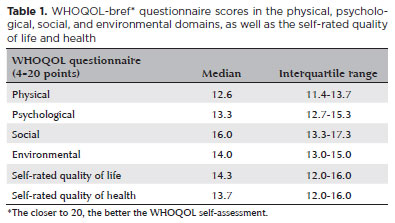
METHODS: COVID-19 survivors who had previously been hospitalized at Hospital das Clínicas de Ribeirão Preto complex underwent an ophthalmological evaluation, which included a dry eye disease questionnaire, break-up time, fluorescein staining, and Schirmer test. We collected the presenting and best-corrected visual acuity, sociodemographic data, personal medical history, and scores from a self-reported quality of life questionnaire (WHOQOL-bref). According to the severity of the acute phase of the disease, the patients were classified into mild-to-moderate, severe, and critical groups.
RESULTS: Ninety-five patients (190 eyes) were evaluated 100 ± 44 days after the onset of COVID-19 symptoms. Of these, 83 patients (87.3%) completed the WHOQOL-bref questionnaire. Ten patients (12.0%) had mild-to-moderate COVID-19, 41 (49.4%) had severe COVID-19, and 32 (38.6%) had critical COVID-19. The median best-corrected visual acuity was logMAR 0 (0-1). Approximately 26.3% patients had a history of dry eye disease or severe dry eye symptoms (frequent or constant ocular dryness and irritation). There was an association between the proportion of patients with dry eye disease and the quality of life (p=0.014) and health (p=0.001). Furthermore, there was a significant trend between the proportion of patients with dry eye disease and how they rated their health and quality of life (p=0.0004 and 0.0027, respectively.
CONCLUSIONS: There is a significant negative correlation between the proportion of patients with dry eye disease and their self-reported quality of life.
Keywords: COVID-19; Coronavirus infections; SARS-CoV-2; Eye diseases; Epidemiology; Ocular surface; Public health
Abstract
PURPOSE: To evaluate the current scenario of rehabilitation services for people with retinal diseases in Brazil.
METHODS: An exploratory study was conducted between February 2023 and June 2023 using a Google Forms questionnaire that was distributed by patient associations.
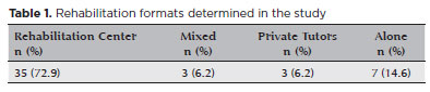
RESULTS: A total of 142 patients, aged 18-80 were included in the study. Forty-eight participants (33.8%) were undergoing rehabilitation, while 94 (66.2%) were not. The main reason for not undergoing rehabilitation was a lack of knowledge about the service (n=41, 43.6%). Healthcare professionals made the most referrals (n=20, 41.7%). Rehabilitation improved the quality of life in 38 (80.9%) participants, and 28 (62.2%) participants were satisfied with the process. There was a statistically significant disparity between patient satisfaction and the locale of rehabilitation implementation. Twenty-three (69.7%) participants who underwent rehabilitation at a specialized center reported satisfaction.
CONCLUSION: The rehabilitation process directly increases the quality of life of individuals with retinal diseases. However, despite the availability of rehabilitation centers in large parts of Brazil, most patients with retinal diseases are not acquainted with the rehabilitation process and do not receive referrals for it. Thus, healthcare providers should increase referrals to rehabilitation centers, and public policies should be formulated to raise awareness among the population regarding the availability of rehabilitation services.
Keywords: Rehabilitation; Quality of life; Retinal diseases; Personal satisfaction; Patient care team; Public policy; Surveys and questionnaires; Brazil
Abstract
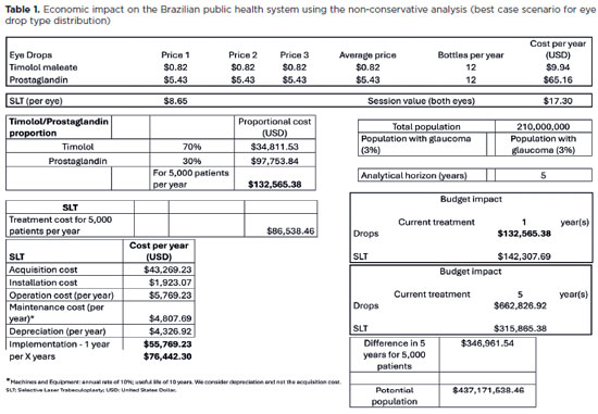
PURPOSE: To evaluate the economic impact of the following initial treatment scenarios for glaucoma on the Brazilian Public Health System (SUS): (1) traditional continuous instillation of hypotensive eye drops and (2) single session of selective laser trabeculoplasty.
METHODS: Economic impact was analyzed in three scenarios, from the least to the most conservative, for a hypothetical cohort of 5,000 individuals with open-angle glaucoma. Thereafter, projections were made on the basis of a glaucoma prevalence of 3% in the 2021 Brazilian population size.
RESULTS: All three scenarios demonstrated that selective laser trabeculoplasty exhibited a significantly lower economic impact than the eye drops on SUS over one and five years. Furthermore, the difference was more than United States Dollar 8 billion at five years when considering 3% of the Brazilian population aged >40 years in 2021.
CONCLUSION: As the initial treatment for primary open-angle glaucoma, selective laser trabeculoplasty exhibited a lower economic impact on SUS than latanoprost and timolol maleate eye drop instillation in all the studied scenarios over one and five-year periods.
Keywords: Glaucoma; Trabeculotomy; Laser therapy; Cost analysis; Health care cost Unified Health System; Brazil
Abstract
PURPOSE: To investigate the clinical benefits of the co-application of bevacizumab and tissue plasminogen activator as adjuncts in the surgical treatment of proliferative diabetic retinopathy.
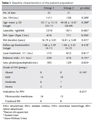
METHODS: Patients who underwent vitrectomy for proliferative diabetic retinopathy complications were preoperatively given intravitreal injection with either bevacizumab and tissue plasminogen activator (Group 1) or bevacizumab alone (Group 2). Primary outcomes were surgery time and number of intraoperative iatrogenic retinal breaks. Secondary outcomes included changes in the best-corrected visual acuity and postoperative complications at 3 months postoperatively.
RESULTS: The mean surgery time in Group 1 (52.95 ± 5.90 min) was significantly shorter than that in Group 2 (79.61 ± 12.63 min) (p<0.001). The mean number of iatrogenic retinal breaks was 0.50 ± 0.59 (0-2) in Group 1 and 2.00 ± 0.83 (0-3) in Group 2 (p<0.001). The best-corrected visual acuity significantly improved in both groups (p<0.001). One eye in each group developed retinal detachment.
CONCLUSION: Preoperative co-application of bevacizumab and tissue plasminogen activator as adjuncts in the surgical treatment of proliferative diabetic retinopathy shortens the surgery time and reduces the number of intraoperative iatrogenic retinal breaks.
Keywords: Diabetic retinopathy; Bevacizumab; Plasminogen activators; Vitrectomy; Operative time
Abstract
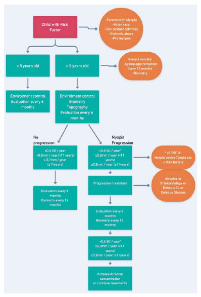
Esta revisão foi baseada na literatura médica e na experiência clínica de um comitê de especialistas membros da Sociedade Brasileira de Oftalmologia Pediátrica e da Sociedade Brasileira de Lentes de Contato e Córnea. Rotineiramente as crianças devem ser submetidas a topografia da córnea no primeiro exame e visitas semestrais com refração cicloplegiada e biometria óptica anual. A progressão da miopia foi definida como um aumento anual no equivalente esférico maior que 0,50 D/ano ou do comprimento axial maior que 0,3 mm (até 10 anos) ou 0,2 mm (mais de 11 anos). Os tratamentos propostos para a progressão são controle ambiental, atropina em baixa concentração, óculos com defocus, lentes de contato ou ortoceratologia, devendo-se considerar associações para casos não controlados. O tratamento deve ser realizado por pelo menos 2 anos. O presente documento é uma diretriz para diagnóstico, tratamento e acompanhamento de crianças pré-míopes e míopes no Brasil.
Keywords: Miopia; Distúrbios pupilares; Progressão da doença; Atropina; Refração ocular; Midriáticos; Lentes de contato; Biometria; Criança; Brasil
 ABO is licensed under a Creative Commons Attribution-NonComercial 4.0 Internacional.
About
Issues
Editorial Board
Submission

Arquivos Brasileiros de Oftalmologia
Official publication of Brazilian Council of Ophthalmology - Conselho Brasileiro de Oftalmologia (CBO)
Rua Casa do Ator, 1.117 - 2nd floor - Zip Code: 04546-004
São Paulo - SP, Brazil
TEL: +55 11 3266-4000
E-mail: [email protected]
