Arq. Bras. Oftalmol. 1999;62 (6 )
:705-711
| DOI: 10.1590/S0004-27491999000600010
Abstract
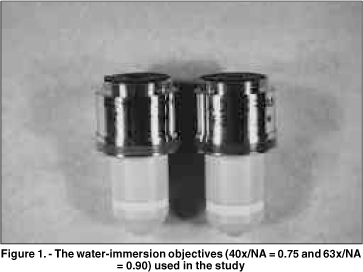
Objetivo: Este estudo prospectivo compara as imagens de microscopia confocal do epitélio corneano do coelho e do homem, obtidas através de 2 objetivas com aberturas numéricas (AN) diferentes. Métodos: Dez olhos de coelhos foram enucleados e fixados através de um suporte pneumático para garantir o melhor desempenho de cada objetiva. Cinco pacientes normais foram selecionados após consentimento. Os olhos de coelhos e dos pacientes foram previamente examinados na lâmpada de fenda. O exame de microscopia confocal (Tomey, Erlangen-Tennenlohe, Alemanha) foi realizado com as objetivas Achroplan 40x/AN = 0,75 e 63x/AN = 0,9 (Zeiss, Oberkochen, Alemanha). Imagens selecionadas do epitélio corneano foram avaliadas qualitativamente com relação ao tamanho, forma e refletividade das células. Resultados: As células no epitélio superficial dos coelhos e dos pacientes, previamente à descamação, tiveram uma refletividade maior que as células adjacentes. Este aspecto foi claramente observado somente com a objetiva 63x/AN = 0,9. As camadas basal e intermediária do epitélio em coelhos foram visualizadas somente através desta objetiva. Estas camadas nos pacientes tornaram-se mais nítidas com a objetiva de abertura numérica maior (63x/AN = 0,9). Conclusão: Uma objetiva de abertura numérica elevada produz melhor resolução dos cortes ópticos, facilitando a análise das camadas do epitélio no coelho e no homem.
Keywords: Corneal epithelium; Confocal microscopy; Epitélio corneano; Microscopia confocal
Arq. Bras. Oftalmol. 2020;83 (4 )
:277-282
| DOI: 10.5935/0004-2749.20200049
Abstract
Objetivo: Este estudo foi realizado para avaliar os resultados do cross-linking corneano acelerado em córneas ceratocônicas com os valores mais baixos de paquimetria <400 μm.
Métodos: O estudo incluiu 28 olhos de 24 pacientes. As acuidades visuais não corrigidas e melhor corrigidas (logMAR), leituras ceratométricas mais planas e íngremes, espessura corneana central no ponto mais fino, aberrações corneanas de mais alta ordem e a sensibilidade ao contraste foram avaliadas antes e em 1, 3, 6, 12 e 24 meses após a realização do do cross-linking.
Resultados: A média da acuidade visual melhor corrigida e a sensibilidade ao contraste aumentaram (p=0,02, p=0,03, respectivamente), enquanto a média da acuidade visual não corrigida não diferiu significativamente (p>0,05) aos 24 meses após o cross-linking, comparada com medidas antes do procedimento. Embora a leitura da média da ceratometria mais plana não tenha apresentado alteração significativa (p=0,58), a leitura ceratométrica mais íngreme diminuiu quando comparada ao seu valor antes do cross-linking (p=0,001). Não foi observada alteração na média da espessura corneana central no ponto mais fino aos 24 meses após o cross-linking em comparação com seu valor antes do procedimento (p=0,12).
Conclusão: O cross-linking corneano acelerado nos olhos ceratocônicos com córneas finas pode interromper a progressão do ceratocone nas córneas mais finas que 400 μm 24 meses após o tratamento.
Keywords: Ceratocone; Paquimetria corneana; Reagentes para ligações cruzadas; Riboblavina/uso terapêutico
Arq. Bras. Oftalmol. 2021;84 (4 )
:324-329
| DOI: 10.5935/0004-2749.20210046
Abstract
OBJETIVO: O ceratocone na população pediátrica apresenta algumas particularidades em relação à população adulta. O maior desafio é devido à doença ser geralmente mais severa e rapidamente progressiva em crianças.
MÉTODOS: Este artigo utiliza uma análise retrospectiva para relatar o uso do crosslinking em jovens menores de 18 anos e sua evolução pelo menos 24 meses após o procedimento. Foram estudados 12 olhos de 10 pacientes, e dados como acuidade visual com e sem correção, ceratometria máxima, espessura corneana, espessura foveal e microscopia endotelial avaliados no pré e pós-operatórios. O crosslinking corneano foi realizado em todos os pacientes pelo mesmo cirurgião.
RESULTADOS: Observou-se uma tendência de redução do valor do Kmax e melhora da acuidade visual corrigida em todos os momentos de pós operatório. Com relação à paquimetria, observou-se afinamento corneano do ponto mais fino, nos primeiros quatro meses de pós-operatório.
CONCLUSÃO: Resultados encorajadores foram obtidos com relação à estabilização da doença, progressão e segurança do procedimento, corroborando com as conclusões de outros autores. A importância do diagnóstico precoce e do acompanhamento a curto prazo do paciente deve ser destacada.
Keywords: Ceratocone/diagnóstico; Ceratocone/tratamento farmacológico; Córnea; Doenças da córnea; Topografia da córnea; Colágeno/metabolismo; Raios ultravioleta; Reagentes para ligações cruzadas/uso terapêutico; Riboflavina/uso terapêutico; Acuidade visual; Adolesc
Arq. Bras. Oftalmol. 2022;85 (4 )
:389-398
| DOI: 10.5935/0004-2749.20220042
Abstract
Objetivos: Explorar sistematicamente as mudanças dinâmicas e a sequência temporal no processo de apoptose de células epiteliais corneanas após excesso de irradiação com ultravioleta B.
Métodos: A radiação ultravioleta B (144 mJ/cm2) foi utilizada para irradiar células epiteliais da córnea de rato durante 2h. A morfologia celular foi observada por meio de microscópio de contraste de interferência diferencial, e os números de diferentes tipos de células apoptóticas foram contados e registrados pelo software ImageJ. A viabilidade celular foi medida pelo método brometo de 3- (4, 5-dimetil-2-tiazolil) -2, 5-difenil-2-H-tetrazólio. A taxa apoptótica celular e a perda do potencial da membrana mitocondrial foram detectadas por meio de análises citométricas de fluxo. Os níveis de expressão de três genes apoptóticos foram medidos por reação em cadeia da polimerase quantitativa em tempo real em diferentes momentos dentro de 0-24 h após a irradiação.
Resultados: Após 144 mJ/cm2 de irradiação com ultravioleta B por 2h, os níveis de expressão de caspase-8 e Bax foram maiores em 0h; o potencial da membrana mitocondrial diminuiu a 0h e permaneceu constante por 6h na cultura subsequente. Às 6h, a caspase-3 foi ativada. A diminuição da viabilidade celular e o aumento da taxa apoptótica atingiu o pico em 6h. A expressão de caspase-3 diminuiu dentro de 12 - 24 h, levando a um declínio na taxa apoptótica e alteração no estágio apoptótico.
Conclusões: As células epiteliais da córnea apresentaram uma apoptose rápida após excesso de irradiação com ultravioleta B, e esse processo foi associado tanto à via extrínseca como à via intrínseca.
Keywords: Irradiação com ultravioleta B; Radiação; Epitélio anterior; Célula epitelial; Sobrevivência celular; Apoptose; Ratos
Arq. Bras. Oftalmol. 2025;88 (1 )
:1-4
| DOI: 10.5935/0004-2749.2023-0056
Abstract
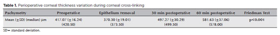
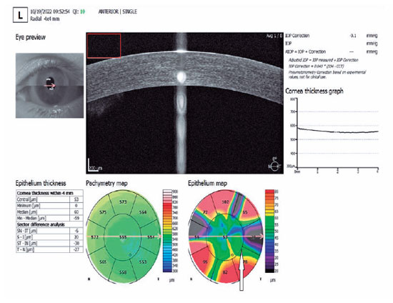
PURPOSE: This study aimed to analyze variations in intraoperative corneal thickness during corneal cross-linking in patients with keratoconus and to investigate its possible correlation with presurgical maximal keratometry (Kmax) and pachymetry.
METHODS: This was a prospective case series. We used a method similar to the Dresden protocol, with the application of hydroxypropyl methylcellulose 0.1% hypo-osmolar riboflavin in corneas between 330 and 400 µm after epithelium removal. Corneal thickness was measured using portable calipers before and immediately after epithelium removal, and 30 and 60 min after the procedure.
RESULTS: The 30 patients in this study were followed up for one year. A statistically significant difference was observed in pachymetry values during the intraoperative period (p<0.0001) and an increase of 3.05 µm (95%CI: 0.56–5.54) for each diopter was seen after epithelium removal (p0.019). We found an average Kmax difference of −2.12 D between men and women (p0.013). One year after treatment, there was a statistically significant reduction in pachymetry (p<0.0001) and Kmax (p0.0170) values.
CONCLUSIONS: A significant increase in pachymetry measurements was seen during the procedure, and most patients showed a regression in Kmax and pachymetry values one year after surgery.
Keywords: Corneal pachymetry; corneal topography; cross-linking reagents/therapeutic use; hypromellose derivatives; keratoconus/surgery; riboflavin/therapeutic use
Arq. Bras. Oftalmol. 2025;88 (3 )
:1-7
| DOI: 10.5935/0004-2749.2023-0309
Abstract
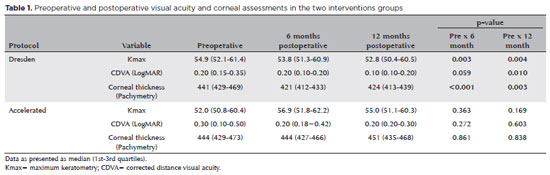
PURPOSE: Keratoconus presents certain peculiarities in pediatric patients when compared with adults. The greatest challenge in children is that the disease is more severe and faster in progression. In this retrospective study, we aimed to compare the accelerated and Dresden protocols for corneal crosslinking in patients aged <18 years who were followed-up for at least 12 months.
METHODS: A total of 36 eyes from 27 patients were included in the study. The best corrected and uncorrected visual acuity, maximal keratometry, corneal thickness, foveal thickness, and endothelial microscopy findings were evaluated at baseline and during the postoperative period at one, three, and six months. Thereafter, the patients were evaluated at one, three, six and twelve months postoperative. Corneal crosslinking was performed in all patients via the Dresden protocol (n=21 eyes) or the accelerated protocol (n=15 eyes). Data between the two groups were compared and XY statistical analysis was used.
RESULTS: Both protocols were effective in halting keratoconus progression. No patient had progression at the 12-month follow-up. A significant reduction in Kmax and improvement in the corrected distance visual acuity were observed only in the Dresden protocol group. Although the Dresden protocol was superior to the accelerated protocol in reducing Kmax (p=0.002), there was no significant difference in corrected distance visual acuity between the two groups.
CONCLUSION: The accelerated protocol is as efficient as the Dresden protocol in stabilizing keratoconus progression. Although the Dresden protocol was superior to the accelerated protocol in reducing the Kmax, it did not produce better clinical results. Thus, the accelerated protocol is an efficient option. Furthermore, considering the advantages of reduced surgical time, the accelerated protocol is effective in halting keratoconus progression in the pediatric age group.
Keywords: Keratoconus; Corneal diseases; Ultraviolet rays; Cross-linking reagents; Visual acuity
Arq. Bras. Oftalmol. 2024;87 (3 )
:1-8
| DOI: 10.5935/0004-2749.2022-0004
Abstract
OBJETIVO: Examinar os efeitos do tratamento de reticulação unilateral do colágeno corneano na acuidade visual e os achados topográficos em olhos não tratados de pacientes com ceratocone progressivo bilateral.
MÉTODOS: Foram rastreados retrospectivamente pacientes com ceratocone progressivo submetidos a tratamento de reticulação. Foram incluídos no estudo 188 olhos não tratados de 188 pacientes tratado unilateralmente com reticulação padrão ou acelerada e que recusaram o procedimento de reticulação no outro olho. A acuidade visual e os achados topográficos dos olhos não tratados foram obtidos no pré- e pós-operatório no 1o, 3o, 6o, 12o, 24o, 30o e 36o mês.
RESULTADOS: As alterações ao longo do tempo foram semelhantes para as variáveis examinadas nos olhos não tratados de pacientes tratados com métodos de reticulação padrão e acelerado (p>0,05). No 12º mês, 136 olhos não tratados (95,8%) estavam estáveis, de acordo com os critérios de progressão. Apenas quatro olhos (8%) mostraram progressão no 24o mês. Nenhuma progressão foi observada nos 16 pacientes que tiveram um acompanhamento de 36 meses.
CONCLUSÕES: O estudo mostrou que os olhos não tratados de pacientes com ceratocone progressivo bilateral não apresentaram taxas de progressão significativas após o tratamento unilateral com reticulação.
Keywords: Topografia da córnea; Reagentes de ligações cruzadas; Ceratocone; Fármacos fotossensibilizantes; Colágeno/uso terapêutico; Fotoquimioterapia/métodos; Acuidade visual
Arq. Bras. Oftalmol. 2024;87 (4 )
:1-6
| DOI: 10.5935/0004-2749.2022-0128
Abstract
Objetivo: Relatar um experimento projetado para determinar alterações anatômicas em córneas porcinas após a colocação de um novo implante depolímero na córnea.
Métodos: Foi utilizado olho de porco ex vivo. Um novo agente modelador biocompatível, de colágeno tipo 1, com 6mm de diâmetro foi moldado com excimer laser em sua face posterior, para criar três formatos planocôncavos. Os implantes foram inseridos dentro de um bolsão, dissecado manualmente, a 200 micrômetros (µm). Foram definidos três grupos de tratamento: grupo A (n=3), teve a profundidade máxima de ablação de70 µm; o grupo B (n=3), profundidade máxima de ablação de 64 µm; e o grupo C (n=3), profundidade máxima de ablação de 104 µm, com buraco central. O grupo controle, D (n=3), foi incluído, com a criação do bolsão estromal, porém sem inserir o material. A avaliação desses olhos foi realizada por tomografia de coerência óptica (OCT) e por tomografia corneana.
Resultados: A tomografia corneana mostrou uma tendência para diminuição da ceratometria média em todos os 4 grupos. A tomografia de coerência óptica mostrou córneas com implantes localizados no estroma anterior e aplanamento visível, enquanto as córneas não mudaram qualitativamente o formato no grupo controle.
Conclusões: O novo implante de biomaterial planocôncavo descrito aqui foi capaz de remodelar a córnea em modelo de animal ex vivo, resultando no aplanamento corneano. Novos estudos são necessários usando modelos animais in vivo para confirmar tais achados.
Keywords: Córnea; Cirurgia da córnea a laser; Substância própria; Proteses e implantes; Lasers de excimer; Materiais biocompatíveis; Animais; Suínos
Arq. Bras. Oftalmol. 2025;88 (5 )
:1-9
| DOI: 10.5935/0004-2749.2024-0326
Abstract
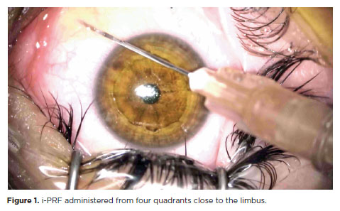
PURPOSE: This study was conducted to investigate the effect of injectable platelet-rich fibrin on the recovery of compromised epithelium due to crosslinking treatment.
METHODS: In this comparative study, the epithelial closure rates and in vivo confocal biomicroscopy results of 26 patients with keratoconus who underwent subconjunctival injection of injectable platelet-rich fibrin near the limbus after epithelium-off corneal crosslinking treatment were compared with those of 25 patients who did not receive the injection of injectable platelet-rich fibrin.
RESULTS: The average time to epithelial defect closure in the injectable platelet-rich fibrin group was 2.76 ± 0.90 days compared to 3.56 ± 0.86 days in the non-injectable platelet-rich fibrin group (p=0.003). At the end of the 1st month, the mean subbasal nerve plexus density was 1.26 ± 1.61 nerves/mm2 in the injectable platelet-rich fibrin group, whereas it was 0.72 ± 0.89 nerves/mm2 in the non-injectable platelet-rich fibrin group (p=0.016). By the 3rd month, the density increased to 3.42 ± 1.13 nerves/mm2 in the injectable platelet-rich fibrin group and 2.36 ± 1.15 nerves/mm2 in the non-injectable platelet-rich fibrin group (p=0.002). Similarly, the anterior stromal keratocyte density at the end of the 1st month was 93.6 ± 33.5 cells/mm2 in the injectable platelet-rich fibrin group compared to 67.3 ± 26.4 cells/mm2 in the non-injectable platelet-rich fibrin group (p=0.001). By the end of the 3rd month, the density increased to 255.2 ± 45.7 cells/mm2 in the injectable platelet-rich fibrin group and 222.1 ± 43.6 cells/mm2 in the non-injectable platelet-rich fibrin group (p=0.011). In the non-injectable platelet-rich fibrin group, one patient developed a sterile infiltrate at the end of the 1st week, whereas no complications were observed in the injectable platelet-rich fibrin group.
CONCLUSION: Subconjunctival injectable platelet-rich fibrin application is an effective and safe method for corneal epithelial healing after crosslinking treatment.
Keywords: Keratoconus; Platelet-rich fibrin; Epithelium; corneal; Corneal crosslinking; Wound healing
Arq. Bras. Oftalmol. 2024;87 (3 )
:1-7
| DOI: 10.5935/0004-2749.2023-0049
Abstract
PURPOSE: To investigate the association of pre-photorefractive keratectomy Schirmer-1 test value with post-photorefractive keratectomy central corneal epithelial thickness, ocular surface disease index score, and uncorrected distance visual acuity.
METHODS: Patients were categorized according to preoperative Schirmer-1 value: the normal Schirmer Group (n=54; Schirmer-1 test value, >10 mm) and the low Schirmer Group (n=52; Schirmer-1 test value, between 6 and 10 mm). We analyzed ablation depth, visual acuity, result of Schirmer-1 test (with anesthesia), tear film break-up time, ocular surface disease index score, central corneal epithelial thickness, and spherical equivalent refraction.
RESULTS: We found significant differences between the groups in Schirmer-1 test value, tear film break-up time, and ocular surface disease index score, both preoperatively and postoperatively (p<0.001). The preoperative central corneal epithelial thicknesses of the two groups were similar (p>0.05). After photorefractive keratectomy, the Schirmer-1 test value and spherical equivalent refraction decreased in both groups (p<0.05), and ocular surface disease index scores and central corneal epithelial thickness values increased in the low Schirmer Group (p<0.001) but not in the normal Schirmer Group (p>0.05). The postoperative central corneal epithelial thicknesses of the low Schirmer Group were significantly higher than those of the normal Schirmer Group (p<0.001). Postoperative uncorrected distance visual acuity did not differ significantly between the two groups (p>0.05).
CONCLUSIONS: In patients with low Schirmer-1 test values before photorefractive keratectomy, the corneal epithelium thickened and ocular surface complaints increased during the postoperative period. However, changes in the corneal epithelium did not affect the postoperative uncorrected distance visual acuity. To reduce postoperative problems on the ocular surface in these patients, we recommend that dry eye be treated before photorefractive keratectomy.
Keywords: Epithelium, corneal; Cornea; Photorefractive keratectomy; Schirmer test; Visual acuity
Arq. Bras. Oftalmol. 2025;88 (3 )
:1-6
| DOI: 10.5935/0004-2749.2024-0207
Abstract
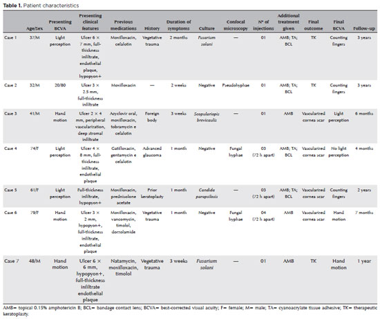
PURPOSE: This study aimed to report the use, efficacy, and safety of intracameral voriconazole as an adjuvant treatment for deep fungal keratitis.
METHODS: This was a prospective case series of seven eyes with fungal keratitis with anterior chamber involvement or a corneal ulcer refractory to conventional topical treatment. In addition to topical treatment with 0.15% amphotericin B eye drops, voriconazole 50 μg/ 0.1 mL
was administered to the anterior chamber of each affected eye up to four times within 72 h. The primary outcome measures were healing (fungal eradication) and the need for therapeutic keratoplasty. Best-corrected visual acuity was a secondary outcome measure.
RESULTS: Three cases were confirmed by confocal microscopy, and four were diagnosed from positive culture tests. At presentation, one patient had a best-corrected visual acuity of 20/80, while all others had hand motion or worse. Four cases received one intracameral injection, two cases received three, and one case received four injections. There were no complications after any of the intracameral voriconazole injections. Four patients had imminent corneal perforations and were treated with cyanoacrylate adhesive and bandage contact lenses. Four patients recovered from the infection, and three underwent therapeutic keratoplasty. The final best-corrected visual acuity was improved in two cases but all patients had a final visual acuity of counting fingers or worse.
CONCLUSION: As an adjuvant treatment for deep fungal keratitis, intracameral voriconazole injection is a feasible option. Although fungal eradication was achieved in all patients, three required therapeutic keratoplasty and all patients had unsatisfactory visual acuity outcomes.
Keywords: Antifungal agents; Fungi; Corneal transplantation; Keratitis; Eye infections, fungal; Voriconazole
Arq. Bras. Oftalmol. 2024;87 (2 )
:1-5
| DOI: 10.5935/0004-2749.2022-0273
Abstract
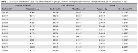
PURPOSE: To determine the absorbance coefficient of the thin porcine cornea to ultraviolet-A radiation (365 nm) submitted for crosslinking.
METHODS: This in vitro, benchtop experiment using cadaver tissue study analyzed 12 porcine corneal lamellas, which were obtained using a microkeratome after mechanical de-epithelization and separated into three thickness groups: 180, 300, and 360 µm. The corneal thickness values were measured by anterior-segment optical coherence tomography. All lamellas had ultraviolet-A (365 nm) absorbance measured with a 96-well plate spectrophotometer using an ultraviolet transparent microplate before riboflavin instillation and pre- and post-crosslinking according to the Dresden protocol.
RESULTS: The ultraviolet absorbance profiles of the 180, 300, and 360 µm groups were obtained as a-coefficients of 12.85, 76.55, and 120.27, respectively. A theoretical formula was calculated though a statistical analysis that demonstrated the correlation between stromal lamellar thickness and ultraviolet absorbance.
CONCLUSIONS: Corneal thickness and ultraviolet-A spectral absorbance of corneal lamellas showed linear correlation. These findings can potentially contribute to the optimization of ultraviolet-A application during crosslinking, making the treatment of corneas with thickness <400 µm safe and personalized energy delivery for each corneal thickness.
Keywords: Ultraviolet light; Spectrophotometry; Crosslinking; Keratoconus; Corneal thickness
 ABO is licensed under a Creative Commons Attribution-NonComercial 4.0 Internacional.
02-tab01tb.jpg)
03-tab01.jpg)
13-tab01tb.jpg)
08-tab01.jpg)
09-fig01.jpg)
17-equ01.jpg)