Arq. Bras. Oftalmol. 2024;87 (1 )
:1-6
| DOI: 10.5935/0004-2749.2021-0536
Abstract
Objetivo: Avaliar os segmentos anterior e posterior em recém-nascidos a termo durante um período de 1,5 anos.
Métodos: Foram analisados recém-nascidos a termo que tiveram os olhos examinados entre junho de 2019 e dezembro de 2020, e os resultados foram registrados retrospectivamente.
Resultados: O estudo foi composto por 2.972 recém-nascidos com média de uma semana de nascimento de 38,7 ± 1,2 semanas e um peso médio ao nascer de 3235 ± 464 g. Os recém-nascidos foram examinados em média pós-natal de 49,3 ± 18,9 dias. Dos recém-nascidos, 185 (6,2%) apresentaram resultados oculares anormais. Os achados oculares anormais mais prevalentes foram hemorragia da retina em 2,3% (n=68) e alterações brancas na retina periférica em 1,9% (n=55) dos recém-nascidos. Casos de patologias de disco óptico (n=20), nevo de coroide (n=10), coloboma iris-coroide (n=5), hemorragia subconjuntival (n=6), alteração pigmentar da retina não específica (n=4), catarata congênita (n=3), Sinequia posterior (n=3), nevo da íris (n=3), opacidade da córnea (n=1), coloboma de coroide (n=1), coloboma de íris (n=1), buftalmos (n=1), anoftalmia (n=1), microftalmia (n=1), hemangioma de pálpebra (n=1) e hemorragia vítrea (n=1) contabilizaram coletivamente cerca de 2% dos recém-nascidos. As patologias que potencialmente prejudicam a visão, detectadas por exame ocular, representaram 1,2% dos recém-nascidos (n=37).
Conclusão: O achado mais prevalente de exames oculares de recém-nascidos neste estudo foi hemorragia da retina. Exames oftalmológicos em recém-nascidos podem ser úteis na identificação de doenças que podem impactar a visão deles, podendo ser curáveis ou levar à ambliopia no longo prazo.
Keywords: Anormalidades do olho/diagnóstico; Hemorragia retiniana; Triagem neonatal; Seleção visual; Humanos; Recém-nascido.
Arq. Bras. Oftalmol. 2020;83 (6 )
:490-496
| DOI: 10.5935/0004-2749.20200090
Abstract
Objetivo: Comparar a espessura central foveal, a da camada de fibras nervosas da retina e a da coróide subfoveal através da tomografia de coerência óptica swept-source em crianças de 5 anos de idade com história de retinopatia da prematuridade (RP) tratada com bevacizumabe intravítreo, ou com fotocoagulação a laser, com crianças em regressão espontânea da retinopatia da prematuridade, e com crianças saudáveis da mesma idade.
Métodos: Um total de 79 crianças foi dividido em quatro grupos. Grupo 1: crianças que receberam tratamento com bevacizumabe intravítreo. Grupo 2: crianças que foram tratadas com fotocoagulação a laser. Grupo 3: crianças que tiveram regressão espontânea da retinopatia da prematuridade . Grupo 4: crianças da mesma idade saudáveis e nascidas a termo. As funções visuais e o status refrativo foram avaliados aos 5 anos de idade. A análise de tomografia de coerência óptica foi feita por um dispositivo do tipo swept-source (DRI-OCT Triton; Topcon, EUA).
Resultados: Haviam 12 crianças (15,2%) no grupo 1, 23 crianças (29,1%) no grupo 2, 30 crianças (38%) no grupo 3 e 14 crianças (17,7%) no grupo 4. A distribuição por sexo foi semelhante em todos os grupos (p=0,420). A acuidade visual com a melhor correção mostrou-se significativamente maior no grupo 4 em comparação com os grupos 1, 2 e 3 (respectivamente, p=0,035, p=0,001 e p=0,001). Os resultados dos erros de refração foram semelhantes em todos os grupos (p=0,119). A espessura foveal central mostrou-se significativamente maior no grupo 2 do que no grupo 1 (p=0,023). Não foram observadas diferenças significativas entre os grupos quanto à espessura da camada de fibras nervosas da retina e à espessura da coroide subfoveal (p>0,05).
Conclusões: Os desfechos visuais funcionais foram melhores nas crianças saudáveis nascidas a termo, em comparação com aqueles observados nas crianças com história de retinopatia da prematuridade tratada ou com regressão espontânea. O tratamento com laser teve um efeito significativo na espessura foveal central em crianças de 5 anos de idade, nascidas prematuras, como revelado pela tomografia de coerência óptica swept-source.
Keywords: Retinopatia da prematuridade/tratamento farmacológico; Tomografia de coerência óptica; Bevacizumab/uso terapêutico; Fotocoagulação; Recém-nascido
Arq. Bras. Oftalmol. 2022;85 (4 )
:364-369
| DOI: 10.5935/0004-2749.20220049
Abstract
Objetivos: Avaliar duas unidades de terapia intensiva neonatais do Paraná e identificar os fatores de risco que levam ao desenvolvimento da retinopatia da prematuridade nestas unidades neonatais.
Metodos: Foi realizado um estudo de coorte, prospectivo, com avaliação dos bebês prematuros examinados no período de 12 meses com idade gestacional ≤32 semanas e/ou com peso de nascimento ≤1500 gramas, internados na unidade de cuidados intensivos neonatais do Hospital do Trabalhador e do Hospital infantil Waldemar Monastier, que recebe neonatos transportados das maternidades de todo o estado do Paraná.
Resultados: A incidência de retinopatia da prematuridade foi maior no Hospital Infantil Waldemar Monastier, entre os prematuros que necessitaram de transporte do local de nascimento para a unidade de cuidados intensivos (52,2% vs 29,6%), Os fatores de risco associados ao desenvolvimento da doença foram; Maior número de dias de internamento, baixa idade gestacional ao nascimento, maior tempo de uso de oxigênio, uso de drogas vasoativas, ausência de uso de corticoide pré-natal, presença de hemorragia intracraniana e qualquer tipo de alteração da glicemia.
Conclusão: Os cuidados neonatais precoces e o transporte do recém-nascido pré- termo podem influenciar a ocorrência e o prognostico da retinopatia da prematuridade.
Keywords: Retinopatia da prematuridade; Recém-nascido; Recém-nascido prematuro; Doenças do prematuro; Cegueira
Arq. Bras. Oftalmol. 2022;85 (2 )
:136-143
| DOI: 10.5935/0004-2749.20220022
Abstract
Objetivo: Estimar a epidemiologia do pterígio; sua correlação com sintomas de olho seco e com potenciais preditores sistêmicos e oculares.
Métodos: Estudo transversal, de base populacional, no qual foram realizadas visitas domiciliares aleatórias a 600 participantes, com 40 anos ou mais de idade, em Ribeirão Preto-SP (n=420) e Cassia dos Coqueiros-SP (n=180), Brasil. Uma entrevista estruturada com um questionário detalhado foi usada para coletar informações sobre demografia e possíveis fatores de risco. Em um segundo momento, participantes aleatórios com pterígio (n=63) ou não (n=110) foram avaliados quanto a alterações na superfície ocular.
Resultados: A frequência de pterígio em Ribeirão Preto foi de 21%; 15.7% entre as mulheres e 32.1% entre os homens (p=0,0002). Em Cássia dos Coqueiros, essa taxa foi de 19.4%; onde 17.3% eram mulheres e 25.5% eram homens (p=0,28). A média de idade naqueles afetados pelo pterígio foi superior à dos participantes sem pterígio, 65,6 ± 10,5 e 61,2 ± 12,0 anos, respectivamente (p=0,02). Houve uma correlação positiva entre o pterígio e história prévia de radioterapia e quimioterapia (p<0,0001 para ambos). Houve maior coloração de fluoresceína na córnea e maior coloração de lissamina verde na conjuntiva em olhos com pterígio (p=0,0003 e 0,0001, respectivamente).
Conclusão: Encontramos uma alta frequência de pterígio em duas populações adultas brasileiras, principalmente em homens e idosos. Danos na superfície ocular e história prévia de radioterapia e/ou quimioterapia foram associados ao pterígio.
Keywords: Pterígio/epidemiologia; Síndrome do olho seco; Prevalência; Fatores de risco
Arq. Bras. Oftalmol. 2025;88 (6 )
:1-9
| DOI: 10.5935/0004-2749.2024-0411
Abstract
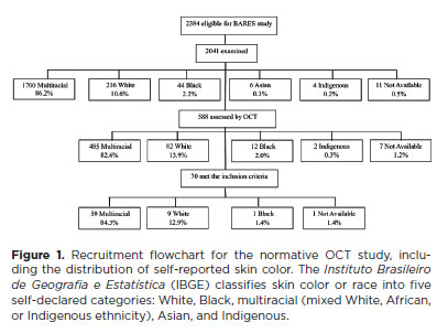
PURPOSE: This study evaluated macular thickness using spectral-domain optical coherence tomography in healthy participants from a population-based eye survey.
METHODS: The Brazilian Amazon Region Eye Survey was a population-based study assessing the prevalence and causes of visual impairment, blindness, and ocular diseases in adults aged ≥45 years from urban and rural areas of Parintins. A subgroup was selected based on inclusion criteria for both eyes: best-corrected visual acuity ≥20/32, normal eye examination results, and no prior ocular surgery. Scans were performed using the iVue optical coherence tomography device. Measurements were taken from the nine subfields defined by the Early Treatment Diabetic Retinopathy Study, examining the full retina as well as the inner and outer retinal layers. Associations of retinal thickness with age and sex were also analyzed. Statistical significance was set at p≤0.05.
RESULTS: In total, 70 healthy participants (25 males), aged 45–65 years (mean=52 ± 5), were included. Mean central foveal thickness was 248.71 ± 18.73 μm. A significant age-related reduction in macular thickness was observed, particularly in the inner superior parafovea (p=0.036), nasal perifovea (p=0.001), superior perifovea (p=0.028), outer layer of inferior parafovea (p=0.049), and the inferior perifovea of the full retina (p=0.029). Males showed significantly greater thickness in the outer layer, especially in the outer parafovea (p=0.004) and perifovea (p<0.0001).
CONCLUSIONS: This study established normative macular thickness values for healthy older adults in the Brazilian Amazon region using spectral-domain optical coherence tomography. Age and sex were found to significantly influence macular thickness and should be considered when interpreting measurements. These data will support future studies of retinal diseases in this population.
Keywords: Retinal diseases/diagnosis; Macula lutea/pathology; Macular degeneration/diagnosis; Diabetic retinopathy/diagnosis; Vision, low; Vision tests; Tomography, optical coherence/methods; Young adult; Cross-sectional studies; Brazil/epidemiology
Arq. Bras. Oftalmol. 2025;88 (6 )
:1-7
| DOI: 10.5935/0004-2749.2025-0083
Abstract
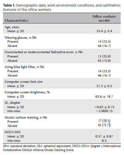
PURPOSE: To examine how ophthalmological features, screen exposure duration, and break habits among office employees affect ocular surface parameters.
METHODS: This single-center cross-sectional study involved two assessments on the same day: one before and one after a visual display terminal task. During the initial assessment, information on screen use was gathered, and refractive error, anterior segment examination, tear breakup time, and Schirmer test measurements were conducted. Participants tracked their screen usage and break durations throughout the day. At the end of the workday, tear breakup time and Schirmer I tests were repeated. Baseline and follow-up results were compared, and regression analysis was performed to identify factors linked to tear breakup time reduction.
RESULTS: The study enrolled 60 female office employees. Their mean screen time was 269.26 ± 70.21 min, with an average break duration of 151.93 ± 46.24 min. Tear breakup time at the second assessment (6.38 ± 2.70) was significantly lower than at baseline (8.62 ± 2.73) (p<0.001), whereas Schirmer test scores showed no significant change (p>0.05). Tear breakup time reduction was noted in 54 participants (90.0%), with a significant association between tear breakup time decrease percentage and screen exposure (p=0.001, r=0.463). Regression analysis showed that uncorrected or undercorrected refractive error was an independent risk factor for a ≥30% tear breakup time reduction, while taking more frequent short breaks (<15 min) acted as a protective factor.
CONCLUSIONS: Taking more frequent short breaks (<15 min) and correcting refractive errors help prevent intra-day tear breakup time decline during visual display terminal use. Structuring breaks to support tear film stability is advisable for occupations that require regular visual display terminal tasks.
Keywords: Tear film; Screen time; Tear breakup time; Office workers; Protective factors; Lacerations; Refractive errors; Risk factors.
Arq. Bras. Oftalmol. 2025;88 (2 )
:1-7
| DOI: 10.5935/0004-2749.2023-0265
Abstract
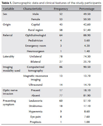
PURPOSE: Although Brazil has a high prevalence of retinoblastoma, there is a lack of epidemiological data on the disease. Thus, in this study, we aimed to evaluate the epidemiological profile of patients diagnosed with retinoblastoma in the ophthalmology department of a pediatric tertiary referral hospital in Ceara, Brazil.
METHODS: A descriptive and cross-sectional study was conducted by retrospectively analyzing the clinical and socioeconomic data from the medical records of pediatric patients followed-up at the hospital between 2007 and 2021. Retinoblastoma was diagnosed on the basis of a fundoscopic or histopathologic examination.
RESULTS: The data of 105 patients were included in the study, and the mean patient age at the time of diagnosis was 1.7 years. Most of the patients were women (50.5%) and hailed from rural areas (57.4%), which was associated with a higher tumor stage. Of the 150 patients, 57.1% initially presented with leukocoria. Ocular hyperemia was associated with more advanced stages of retinoblastoma (p=0.004). Bilateral involvement was observed in 25.7% of the patients and at a significantly younger age (p=0.009). The presence of retinal detachment, vascularized lesions, and vitreous seeds significantly increased the likelihood of requiring enucleation.
DISCUSSION: This study presents an epidemiological description of retinoblastoma in Brazil, which highlights the significance of early detection. Delayed diagnosis is associated with a poorer visual prognosis and higher mortality rate, particularly in patients with unilateral disease. Risk factors for a more severe disease were retinal detachment, vascularized lesions, and vitreous seeds. The correlation between histopathological features and clinical outcomes was limited.
CONCLUSION: Further studies are required to assess the influence of ocular hyperemia, fundoscopic assessment, and histopathologic findings on the prognosis of retinoblastoma. Moreover, it is critical to devise interventions to reduce the time-to-diagnosis in rural areas.
Keywords: Retinoblastoma; Retinal neoplasms; Epidemiology; Prevalence; Risk factors; Delayed diagnosis; Child
Arq. Bras. Oftalmol. 2025;88 (3 )
:1-3
| DOI: 10.5935/0004-2749.2023-0290
Abstract
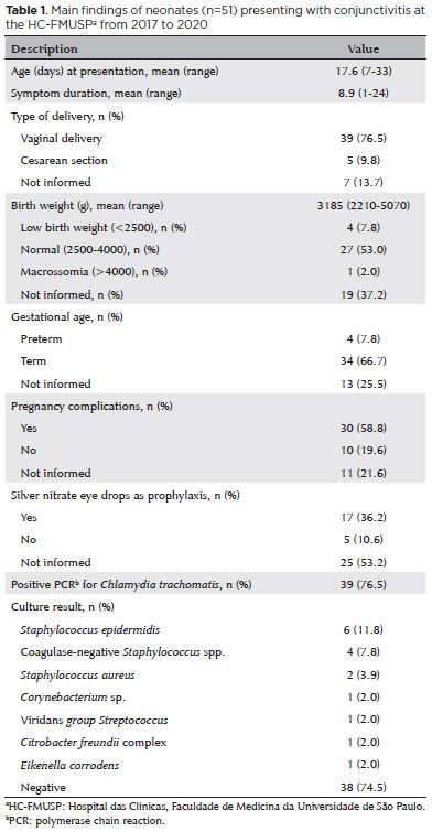
PURPOSE: The microbiology pattern of neonatal conjunctivitis has changed over time, and the incidence of gonococcal conjunctivitis is almost nil. This study aimed to determine the etiology of neonatal conjunctivitis cases referred to a tertiary health center in Brazil.
METHODS: From 2017 to 2020, conjunctival swabs were taken from neonates with clinical signs of conjunctivitis and tested with bacterial culture and polymerase chain reaction for Neisseria gonorrhoeae and Chlamydia trachomatis.
RESULTS: A total of 51 neonates were included in the 3-year study. Chlamydial conjunctivitis was diagnosed in 39 (76.5%) patients, and microbial growth was detected in 13 (25.5%) patients. The most isolated bacterium was Staphylococcus epidermidis (n=6, 11.8%), followed by other coagulase-negative Staphylococcus species (n=4, 7.8%) and S. aureus (n=2, 3.9%). One S. aureus isolate was resistant to oxacillin. There were no cases of gonococcal conjunctivitis. Ten (19.6%) patients showed polymerase chain reaction-negative C. trachomatis and negative bacterial culture test results.
CONCLUSION: Findings show that C. trachomatis is the most common pathogen causing neonatal conjunctivitis. The high prevalence of C. trachomatis infection highlights the importance of screening and treating pregnant woman.
Keywords: Conjunctivitis; Infant, newborn, diseases; Ophthalmia neonatorum; Chlamydia infections; Sexually transmitted diseases
Arq. Bras. Oftalmol. 2026;89 (3 )
:1-14
| DOI: 10.5935/0004-2749.2025-0248
Abstract
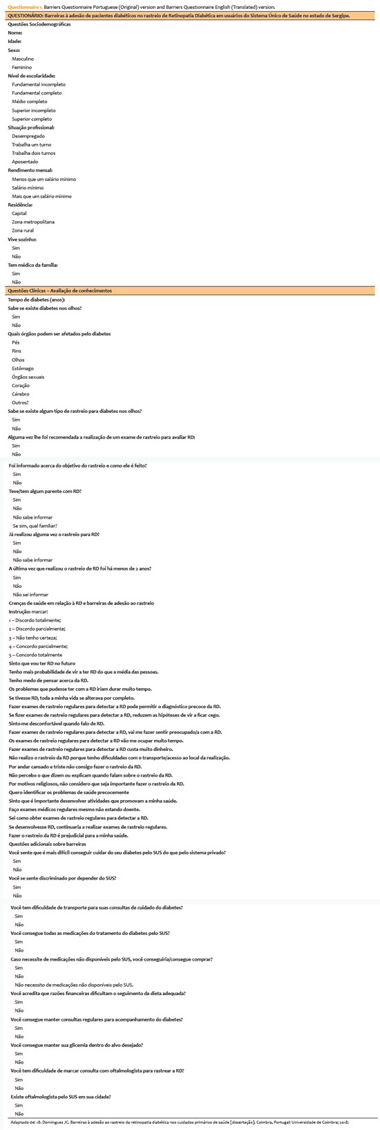
PURPOSE: This study aimed to identify barriers to diabetic retinopathy screening among a socioeconomically vulnerable urban population in northeast Brazil.
METHODS: A cross-sectional study was conducted during a diabetic retinopathy screening campaign at primary healthcare units. Ninety-five patients with diabetes underwent retinal examinations and completed a structured interview. Clinical, demographic, and socioeconomic data were collected.
RESULTS: The study population consisted predominantly of older adults (mean age: 60.7 ± 10.5 years), with a high prevalence of type 2 diabetes (99.0%) and low educational attainment. Most participants were economically inactive (81.1%) and reported low income (83.2%). Diabetic retinopathy and maculopathy were highly prevalent, affecting 50.0% and 22.9% of participants, respectively. Longer duration of diabetes was significantly associated with greater awareness of diabetic retinopathy (p=0.035), higher HbA1c levels (p<0.001), and increased prevalence of diabetic retinopathy (p=0.013) and maculopathy (p=0.002). Notably, 33.3% of participants reported difficulties attending medical appointments for diabetes management. In addition, 78.1% experienced challenges scheduling ophthalmologic evaluations, and 76.3% reported that no ophthalmologist was available in their city through the public healthcare system. Financial constraints also limited adherence to recommended dietary practices (90.4%) and impaired glycemic control, with more than half of participants reporting difficulty maintaining target glucose levels.
CONCLUSION: Major barriers to diabetic retinopathy screening included limited awareness of the importance of screening, financial hardship, and transportation challenges. Targeted educational initiatives and structural interventions such as expanded screening programs incorporating telemedicine and subsidized transportation—may improve screening adherence among vulnerable populations.
Keywords: Diabetic retinopathy; Mass screening; Health services accessibility; Health knowledge, attitudes, practices; Socioeconomic factors
Arq. Bras. Oftalmol. 2025;88 (3 )
:1-6
| DOI: 10.5935/0004-2749.2024-0170
Abstract
PURPOSE: To assess the sensitivity and specificity of the retinopathy of prematurity score (ROPScore) and weight, insulin-like growth factor-1, retinopathy of prematurity algorithm in predicting the risk of developing severe retinopathy of prematurity (prethreshold type 1) in a sample of preterm infants in Brazil.
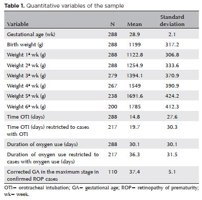
METHODS: Retrospective analysis of medical records of preterm infants (n=288) with birth weight of ≤1500 g and/or gestational age of 23-32 weeks in a neonatal unit in Southern Brazil from May 2013 to December 2020 (92 months).
RESULTS: The incidence of confirmed severe retinopathy of prematurity was 6.6%. ROPScore showed a 100% sensitivity, 44.6% specificity (95% confidence interval [CI] 38.7-50.6), 11.3% positive predictive value (95% CI 6.5-16.1), and 100% negative predictive value in predicting severe retinopathy of prematurity. The weight, insulin-like growth factor-1, retinopathy of prematurity algorithm demonstrated a 78.9% sensitivity (95% CI 60.6-97.3), 51.3% specificity (95% CI 45.3-57.3), 10.3% positive predictive value (95% CI 5.3-15.2), and 97.2% negative predictive value (95% CI 94.5-99.9).
CONCLUSION: ROPScore identified all patients at risk for severe retinopathy of prematurity. These findings support incorporating ROPScore into Brazilian guidelines to optimize retinopathy of prematurity screening and reduce unnecessary ophthalmologic examinations. Weight, insulin-like growth factor-1, retinopathy of prematurity's suboptimal performance in this Brazilian sample highlights the need for country-specific algorithm adjustments.
Keywords: Retinopathy of prematurity; ROPScore, WINROP; Prediction algorithm; Infant, premature
Arq. Bras. Oftalmol. 2024;87 (4 )
:1-6
| DOI: 10.5935/0004-2749.2023-0200
Abstract
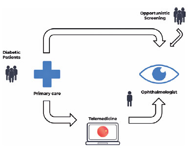
PURPOSE: Timely screening and treatment are essential for preventing diabetic retinopathy blindness. Improving screening workflows can reduce waiting times for specialist evaluation and thus enhance patient outcomes. This study assessed different screening approaches in a Brazilian public healthcare setting.
METHODS: This retrospective study evaluated a telemedicine-based diabetic retinopathy screening implemented during the COVID-19 pandemic and compared it with in-person strategies. The evaluation was conducted from the perspective of a specialized referral center in an urban area of Central-West Brazil. In the telemedicine approach, a trained technician would capture retinal images by using a handheld camera. These images were sent to specialists for remote evaluation. Patient variables, including age, gender, duration of diabetes diagnosis, diabetes treatment, comorbidities, and waiting time, were analyzed and compared.
RESULTS: In total, 437 patients with diabetes mellitus were included in the study (mean age: 62.5 ± 11.0 years, female: 61.7%, mean diabetes duration: 15.3 ± 9.7 years, insulin users: 67.8%). In the in-person assessment group, the average waiting time between primary care referral and specialist evaluation was 292.3 ± 213.9 days, and the referral rate was 73.29%. In the telemedicine group, the average waiting time was 158.8 ± 192.4 days, and the referral rate was 29.38%. The telemedicine approach significantly reduced the waiting time (p<0.001) and significantly lowered the referral rate (p<0.001).
CONCLUSION: The telemedicine approach significantly reduced the waiting time for specialist evaluation in a real-world setting. Employing portable retinal cameras may address the burden of diabetic retinopathy, especially in resource-limited settings.
Keywords: Telemedicine/methods; Diabetic retinopathy; Diagnostic screening programs; Vision screening; Practice patterns, physicians
 ABO is licensed under a Creative Commons Attribution-NonComercial 4.0 Internacional.
08-fig01tb.jpg)
05-fig01tb.jpg)
06-tab01.jpg)
08-tab01tb.jpg)
08-fig01.jpg)
03-fig01.jpg)
12-fig01.jpg)