Showing of 1 until 15 from 115 result(s)
Search for: Visual acuity; Cross-sectional studies; School health; Health education
10-fig01.jpg)
Abstract
Objetivo: Avaliar os resultados visuais, satisfação e qualidade de vida de pacientes atendidos em um hospital escola pelo Sistema Único de Saúde, submetidos a implante bilateral de lente intraocular multifocal difrativa.
Métodos: Estudo tipo série de casos com intervenção, incluindo 20 pacientes submetidos a implante bilateral da lente intraocular multifocal difrativa EyeDiff® (Eyeol UK, Dunstable, UK). Os critérios de exclusão foram astigmatismo corneano >1,5 dioptria cilíndrica, cirurgia ou doença ocular prévias e complicações intraoperatórias ou pós-operatórias. Os pacientes foram avaliados após 1, 3 e 6 meses da cirurgia. Foram avaliadas a acuidade visual monocular e binocular para longe, intermediário e perto sob condições fotópica e mesópica, sensibilidade ao contraste monocular sob condições fotópicas, curva de defocus e questionário para avaliação da qualidade de vida.
Resultados: A acuidade visual para longe corrigida monocular foi de 0,3 logMAR ou melhor e a acuidade visual para perto com correção para longe foi J3 ou melhor em todos os olhos, sob condições fotópicas. A acuidade visual binocular para perto com a correção para longe foi J1 em todos os casos. A sensibilidade ao contraste estava no nível mínimo de normalidade para frequências espaciais baixas e altas e abaixo dos limites normais para frequência espacial intermediária. O questionário de qualidade de vida mostrou que os pacientes apresentavam altos níveis de satisfação.
Conclusão: O implante bilateral da lente intraocular multifocal EyeDiff® proporcionou boa acuidade visual e qualidade de vida, e independência de óculos aos pacientes. A acuidade visual e a sensibilidade ao contraste melhoraram progressivamente entre um e seis meses de pós-operatório.
Keywords: Acuidade visual; Qualidade de vida; Satisfação do paciente; Implante de lente intraocular; Sistema Único de Saúde.
Abstract
PURPOSE: To analyze the quality of life and treatment adherence of patients with glaucoma at different disease stages, considering factors such as sex, visual acuity, disease severity, and treatment characteristics.
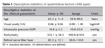
METHODS: This cross-sectional study included 174 patients (346 glaucomatous eyes) recruited from clinical records and routine follow-ups at a specialized ophthalmology center. Their mean age was 39–90 years, and 60.9% of them were women. Their quality of life and adherence were assessed using the NEI-VFQ25 and MMAS-8 questionnaires, respectively. Complementary tests included 24:2 visual field test, retinography, and optical coherence tomography. Patients diagnosed with glaucoma for at least 6 months were included, whereas pregnant patients and those with ocular diseases were excluded.
RESULTS: Among the participants, 59.2% adhered to the treatment whereas 40.8% showed low adherence. The mean quality of life score was 81.87. Patients with low adherence had slightly higher quality of life scores (mean 83.1) than those with good adherence (mean 81.0), but the difference was not statistically significant. Disease severity was associated with increased optic nerve cupping, reduced thickness of the nerve fiber and ganglion cell layers, and great visual field loss. No significant correlation was observed between adherence and quality of life, indicating the independence of these factors and the influence of psychological or social elements.
CONCLUSION: The absence of a correlation between quality of life and treatment adherence highlights the need for tailored interventions for psychological and social aspects. These findings indicate the importance of a comprehensive approach to managing glaucoma, preserving visual function, strengthening doctor–patient relationships, and considering psychosocial factors to enhance quality of life and treatment adherence.
Keywords: Glaucoma; Quality of life; Patient health questionnaire; Patient acuity; Antiglaucoma agents; Visual acuity; Treatment adherence and compliance; Surveys and questionnaires
14-fig01tb.jpg)
Abstract
Objetivos: Relatar a distribuição dos motivos de encaminhamento de crianças para ambulatório de glaucoma infantil em um serviço oftalmológico terciário.
Métodos: Dados médicos de pacientes menores que 18 anos encaminhados para ambulatório de glaucoma infantil na cidade de São Paulo, Brasil, de 2012 a 2018 foram retrospectivamente analisados. Os dados incluíram os motivos de encaminhamento, a idade, a origem e quem detectou a alteração ocular. Para definição diagnóstica, a classificação do Childhood Glaucoma Research Network foi usada.
Resultados: 563 olhos de 328 pacientes foram incluídos no estudo. O diagnóstico de glaucoma foi confirmado em 162 crianças (49%). 83 (25%) pacientes tiveram diagnóstico de glaucoma descartado, e 83 (25%) continuaram em acompanhamento como glaucoma suspeito. Os principais motivos de encaminhamento foram relação escavação-disco >0,5 ou assimetria ≥0,2 (24%), pressão intraocular >21 mmHg (21%) e opacidade corneana (15%). No período neonatal, os motivos de encaminhamento foram opacidade corneana, buftalmo, lacrimejamento e fotofobia. Após o período neonatal, além desses sinais externos, outros sinais foram também motivos de encaminhamento, como escavação-disco >0,5 ou assimetria ≥0,2, pressão intraocular >21 mmHg e miopização. Os encaminhamentos ocorreram por profissionais de saúde em 69% e preocupação dos pais em 30%. Os pais foram os primeiros a identificar as alterações e procurar cuidado médico em 97% dos casos de glaucoma congênito primário.
Conclusões: Os motivos de encaminhamento de crianças para um serviço de glaucoma de glaucoma terciário foram determinados e diferem em diferentes faixas etárias e grupos. Os autores reforçam a necessidade de alertar diferentes grupos para os sinais mais comuns, a fim de evitar, não só o adiamento do diagnóstico, o que impacta no prognóstico, mas também despender recursos importantes direcionados ao enfrentamento dessas doenças, com encaminhamentos imprecisos.
Keywords: Glaucoma/congênito; Glaucoma/fisiopatologia; Opacidade da córnea; Criança; Acuidade visual; Encaminhamento e consulta; Serviços de saúde ocular
05-tab01.jpg)
Abstract
Objetivo: Avaliar a prevalência de transtornos de depressão e ansiedade em pacientes com glaucoma e identificar fatores de riscos associados.
Métodos: Estudo transversal em pacientes com glaucoma, avaliados durante Agosto de 2016 e Agosto de 2017 no Hospital das Clínicas da Universidade de Campinas e no Hospital Oftalmológico de Brasília. Todos pacientes foram submetidos à exame oftalmológico completo para confirmar o diagnóstico de glaucoma. Todos pacientes preencheram o questionário “Hospital Anxiety and Depression Scale”.
Resultados: Foram incluídos 129 pacientes no estudo, sendo 74 homens (57.36%) e 55 (42.64%) mulheres, 90 pacientes eram brancos (69.77%) e 38 (29.46%) eram negros. A idade média foi de 70.14 ± 15.8 anos. O estudo demonstrou uma prevalência de 10.08% de transtornos depressivo e/ou ansiedade.
A regressão logística demonstrou que mulheres apresentam
maior risco de desenvolver transtornos depressivos e/ou ansiedade (Risco relativo: 5.25, p=0.015), assim como pacientes com maior número de co-morbidades clínicas (Risco relativo: 2.82, p=0.038).
Conclusão: Uma proporção significativa dos pacientes com glaucoma podem apresentar transtornos de depressão e/ou ansiedade. Pacientes com glaucoma do sexo feminino e que apresentem maiores co-morbidades clínicas apresentam maior risco de apresentar esses transtornos.
Keywords: Glaucoma; Depressão/epidemiologia; Ansiedade/epidemiologia; Estudos transversais
05-tab01tb.jpg)
Abstract
Objetivo: Avaliar as razões para não comparecimento à clínica oftalmológica da universidade após triagem oftalmológica realizada usando uma unidade móvel oftalmológica que fornece exame oftalmológico para comunidades não assistidas em uma região do Brasil.
Métodos: Foi realizado um estudo observacional prospectivo no ano de 2017/2018 para avaliar as razões que fizeram com que os indivíduos triados usando uma unidade móvel oftalmológica e referenciados para a clínica oftalmológica da universidade não comparecessem à consulta agendada. A triagem foi feita em 10 municípios da região centro-oeste do estado de São Paulo, Brasil. Todos os 1.928 participantes fizeram o exame oftalmológico sem custo e 37,1% deles necessitaram de encaminhamento para a clínica oftalmológica da universidade para exames especializados ou cirurgias. O estudo usou duas ferramentas: (1) análise comparativa entre os dados dos indivíduos encaminhados que compareceram ao agendamento com os que não compareceram; (2) busca ativa dos indivíduos que não compareceram à consulta agendada, aplicando-se um questionário para avaliar os motivos para o não comparecimento.
Resultados: Fatores como idade, sexo, distância entre a cidade de origem e o hospital universitário, número de oftalmologistas na cidade de procedência, renda familiar média e acuidade visual não influenciaram no comparecimento ao encaminhamento. Catarata foi a maior causa para o encaminhamento (350 casos). O não comparecimento foi maior nos portadores de glaucoma/glaucoma suspeitos (54,1%), estrabismo (45%) e afecções do segmento anterior (33,6%). Muitos indivíduos que não compareceram ao serviço de referência procuraram por outro local para o atendimento oftalmológico.
Conclusão: O não comparecimento para tratamento oftalmológico sem custo depende de fatores relacionados ao paciente ou à falta de conhecimento das próprias condições oftalmológicas. Campanhas educativas nas comunidades assistidas devem ser feitas para alcançar maior comparecimento às consultas e melhor prevenir a cegueira evitável.
Keywords: Serviços de saúde ocular; Unidades móveis de saúde; Acesso aos serviços de saúde; Pacientes desistentes do tratamento; Promoção da saúde
Abstract
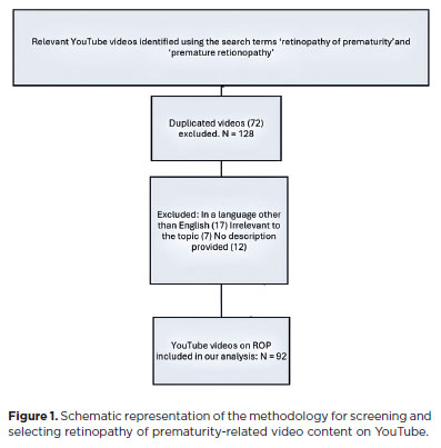
PURPOSE: This study aimed to evaluate the quality and reliability of YouTube videos as an educational resource about retinopathy of prematurity.
METHODS: Videos were sourced from YouTube using the search terms "retinopathy of prematurity" and "premature retinopathy" with the default settings. Each video was assessed on the following metrics: views, likes, dislikes, comments, upload source, country of origin, view ratio, like ratio, and video power index. The quality and reliability of the videos were evaluated by two independent researchers using the DISCERN questionnaire, the JAMA benchmarks, the Global Quality Score scale, the Health on the Net Code of Conduct, and the Ensuring Quality Information for Patients scale.
RESULTS: The study assessed 92 videos, the majority of which (42 videos, 45.7%) originated from the United States. Most of the videos focused on screening, pathophysiology, and diagnosis of retinopathy of prematurity (61.9%). The primary contributors were medical organizations (19 videos, 20.6%), nonacademic health channels (19 videos, 20.6%), and physicians (15 videos, 16.3%). Significant differences were found between the DISCERN (p=0.003), JAMA (p=0.001), Global Quality Score (p=0.003), Health on the Net Code of Conduct (p=0.006), and Ensuring Quality Information for Patients (p=0.001) scores among different video sources. However, the key video metrics did not differ. Using the DISCERN and Global Quality Score scales, the overall YouTube video content on retinopathy of prematurity was rated as moderate in quality. Using the Health On the Net Code of Conduct and Ensuring Quality Information for Patients scales, it was rated as high quality. Strong correlations were observed between the scores on all of the scales (p<0.001).
CONCLUSION: Videos from medical organizations and healthcare centers were of a higher quality than those from nonmedical sources. Despite the varied foci of each evaluation scale, the strong correlation between them indicates that they provide reliable and comprehensive assessments of the quality of informational content.
Keywords: Retinopathy of prematurity; YouTube; Information dissemination/methods; Online education; Internet access; Social media/instrumentation; Information seeking behavior; Internet/statistics & numerical data; Consumer health information; Social networking
08-fig01.jpg)
Abstract
Objetivo: Determinar o impacto do uso de unidade móvel no acesso à saúde ocular e avaliar o perfil da população que necessita de cuidados oftalmológicos, as doenças oculares mais frequentes e o tratamento. Métodos: Estudo transversal realizado em 14 municípios da região sudoeste do Estado de São Paulo utilizando uma unidade móvel oftalmológica. Os participantes eram usuários do Sistema Único de Saúde que procuraram atendimento oftalmológico, sem restrição quanto a idade, gênero ou condição socioeconômica. Os dados foram transferidos para a tabela Excel para análise estatística. Resultados: Participaram do estudo 6.878 pessoas, com média de idade de 44 anos (variação de 4 meses a 96 anos) e 65,5% eram mulheres. Erros refrativos estavam presentes em 78,6% dos participantes, catarata em 9,6% e pterígio em 8,3%. Para 60% foram prescritos óculos, para 10% foi mantida a correção óptica em uso e para 28% foram necessárias apenas orientações. Exames especializados ou procedimentos cirúrgicos foram indicados para 18,1% dos casos que foram encaminhados para tratamento em serviço terciário. Dentre os pacientes referenciados, 36,4% necessitavam de cirurgia oculoplástica ou para tratar afecções externas do olho e 31,8%, de cirurgia de catarata. Conclusão: A grande maioria dos pacientes que procurou atendimento na unidade móvel necessitava de prescrição de óculos. A unidade móvel oftalmológica possui alto grau de resolutividade para os problemas oculares, com oportunidade de tratar os erros refrativos e referenciar os pacientes que necessitam de atendimento especializado, geralmente relacionado a condições cirúrgicas. Unidades móveis podem ser uma alternativa aos cuidados oftalmológicos básicos, melhorando o acesso, atuando na promoção da saúde ocular e prevenindo a cegueira.
Keywords: Unidades móveis de saúde; Saúde ocular; Transtornos da visão; Erros de refração; Óculos; Cegueira/prevenção & controle
Abstract
PURPOSE: This study evaluated macular thickness using spectral-domain optical coherence tomography in healthy participants from a population-based eye survey.
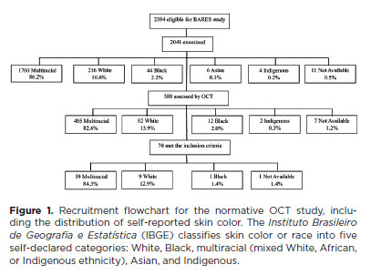
METHODS: The Brazilian Amazon Region Eye Survey was a population-based study assessing the prevalence and causes of visual impairment, blindness, and ocular diseases in adults aged ≥45 years from urban and rural areas of Parintins. A subgroup was selected based on inclusion criteria for both eyes: best-corrected visual acuity ≥20/32, normal eye examination results, and no prior ocular surgery. Scans were performed using the iVue optical coherence tomography device. Measurements were taken from the nine subfields defined by the Early Treatment Diabetic Retinopathy Study, examining the full retina as well as the inner and outer retinal layers. Associations of retinal thickness with age and sex were also analyzed. Statistical significance was set at p≤0.05.
RESULTS: In total, 70 healthy participants (25 males), aged 45–65 years (mean=52 ± 5), were included. Mean central foveal thickness was 248.71 ± 18.73 μm. A significant age-related reduction in macular thickness was observed, particularly in the inner superior parafovea (p=0.036), nasal perifovea (p=0.001), superior perifovea (p=0.028), outer layer of inferior parafovea (p=0.049), and the inferior perifovea of the full retina (p=0.029). Males showed significantly greater thickness in the outer layer, especially in the outer parafovea (p=0.004) and perifovea (p<0.0001).
CONCLUSIONS: This study established normative macular thickness values for healthy older adults in the Brazilian Amazon region using spectral-domain optical coherence tomography. Age and sex were found to significantly influence macular thickness and should be considered when interpreting measurements. These data will support future studies of retinal diseases in this population.
Keywords: Retinal diseases/diagnosis; Macula lutea/pathology; Macular degeneration/diagnosis; Diabetic retinopathy/diagnosis; Vision, low; Vision tests; Tomography, optical coherence/methods; Young adult; Cross-sectional studies; Brazil/epidemiology
Abstract
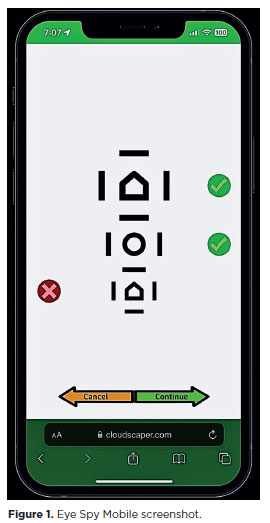
PURPOSE: This cross-sectional study compared best-corrected visual acuity obtained using Cloudscaper symbols, a novel optotype developed according to ETDRS specifications for children's virtual screening, with that obtained using LEA symbols.
METHODS: A total of 560 children aged 3-16 yr underwent visual acuity test with both Cloudscaper symbols and LS. The test application was standardized using the EyeSpy algorithm. Additionally, 147 participants were tested with the standard Snellen E paper chart. Paired t tests were performed to assess the clinical significance of logMAR visual acuity differences.
RESULTS: The mean logMAR visual acuity with LEA symbols was 0.12 (standard deviation [SD]=0.18; range, -0.10 to 0.80), while with Cloudscaper symbols it was 0.18 (SD=0.19; range, -0.10 to 0.80). The mean difference between Cloudscaper symbols and LEA symbols was 0.099 logMAR (approximately 0.5 optotypes; SD=0.08; range, 0.0-0.14; p<0.0001). Cloudscaper symbols slightly underestimated visual acuity compared to LEA symbols. Visual acuity measured by both methods was highly correlated (Spearman's r=0.74, p<0.0001). The mean visual acuity difference between Cloudscaper symbols and the Snellen E chart was 0.0045 (p=0.805; 95% confidence interval [95% CI]), whereas the difference between LEA symbols and Snellen E was 0.0883 (p<0.001; 95% CI).
CONCLUSIONS: Cloudscaper symbols provide a reliable tool for visual screening in children. Although they slightly underestimate visual acuity compared to LEA symbols – a finding also reported when comparing ETDRS letters with LEA symbols – Cloudscaper symbols show strong agreement with Snellen E chart measurements. This suggests that Cloudscaper symbols allow precise visual acuity assessment comparable to the gold standard.
Keywords: Vision screening; Vision tests; Visual acuity; Mobile applications; Eye health; Child health; Diagnostic techniques, Ophthalmological; Child; Preschool child; Adolescent

Abstract
PURPOSE: This pilot study evaluated the diagnostic accuracy of a deep learning model for detecting pterygium in anterior segment photographs taken using smartphones in the Brazilian Amazon. The model’s performance was benchmarked against assessments made by experienced ophthalmologists, considered the clinical gold standard.
METHODS: In this cross-sectional study, 38 participants (76 eyes) from Barcelos, Brazil, were enrolled. Trained nonmedical health workers captured high-resolution anterior segment images using smartphones. These images were analyzed using a deep learning model based on the MobileNet-V2 convolutional neural network. Diagnostic metrics–including sensitivity, specificity, accuracy, positive predictive value, negative predictive value, and area under the receiver operating characteristic curve–were calculated and compared with the ophthalmologists’ evaluations.
RESULTS: The deep learning model achieved a sensitivity of 91.43%, specificity of 90.24%, positive predictive value of 88.46%, negative predictive value of 92.79%, and an area under the curve of 0.91. Logistic regression revealed no statistically significant association between pterygium and demographic variables such as age or gender.
CONCLUSIONS: The deep learning model demonstrated high diagnostic performance in identifying pterygium in a remote Amazonian population. These preliminary findings support the potential use of artificial intelligence–based tools to facilitate early detection and screening in underserved regions, thereby enhancing access to ophthalmic care.
Keywords: Pterygium/diagnostic imaging; Smartphone; Diagnostic techniques, ophthalmological; Deep learning; Telemedicine; Artificial intelligence; Cross-sectional studies; Brazil/epidemiology
Abstract
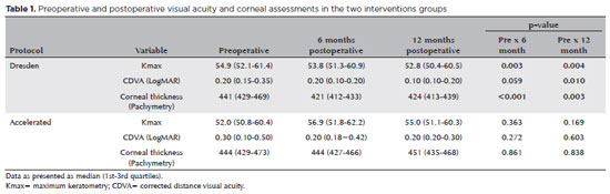
PURPOSE: Keratoconus presents certain peculiarities in pediatric patients when compared with adults. The greatest challenge in children is that the disease is more severe and faster in progression. In this retrospective study, we aimed to compare the accelerated and Dresden protocols for corneal crosslinking in patients aged <18 years who were followed-up for at least 12 months.
METHODS: A total of 36 eyes from 27 patients were included in the study. The best corrected and uncorrected visual acuity, maximal keratometry, corneal thickness, foveal thickness, and endothelial microscopy findings were evaluated at baseline and during the postoperative period at one, three, and six months. Thereafter, the patients were evaluated at one, three, six and twelve months postoperative. Corneal crosslinking was performed in all patients via the Dresden protocol (n=21 eyes) or the accelerated protocol (n=15 eyes). Data between the two groups were compared and XY statistical analysis was used.
RESULTS: Both protocols were effective in halting keratoconus progression. No patient had progression at the 12-month follow-up. A significant reduction in Kmax and improvement in the corrected distance visual acuity were observed only in the Dresden protocol group. Although the Dresden protocol was superior to the accelerated protocol in reducing Kmax (p=0.002), there was no significant difference in corrected distance visual acuity between the two groups.
CONCLUSION: The accelerated protocol is as efficient as the Dresden protocol in stabilizing keratoconus progression. Although the Dresden protocol was superior to the accelerated protocol in reducing the Kmax, it did not produce better clinical results. Thus, the accelerated protocol is an efficient option. Furthermore, considering the advantages of reduced surgical time, the accelerated protocol is effective in halting keratoconus progression in the pediatric age group.
Keywords: Keratoconus; Corneal diseases; Ultraviolet rays; Cross-linking reagents; Visual acuity
Abstract
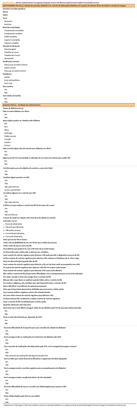
PURPOSE: This study aimed to identify barriers to diabetic retinopathy screening among a socioeconomically vulnerable urban population in northeast Brazil.
METHODS: A cross-sectional study was conducted during a diabetic retinopathy screening campaign at primary healthcare units. Ninety-five patients with diabetes underwent retinal examinations and completed a structured interview. Clinical, demographic, and socioeconomic data were collected.
RESULTS: The study population consisted predominantly of older adults (mean age: 60.7 ± 10.5 years), with a high prevalence of type 2 diabetes (99.0%) and low educational attainment. Most participants were economically inactive (81.1%) and reported low income (83.2%). Diabetic retinopathy and maculopathy were highly prevalent, affecting 50.0% and 22.9% of participants, respectively. Longer duration of diabetes was significantly associated with greater awareness of diabetic retinopathy (p=0.035), higher HbA1c levels (p<0.001), and increased prevalence of diabetic retinopathy (p=0.013) and maculopathy (p=0.002). Notably, 33.3% of participants reported difficulties attending medical appointments for diabetes management. In addition, 78.1% experienced challenges scheduling ophthalmologic evaluations, and 76.3% reported that no ophthalmologist was available in their city through the public healthcare system. Financial constraints also limited adherence to recommended dietary practices (90.4%) and impaired glycemic control, with more than half of participants reporting difficulty maintaining target glucose levels.
CONCLUSION: Major barriers to diabetic retinopathy screening included limited awareness of the importance of screening, financial hardship, and transportation challenges. Targeted educational initiatives and structural interventions such as expanded screening programs incorporating telemedicine and subsidized transportation—may improve screening adherence among vulnerable populations.
Keywords: Diabetic retinopathy; Mass screening; Health services accessibility; Health knowledge, attitudes, practices; Socioeconomic factors
08-tab01.jpg)
Abstract
OBJETIVO: Examinar os efeitos do tratamento de reticulação unilateral do colágeno corneano na acuidade visual e os achados topográficos em olhos não tratados de pacientes com ceratocone progressivo bilateral.
MÉTODOS: Foram rastreados retrospectivamente pacientes com ceratocone progressivo submetidos a tratamento de reticulação. Foram incluídos no estudo 188 olhos não tratados de 188 pacientes tratado unilateralmente com reticulação padrão ou acelerada e que recusaram o procedimento de reticulação no outro olho. A acuidade visual e os achados topográficos dos olhos não tratados foram obtidos no pré- e pós-operatório no 1o, 3o, 6o, 12o, 24o, 30o e 36o mês.
RESULTADOS: As alterações ao longo do tempo foram semelhantes para as variáveis examinadas nos olhos não tratados de pacientes tratados com métodos de reticulação padrão e acelerado (p>0,05). No 12º mês, 136 olhos não tratados (95,8%) estavam estáveis, de acordo com os critérios de progressão. Apenas quatro olhos (8%) mostraram progressão no 24o mês. Nenhuma progressão foi observada nos 16 pacientes que tiveram um acompanhamento de 36 meses.
CONCLUSÕES: O estudo mostrou que os olhos não tratados de pacientes com ceratocone progressivo bilateral não apresentaram taxas de progressão significativas após o tratamento unilateral com reticulação.
Keywords: Topografia da córnea; Reagentes de ligações cruzadas; Ceratocone; Fármacos fotossensibilizantes; Colágeno/uso terapêutico; Fotoquimioterapia/métodos; Acuidade visual
11-tab01tb.jpg)
Abstract
OBJETIVO: Descrever as características clínicas e os fatores associados à presença de ceratite em pacientes com corpos estranhos na córnea em uma população colombiana.
MÉTODOS: Trata-se de um estudo transversal baseado na revisão dos registros clínicos de pacientes com corpos estranhos na córnea admitidos em um departamento de emergência em Cali, Colômbia, entre junho de 2018 e junho de 2019. O desfecho primário foi a presença de ceratite diagnosticada através de critérios clínicos. Foram utilizados modelos de regressão logística univariada e multivariada para identificar os fatores associados.
RESULTADO: Neste estudo, foi analisado um total de 381 corpos estranhos na córnea em 372 pacientes (idade média: 40,0 anos, intervalo interquartil: 29,0-53,0; sexo masculino: 94,7% [352 casos]). Noventa e cinco casos desenvolveram ceratite (24,9%, intervalo de confiança de 95% — IC 95%: 20,8%-29,5%). Na análise multivariada, para idade ≤30 anos (razão de chances — RC: 2,15, IC 95%: 1,06-4,36), o achado de flare aquoso (RC: 2,81, IC 95%: 1,39-5,66]) e a presença de corpo estranho na periferia da córnea (RC: 2,05, IC 95%: 1,19-3,50) foram associados a um risco aumentado de ceratite. Sexo, tempo entre a lesão e a internação, e edema da córnea não foram relacionados à ceratite (p>0,05).
CONCLUSÃO: Há uma proporção elevada de ceratite em casos de corpos estranhos na córnea em Cali, Colômbia. Os três fatores associados à ceratite foram a idade, o achado de flare aquoso e a presença de corpo estranho na periferia da córnea.
Keywords: Corpos estranhos no olho; Lesões da córnea; Ceratite/epidemiologia; Estudos transversais; Colômbia.
Abstract
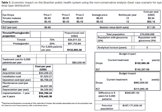
PURPOSE: To evaluate the economic impact of the following initial treatment scenarios for glaucoma on the Brazilian Public Health System (SUS): (1) traditional continuous instillation of hypotensive eye drops and (2) single session of selective laser trabeculoplasty.
METHODS: Economic impact was analyzed in three scenarios, from the least to the most conservative, for a hypothetical cohort of 5,000 individuals with open-angle glaucoma. Thereafter, projections were made on the basis of a glaucoma prevalence of 3% in the 2021 Brazilian population size.
RESULTS: All three scenarios demonstrated that selective laser trabeculoplasty exhibited a significantly lower economic impact than the eye drops on SUS over one and five years. Furthermore, the difference was more than United States Dollar 8 billion at five years when considering 3% of the Brazilian population aged >40 years in 2021.
CONCLUSION: As the initial treatment for primary open-angle glaucoma, selective laser trabeculoplasty exhibited a lower economic impact on SUS than latanoprost and timolol maleate eye drop instillation in all the studied scenarios over one and five-year periods.
Keywords: Glaucoma; Trabeculotomy; Laser therapy; Cost analysis; Health care cost Unified Health System; Brazil
 ABO is licensed under a Creative Commons Attribution-NonComercial 4.0 Internacional.
About
Issues
Editorial Board
Submission
Arquivos Brasileiros de Oftalmologia
Official publication of Brazilian Council of Ophthalmology - Conselho Brasileiro de Oftalmologia (CBO)
Rua Casa do Ator, 1.117 - 2nd floor - Zip Code: 04546-004
São Paulo - SP, Brazil
TEL: +55 11 3266-4000
E-mail: [email protected]