Arq. Bras. Oftalmol. 2023;86 (5 )
:1-6
| DOI: 10.5935/0004-2749.20230064
Abstract
Objetivo: Avaliar a resposta tecidual e clínica a um implante orbitário de polimetilmetacrilato, oco e multiperfurado em sua porção posterior em modelo animal após evisceração.
Métodos: Dezesseis coelhos da raça Nova Zelândia foram submetidos à evisceração do globo ocular direito. Todos receberam implante oco de polimetilmetacrilato de 12 mm de diâmetro, multiperfurado em sua semiesfera posterior. O estudo foi dividido em avaliação clínica e histopatológica. A avaliação clínica foi diária até 14 dias pós-evisceração e, a cada sete dias, até completar 180 dias. Os animais foram divididos em grupos de quatro animais e cada um foi submetido à exenteração com 07, 30, 90 e 180 dias e depois à eutanásia. A análise histopatológica teve por fim caracterizar o padrão inflamatório, a estrutura do colágeno e o grau de neovascularização. Para isso, além da tradicional coloração pela hematoxilina-eosina, utilizou-se o corante Picrosirius Red (PSR) e imuno-histoquímica com o marcador CD 34.
Resultados: Não houve sinais de infecção, afinamento conjuntival ou escleral, exposição ou extrusão do implante em nenhum animal durante o estudo. Já no sétimo dia, o tecido neoformado migrou para dentro do implante formando uma rede fibrovascular através dos canais posteriores. A resposta inflamatória diminuiu ao longo do tempo avaliado e não foram encontradas células gigantes multinucleadas.
Conclusão: O implante analisado permite a sua integração aos tecidos orbitários pelo crescimento fibrovascular em seu interior. Os autores acreditam que este modelo de implante orbital pode fazer parte de testes com humanos.
Keywords: Implantes orbitários; Polimetilmetacrilato; Evisceração ocular; Anoftalmia; Procedimentos cirúrgicos oftalmológicos; Coelhos.
Arq. Bras. Oftalmol. 2022;85 (4 )
:351-358
| DOI: 10.5935/0004-2749.20220051
Abstract
Objetivo: Desenvolver um aplicativo (TopEye) na plataforma iOS para dispositivos móveis que possibilite a captação e interpretação do mapa de cores gerados por qualquer topógrafo corneano através da inteligência artificial (IA).
Métodos: A execução, acompanhamento e avaliação do projeto foi utilizada a metodologia Scrum, processo de desenvolvimento interativo e incremental para gerenciamento de projetos e desenvolvimento ágil de software. O banco de padrões de diagnóstico gerado consiste em 1172 exemplos, divididos em: 275 padrões esféricos, 302 regulares simétricos, 295 regulares assimétricos e 300 irregulares (ceratocone). Para o desenvolvimento da inteligência artificial do aplicativo, foi estabelecido o treinamento da rede com 240 imagens de cada tipo de padrão, totalizando 960 (81,91%) padrões. O restante das imagens, 212 (18,09%), foram utilizadas para testar o aplicativo e usadas para gerar os resultados. O processo é semiautomático, assim a captação da imagem topográfica é realizada com smartphone, o examinador realiza o contorno do relevo corneano manualmente para em seguida a rede neural realizar o diagnóstico.
Resultados: O aplicativo diagnosticou 201 (94,81%) imagens corretamente. De um total de 212 imagens, o algoritmo errou a classificação de apenas 11 (5,19%). A principal ocorrência de erro foi na distinção das classes simétrica e assimétrica. No rastreio do ceratocone o aplicativo alcançou 95,00% de sensibilidade e 98,68% especificidade.
Conclusão: O trabalho resultou na obtenção de um aplicativo eficiente na captura da imagem topográfica pela câmera do smartphone e na interpretação da mesma através da inteligência artificial aplicada.
Keywords: Dispositivos móveis; Inteligência artificial; Topografia corneana; Astigmatismo
Arq. Bras. Oftalmol. 2018;81 (1 )
:42-6
| DOI: 10.5935/0004-2749.20180011
Abstract
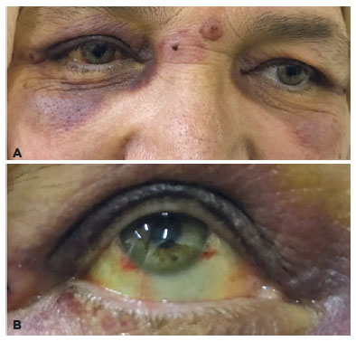
Objetivo: Compartilhar os resultados dos pacientes submetidos à rotação de retalho tarsal anterior, combinados com a reposição lamelar anterior devido à entrópio cicatricial da pálpebra superior e determinar a eficácia e a confiabilidade desta técnica cirúrgica.
Métodos: Foram incluídos neste estudo quinze olhos de 11 pacientes em quem realizamos cirurgia de rotação de retalho tarsal anterior combinada com reposição lamelar anterior devido ao entrópio cicatricial. Os registros médicos dos pacientes foram analisados retrospectivamente e as causas da entrópio cicatricial, bem como os achados do exame oftalmológico pré-operatório e pós-operatório foram registrados. A integridade anatômica e funcional da pálpebra foi considerada como sucesso cirúrgico.
Resultados: A idade média foi de 59,81 ± 18 anos. O período médio de seguimento foi de 21,72 ± 14 meses (intervalo 5-43 meses). As causas da entrópio cicatricial foram o desenvolvimento de cicatrizes pós-operatórias devido a eletrólises múltiplas para triquíase e/ou distiquiase em 7 olhos, tracoma em 6 olhos e trauma em 2 olhos. Todos os pacientes foram tiveram irritação e lacrimejamento pré-operatório, enquanto que 10 pacientes apresentavam opacidade e erosão da córnea e 1 paciente apresentava apenas erosão epitelial. O sucesso total anatômico e funcional foi alcançado em todos os casos.
Conclusão: A rotação do retalho tarsal anterior combinada com a reposição lamelar anterior no reparo da entrópio cicatricial é um procedimento cirúrgico alternativo efetivo e confiável.
Keywords: Tracoma/complicações; Pálpebras/cirurgia; Entrópio/cirurgia; Cicatriz; Retalhos cirúrgicos; Procedimentos cirúrgicos oftalmológicos/métodos
Arq. Bras. Oftalmol. 2023;86 (4 )
:365-371
| DOI: 10.5935/0004-2749.20230043
Abstract
Objetivo: Avaliar as alterações da superfície ocular em pacientes com Rosácea, e comparar com grupo controle.
Métodos: Noventa e três indivíduos foram selecionados para este estudo transversal, observacional e não intervencionista, dividido em dois grupos: rosácea (n=40) e controles (n=53). Foram avaliados parâmetros objetivos da superfície ocular (hiperemia conjuntival, estabilidade e volume do filme lacrimal, disfunção da glândula meibomiana, doença do olho seco, coloração da superfície ocular) e comparado indivíduos saudáveis com pacientes com rosácea.
Resultados: 69,23% dos indivíduos com rosácea eram mulheres, com média de idade de 47,34 ± 12,62 anos. Em comparação com controles pareados, não foram evidenciadas diferenças estatisticamente significativas em relação à acuidade visual (p=0,987) e parâmetros do filme lacrimal (altura do menisco lacrimal (p=0,338), tempo de ruptura do filme lacrimal não invasivo (p=0,228), tempo invasivo de ruptura (p=0,471) e teste de Schirmer (p=0,244), bem como hiperemia conjuntival (p=0,106) e coloração com fluoresceína (p=0,489). Associação significativa foi encontrada na avaliação da meibografia (p=0,026), integridade da camada mucosa (p=0,015) e sintomas de superfície ocular (p<0,0001). Pacientes com rosácea também apresentaram alterações importantes na borda palpebral: expressibilidade glandular (p<0,001), padrão de secreção glandular (p<0,001) e telangiectasia (p<0,001).
Conclusão: A disfunção da glândula de Meibômio está frequentemente associada a condições dermatológicas e é caracterizada por achados morfológicos na meibografia, bem como comprometimento da secreção lipídica que leva ao olho seco evaporativo e alterações da superfície ocular e inflamação.
Keywords: Rosácea/complicações; Disfunção da glândula tarsal; Túnica conjuntiva; Síndromes do olho seco; Técnicas de diagnóstico oftalmológico.
Arq. Bras. Oftalmol. 2025;88 (4 )
:1-6
| DOI: 10.5935/0004-2749.2024-0083
Abstract
PURPOSE: We developed an artificial intelligence program for calculating intraocular lenses and analyzed its accuracy rate via ultrasonic biometry. This endeavor is aimed at enhancing precision and efficacy in the selection of intraocular lenses, particularly in cases where optical biometry is unavailable.
METHODS: Data was collected from the Hospital de Clínicas de Porto Alegre, which included cases of phacoemulsification with intraocular lens implantation, in which the lens selection was based on ultrasonic biometry. The program, implemented in Python, Java, and PHP, employs the ridge regression method. Two design options were developed: a basic model, which uses only keratometry variables (K1 and K2), axial size and final target refraction in the spherical equivalent, and an advanced model, which incorporates preoperative refraction and the patient's age. The Universal Barrett II formula was used to compare both models.
RESULTS: The sample consisted of 486 eyes from 313 patients, with 350 eyes used for program training and 136 for program validation. The spherical equivalent hit rates, with a variation of ±0.5 D, were 86% and 87.5% for the basic and advanced models, respectively, with no statistically significant difference between them. With the Barret Universal II formula, the success rate was 69%, which was significantly different from the values of the two aforementioned models (p<0.0001). The system was better for medium and long eyes but worse for short eyes (<=22.00 mm).
CONCLUSION: The developed artificial intelligence program was superior to the Barrett formula in terms of performance, in the general context and within the subgroup of patients with longer eyes. This innovation can considerably contribute to the selection of intraocular lenses, particularly in cases where optical biometry is unavailable.
Keywords: Biometry; Intraocular lens; Cataract; Artificial intelligence
Arq. Bras. Oftalmol. 2026;89 (1 )
:1-8
| DOI: 10.5935/0004-2749.2025-0025
Abstract
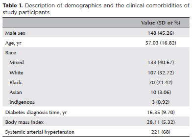
PURPOSE: Diabetic retinopathy screening in low- and middle-income countries is limited by restricted access to specialized care. Portable retinal cameras offer a practical alternative; however, image quality – affected by mydriasis – directly influences the performance of artificial intelligence models. This study evaluated the effect of mydriasis on image gradability and AI-based diabetic retinopathy detection in real-world, resource-limited settings.
METHODS: The proportions of gradable images were compared between mydriatic and non-mydriatic groups. Generalized estimating equations were used to identify factors associated with image gradability, including age, sex, race, diabetes duration, and systemic hypertension. A ResNet-200d model was trained on the mobile Brazilian Ophthalmological dataset and externally validated on both mydriatic and non-mydriatic images. Model performance was evaluated using accuracy, F1 score, area under the curve, and confusion matrix metrics. Sensitivity differences were assessed using the McNemar test, and area under the curves were compared using DeLong's test. The Youden index was used to determine optimal classification thresholds. Agreement between macula- and disc-centered images was analyzed using Cohen's κ.
RESULTS: The mydriatic group demonstrated a higher proportion of gradable images compared with the non-mydriatic group (82.1% vs. 55.6%; p<0.001). In non-mydriatic images, lower gradability was associated with systemic hypertension, older age, male sex, and longer diabetes duration. The AI model achieved better performance in mydriatic images (accuracy, 85.15%; area under the curve, 0.94) than in non-mydriatic images (accuracy, 79.68%; area under the curve, 0.93). The McNemar test showed a significant difference in sensitivity (p=0.0001), whereas DeLong's test revealed no significant difference in area under the curve (p=0.4666). The Youden index indicated that optimal classification thresholds differed based on mydriasis status. Agreement between image fields was moderate to substantial and improved with mydriasis.
CONCLUSION: Mydriasis significantly improves image gradability and enhances AI performance in diabetic retinopathy screening. Nonetheless, in low- and middle-income countries where pharmacologic dilation may be impractical, optimizing model calibration and thresholding for non-mydriatic images is essential to ensure effective AI implementation in real-world clinical environments.
Keywords: Artificial intelligence; Bias; Diabetic retinopathy; Portable camera; Retina
Arq. Bras. Oftalmol. 2025;88 (2 )
:1-7
| DOI: 10.5935/0004-2749.2023-0215
Abstract
PURPOSE: To compare the refractive prediction error of Hill-radial basis function 3.0 with those of 3 conventional formulas and 11 combination methods in eyes with short axial lengths.
METHODS: The refractive prediction error was calculated using 4 formulas (Hoffer Q, SRK-T, Haigis, and Hill-RBF) and 11 combination methods (average of two or more methods). The absolute error was determined, and the proportion of eyes within 0.25-diopter (D) increments of absolute error was analyzed. Furthermore, the intraclass correlation coefficients of each method were computed to evaluate the agreement between target refractive error and postoperative spherical equivalent.
RESULTS: This study included 87 eyes. Based on the refractive prediction error findings, Hoffer Q formula exhibited the highest myopic errors, followed by SRK-T, Hill-RBF, and Haigis. Among all the methods, the Haigis and Hill-RBF combination yielded a mean refractive prediction error closest to zero. The SRK-T and Hill-RBF combination showed the lowest mean absolute error, whereas the Hoffer Q, SRK-T, and Haigis combination had the lowest median absolute error. Hill-radial basis function exhibited the highest intraclass correlation coefficient, whereas SRK-T showed the lowest. Haigis and Hill-RBF, as well as the combination of both, demonstrated the lowest proportion of refractive surprises (absolute error >1.00 D). Among the individual formulas, Hill-RBF had the highest success rate (absolute error ≤0.50 D). Moreover, among all the methods, the SRK-T and Hill-RBF combination exhibited the highest success rate.
CONCLUSIONS: Hill-radial basis function showed accuracy comparable to or surpassing that of conventional formulas in eyes with short axial lengths. The use and integration of various formulas in cataract surgery for eyes with short axial lengths may help reduce the incidence of refractive surprises.
Keywords: Cataract; Lenses, intraocular; Axial length, eye; Refractive errors; Artificial intelligence
Arq. Bras. Oftalmol. 2024;87 (5 )
:1-5
| DOI: 10.5935/0004-2749.2022-0245
Abstract
Objetivo: Recentemente, o ácido hialurônico foi proposto como promissor no tratamento do ectrópio cicatricial adquirido da pálpebra inferior. No entanto, não foram feitas avaliações quantitativas para confirmar este efeito, motivo que levou a realização do presente estudo que visou avaliar o efeito do ácido hialurônico no tratamento do ectrópio cicatricial adquirido da pálpebra inferior.
Métodos: Oito portadores de ectrópio
cicatricial adquirido da pálpebra inferior (13 pálpebras) foram tratados com uma única dose de 1 mL de ácido hialurônico, injetada na área pré-septal da pálpebra inferior. Os sintomas e o exame biomicroscópico foram realizados antes e 30 dias após a injeção do ácido hialurônico. A análise quantitativa da posição palpebral inferior (com e sem tração palpebral) foi determinada antes e 30 dias após a injeção do ácido hialurônico por meio de fotografias que foram analisadas usando o programa ImageJ.
Resultados: Todos os pacientes apresentaram melhora parcial dos sintomas. A posição da pálpebra inferior foi elevada significativamente após a injeção do ácido hialurônico, com redução significativa dos ângulos medial e lateral, da distância entre o reflexo pupilar e a margem da pálpebra inferior, da área de fissura palpebral total e da área medial. No entanto, sinais de inflamação da margem palpebral e ceratite puntata da córnea persistiram.
Conclusão: O ácido hialurônico injetado na área pré-septal da pálpebra inferior, melhorou os sintomas do ectrópio cicatricial adquirido da pálpebra inferior e elevou significativamente a posição da pálpebra inferior. Estudos com maior número de participantes e período de acompanhamento mais longo são necessários para melhor determinar os efeitos das injeções de ácido hialurônico a longo prazo no tratamento do ectrópio cicatricial adquirido da pálpebra inferior.
Keywords: Ectrópio; Cicatriz; Pálpebras; Anormalidades da pele; Ácido hialurônico; Preenchedores dérmicos; Injeções
Arq. Bras. Oftalmol. 2026;89 (3 )
:1-9
| DOI: 10.5935/0004-2749.2025-0312
Abstract
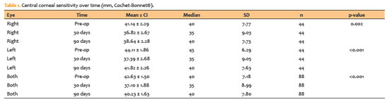
PURPOSE: To quantitatively assess changes in central corneal sensitivity after phacoemulsification and to characterize recovery patterns up to 90 days using standardized esthesiometry.
METHODS: This prospective observational study included 44 patients (88 eyes) undergoing uncomplicated phacoemulsification with intraocular lens implantation. Central corneal sensitivity was measured using a Cochet-Bonnet® esthesiometer preoperatively and at 30 and 90 days postoperatively. Repeated-measures data were analyzed using Friedman and Wilcoxon signed-rank tests (p<0.05). Inter-eye differences were assessed with a paired Wilcoxon test. Individual changes from baseline (Δ30, Δ90) were calculated, and 90-day recovery was categorized according to thresholds aligned with the 5-mm device resolution. Spearman correlation was used to explore associations between age and Δ90.
RESULTS: Corneal sensitivity decreased after surgery. In right eyes, mean sensitivity declined from 41.14 ± 7.77 mm at baseline to 36.82 ± 9.03 mm at 30 days and partially recovered to 38.64 ± 7.73 mm at 90 days. In left eyes, sensitivity decreased from 44.11 ± 6.29 mm to 37.39 ± 9.05 mm at 30 days and recovered to 41.82 ± 7.63 mm at 90 days. Left eyes showed higher sensitivity than right eyes at baseline (p=0.023) and at 90 days (p=0.018). At 90 days, complete or near-complete recovery (within ± 5 mm of baseline) occurred in 73.2% of right eyes and 78.0% of left eyes, while improvement above baseline (≥ +5 mm) occurred in 7.3% and 4.9%, respectively. Age showed weak, nonsignificant correlations with Δ90 (p=−0.14 to −0.19; p>0.2).
CONCLUSION: Phacoemulsification with a 2.75-mm clear corneal incision leads to a temporary reduction in central corneal sensitivity, with partial recovery by 90 days. Recovery patterns vary among individuals, highlighting the value of postoperative sensitivity monitoring to identify atypical trajectories and guide ocular surface care during visual rehabilitation.
Keywords: Phacoemulsification; Cornea/innervation; Ophthalmic nerve/physiology; Optometry/instrumentation; Diagnostic techniques, ophthalmological; Neural regeneration; Visual rehabilitation.
Arq. Bras. Oftalmol. 2024;87 (4 )
:1-7
| DOI: 10.5935/0004-2749.2023-0026
Abstract
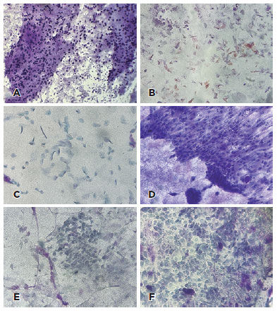
PURPOSE: To describe cellular alterations detected by impression cytology of the ocular surface in patients with xeroderma pigmentosum. The secondary objective was to assess the reliability of impression cytology in diagnosing ocular surface squamous neoplasia.
METHODS: Patients with xeroderma pigmentosum underwent a single-day complete ophthalmological examination and impression cytology for ocular surface evaluation using 13 mm diameter mixed cellulose esters membrane filters and combined staining with Periodic Acid Schiff, Hematoxylin and Eosin, and Papanicolaou stains followed by microscopic analysis. The cytological findings were correlated with the clinical diagnosis. The impression cytology findings at baseline and one-year follow-up were correlated with the clinical course (no tumor, treated tumor, residual tumor recurrent tumor, new tumor).
RESULTS: Of the 42 patients examined, impression cytology was performed in 62 eyes of 34 participants (65% females). The mean age of patients was 29.6 ± 17 years (range 7-62). Fifteen eyes had a clinical diagnosis of ocular surface squamous neoplasia. Impression cytology showed goblet cells (47, 75%), inflammatory cells (12, 19%), keratinization (5, 8%), and squamous metaplasia (30, 48%). Impression cytology was positive for atypical cells in 18 patients (12 with and 6 without ocular surface squamous neoplasia). The sensitivity, specificity, positive predictive value, and negative predictive value of impression cytology (at baseline) for diagnosis of ocular surface squamous neoplasia were 80%, 87%, 67%, and 93%, respectively, using clinical diagnosis of ocular surface squamous neoplasia as the reference standard.
CONCLUSION: Impression cytology has a moderate positive predictive value for the diagnosis of ocular surface squamous neoplasia in patients with xeroderma pigmentosum. However, the lack of detection of atypical cells on impression cytology has a high negative predictive value for ocular surface squamous neoplasia. Integration of impression cytology in the long-term management of high-risk patients, such as patients with xeroderma pigmentosum, can avoid unnecessary diagnostic biopsies.
Keywords: Xeroderma pigmentosum; Eye neoplasms; Conjunctiva/cytology; Cornea/cytology; Cytological techniques
Arq. Bras. Oftalmol. 2024;87 (4 )
:1-5
| DOI: 10.5935/0004-2749.2023-0143
Abstract
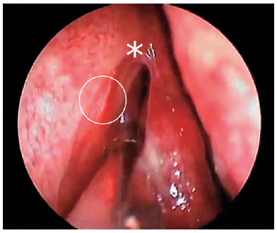
PURPOSE: The purpose of this study is to assess the long-term outcomes of modified transcanalicular diode laser dacryocys torhinostomy in a large cohort of patients affected by primary acquired nasolacrimal duct obstruction.
METHODS: This study, conducted from January 17 to June 2022, encompassed 141 patients (159 procedures) who underwent modified transcanalicular diode laser dacryocystorhinostomy (MT-DCR). The procedure employed an 810-nm diode laser. Patients were monitored for at least a year after the intervention. Anatomical success was determined by ostium patency upon irrigation, while functional success referred to epiphora resolution. Parameters studied included patient demographics, procedure duration, complications, and both anatomical and functional success. Statistical analysis was performed using the Statistical Package for the Social Sciences software, with results considered significant at a 95% confidence interval (p≤0.05).
RESULTS: A total of 159 lacrimal drainage systems (141 patients: 112 women and 29 men) were included in this study. Among them, 18 underwent bilateral procedures. The average patient age was 58 years (range: 34-91 years), and the average surgical duration was 24 minutes (range: 18-35 minutes). One year after the surgery, MT-DCR exhibited anatomical and functional success rates of 84.9% (135/159) and 83% (132/159), respectively.
CONCLUSION: MT-DCR achieved an anatomical success rate of 84.9%, reflecting an excellent outcome. However, further extensive studies with larger sample sizes and longer follow-up periods are necessary to substantiate these findings.
Keywords: Lacrimal duct obstruction; Nasolacrimal duct/surgery; Dacryocystorhinostomy; Lacrimal apparatus diseases; Laser therapy/methods; Lasers, semiconductor/therapeutic use; Regeneration
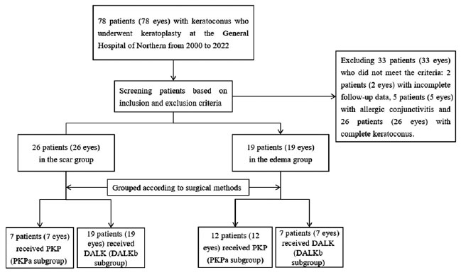
Arq. Bras. Oftalmol. 2024;87 (4 )
:1-8
| DOI: 10.5935/0004-2749.2023-0144
Abstract
PURPOSE: To assess the outcomes of deep anterior lamellar keratoplasty or penetrating keratoplasty at the scar and the edema stages.
METHODS: Forty-five patients (45 eyes) with keratoconus scar stage (scar group, n=26; penetrating keratoplasty a subgroup, n=7; deep anterior lamellar keratoplasty b subgroup, n=19) and keratoconus edema stage (edema group, n=19; penetrating keratoplasty c subgroup, n=12; deep anterior lamellar keratoplasty d group, n=7) who received penetrating keratoplasty or deep anterior lamellar keratoplasty from 2000 to 2022 were retrospectively studied. At 1, 6, and 12 months after surgery, the best-corrected visual acuity, astigmatism, spherical equivalent, corneal endothelial cell density, and complications were analyzed.
RESULTS: The best-corrected visual acuity and average corneal endothelial cell loss rate were not significantly different between the scar and edema groups (p>0.05). At 6 and 12 months after surgery, the astigmatism and spherical equivalent in the scar group were significantly lower than those in the edema group (p<0.05). The spherical equivalent of the deep anterior lamellar keratoplasty b subgroup was lower than that of the penetrating keratoplasty a subgroup in the scar group 6 months after surgery (p<0.05). In the edema group, there was no significant difference in spherical equivalent between subgroups (p>0.05). There were no significant differences in best-corrected visual acuity and astigmatism between subgroups within the two groups (p>0.05). In comparison to the scar group, the edema group experienced more complications. According to a survival analysis, there was no statistically significant difference between the scar group and the edema group regarding the progression of vision.
CONCLUSIONS: In terms of the outcomes and prognosis for vision after keratoplasty with edema stage and scar stage, deep anterior lamellar keratoplasty may be as effective as penetrating keratoplasty.
Keywords: keratoconus; Edema; Cicatrix; keratoplasty, penetrating; Corneal transplantation; Astigmatism; Corneal endothelial cell loss; Endothelial cells
 ABO is licensed under a Creative Commons Attribution-NonComercial 4.0 Internacional.
06-fig01.jpg)
08-fig01.jpg)
12-tab01.jpg)

07-fig01.jpg)
11-fig01tb.jpg)