Showing of 1 until 14 from 87 result(s)
Search for: Health care costs; Anesthesia, local; Cataract extraction; Phacoemulsification
01-ane01.jpg)
Abstract
OBJETIVO: Avaliar a relação entre a incidência de complicações e reações emocionais durante a cirurgia de catarata sob anestesia tópica em pacientes funcionalmente monoculares.
MÉTODOS: Estudo prospectivo, transversal, caso-controle de vinte e dois pacientes monoculares e dezenove controles pareados por idade e sexo . Dados demográficos foram analisados: idade, sexo e escolaridade. As cirurgias foram realizadas pelo mesmo cirurgião e durante o procedimento os sinais vitais dos pacientes (como pressão arterial sistêmica e frequência cardíaca) e eventos cirúrgicos (duração da cirurgia, movimentos corporais, sinais de aumento da pressão vítrea, dificuldade de realização da capsulorrexis e complicações) foram coletados. A acuidade visual pré e pós foi analisada. A distribuição normal dos dados foi confirmada com o teste de Shapiro-Wilk. Os dados foram expressos como média ± DP e porcentagem. A comparação dos diferentes testes clínicos entre os grupos foi realizada utilizando Student’s t-test e ANOVA com correção de Bonferroni. O qui-quadrado foi usado para comparar dados demográficos. Valor de p<0,05 foi considerado estatisticamente significante.
RESULTADOS: Este estudo incluiu vinte e dois olhos de 22 pacientes funcionalmente monoculares (6 homens e 13 mulheres) e dezenove olhos de 19 controles (11 homens e 11 mulheres). A média de idade foi de 73,05 ± 13,31 anos nos indivíduos monoculares e 69,74 ± 16,81 no controle. Considerando-se os sinais vitais não houve diferença significativa entre os grupos (p>0,05). Durante o procedimento, a percepção do cirurgião em relação aos movimentos excessivos de olho, pálpebra ou cabeça em ambos os grupos foi semelhante, assim como sinais de aumento da pressão vítrea (p=0,2 e p=0,1, respectivamente).
CONCLUSÃO: Este estudo sugere que é seguro realizar a extração de catarata com anestesia tópica em pacientes funcionalmente monoculares. Esses pacientes aparentemente se comportam de maneira semelhante aos pacientes binoculares.
Keywords: Facoemulsificação/psicologia; Capsulorrexe; Anestésicos locais; Acuidade visual
03-fig01.jpg)
Abstract
OBJETIVO: Avaliar a influência da dinâmica pupilar na curva de desfoco de olhos implantados com lente intraoculares multifocais difrativas.
MÉTODOS: Estudo prospectivo e randomizado realizado na Faculdade de Medicina de Ribeirão Preto - Universidade de São Paulo - Departamento de Oftalmologia. Trinta e oito pacientes foram aleatoriamente designados para receber bilateralmente lentes intraoculares SN6AD1 (n=20) (mfIOL) ou SN60WF (n=18) (aIOL). Além da acuidade visual para longe e perto, corrigida e não corrigida, e curva de desfoco, foi ainda realizada pupilometria dinâmica. A área sob a curva de desfoco foi calculada usando um modelo polinomial empírico.
RESULTADOS: Um total de 16 e 17 pacientes (n=32 e 34 olhos) completaram 1 ano de seguimento nos grupos mfIOL e aIOL, respectivamente. Não houve diferenças significativas entre grupos para as acuidades visuais seja para longe ou perto. As curvas de desfoco do grupo mfIOL mostraram um pico duplo; enquanto o SN60WF mostrou apenas um pico, típico para uma lente intraoculares monofocal. A média da área sob a curva de desfoco do grupo aIOL foi (4,66 ± 1,51 logMAR.dp), e essa é estatisticamente significante diferente da métrica do grupo mfIOL (1,99 ± 1,31 logMAR.dp). A pupila na contração máxima após a exposição a um flash de 30 cd/m2 por 1 segundo foi significativamente correlacionada com uma melhor área de foco no grupo mfIOL (r=0,54; p=0,0017), essa relação não foi observada para o grupo aIOL.
CONCLUSÃO: Estes dados indicam que quanto menor a pupila durante contração, melhor é a área sob a curva de desfoco e, portanto, o desempenho visual dos olhos implantados com essa mfIOL. Esta correlação não foi encontrada para lentes intraoculares monofocais.
Keywords: Lentes intraoculares multifocais; Pupila/fisiologia, Catarata; Facoemulsificacão
03-tab01tb.jpg)
Abstract
Objetivo: Avaliar e comparar a variação do diâmetro pupilar antes e após a cirurgia de catarata por facoemulsificação convencional versus cirurgia de catarata assistida por laser de femtossegundo, usando o LDV Z8 (Ziemer Ophtalmic). Também avaliamos a relação entre o diâmetro pupilar com o tempo da cirurgia e o tempo de ultrassom.
Métodos: Estudo comparativo prospectivo, realizado no Centro de Excelência em Oftalmologia, Brasil. Foram incluídos 79 olhos de 67 pacientes com opacidade nuclear. Os mesmos foram divididos em Grupo Controle, que foi submetido a cirurgia de catarata com facoemulsificação manual, e Grupo Estudo, com catarata assistida por laser de femtossegundo. Todas as cirurgias foram realizadas pelo mesmo cirurgião experiente. Todos os pacientes receberam antiinflamatório não esteróide tópico no dia anterior à cirurgia e o mesmo colírio midriático no pré-operatório. Para quantificar o tamanho da pupila, as medidas foram realizadas usando um compasso cirúrgico: anterior ao procedimento de facoemulsificação e ao final da cirurgia. No grupo de estudo, medidas após o laser foram adicionadas. O tempo cirúrgico e o tempo de facoemulsificação também foram analisados.
Resultados: Não foi encontrada diferença significativa entre o tamanho da pupila pré-femto x pré-faco (8,69 ± 0,44 mm x 8,63 ± 0,72 mm; p=0,643), bem como o tamanho da pupila no final da cirurgia (7,96 ± 0,98 mm x 7,78 ± 0,95 mm; p=0,480) e o tempo médio de cirurgia (p=0,780). No entanto, no grupo de catarata assistida por laser de femtossegundo, houve um aumento transitório do diâmetro pupilar após o laser, indicando uma tendência para maior variação no grupo femto.
Conclusões: Embora o diâmetro pupilar fosse semelhante ao final da cirurgia, o grupo com catarata assistida por laser de femtossegundo apresentou maior variação intraoperatória da pupila. Portanto, para uma cirurgia de catarata assistida por laser de femtossegundo mais eficiente e segura, o cirurgião deve estar ciente do tamanho do diâmetro pupilar antes do procedimento.
Keywords: Catarata; Miose; Facoemulsificação; Laser; Pupila

Abstract
Objetivos: Avaliar a percepção do risco de exposição da Doença de Coronavírus 2019 (COVID-19), conhecimento sobre medidas de proteção pessoal entre os profissionais de oftalmologia latino-americanos e priorização de pacientes com Covid-19.
Métodos: Pesquisa anônima voluntária autoadministrada (formulários do Google Drive) distribuída por mensagem de texto para profissionais de oftalmologia em 1º a 5 de maio de 2020.
Resultados: Trezentos e setenta e um profissionais completaram a pesquisa (taxa de resposta de 45%), composta por 118 residentes (27,6%), 111 oftalmologistas (40,5%) e 142 subespecialistas (32,8%). 106 profissionais (32,6%) sentiram-se em alto risco de adquirir o COVID-19 no trabalho. 273 (69,1%) acreditavam que as diretrizes atuais não são suficientes para identificar os pacientes com COVID-19. 265 (59,5%) não tinham treinamento para usar os equipamentos de proteção individual (EPI) e, mesmo com seu uso correto, 341 (91,5%) ainda se sentiram em risco de adquirir COVID-19. 80% consideraram que a equipe de trabalho não tem conhecimento de protocolos nacionais para o atendimento aos pacientes com COVID-19. Apenas 9 dos profissionais (2%) consideraram mudar a profissão para minimizar o risco de contágio por COVID-19.
Conclusão: Esta pesquisa mostra a escassez de pessoal e treinamento específico que os praticantes de oftalmologia na América Latina enfrentam em sua prática diária. Essas preocupações e ansiedade parecem ser as mesmas em todo o mundo com a pandemia de COVID- 19. É importante reforçar a confiança dos profissionais de oftalmologia nas diretrizes atuais de atendimento ao paciente com COVID-19 e também disponibilizar programas de treinamento sobre o uso de EPI e também itens de EPI disponíveis em todos os momentos para garantir a qualidade do atendimento e a disseminação mínima da doença.
Keywords: Pandemias; Oftalmologia; Pesquisas sobre serviços de saúde; América Latina; SARS-CoV2; Infecções por coronavirus; COVID-19
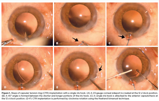
Abstract
PURPOSE: To evaluate the effect of using a single iris retractor, affixed to the anterior capsulorhexis at the 12 o'clock position, on the ease of capsular tension ring implantation.
METHODS: This prospective comparative study comprised 37 patients with zonular weakness attributed to pseudoexfoliation syndrome who underwent capsular tension ring implantation during cataract surgery. In Group 1, a single iris retractor was inserted into the anterior capsulorhexis at the 12 o'clock position. Group 2 did not receive this intervention. Zonular weakness was graded on a scale of 1–5, and the subjective difficulty of capsular tension ring implantation was categorized as easy, medium, or difficult.
RESULTS: Group 1 and 2 comprised 20 and 17 patients, respectively. There were no significant differences between the groups in age, sex distribution, and presence of glaucoma (p=0.53, p=0.28, and p=1.00, respectively). The mean zonular weakness score was significantly higher in Group 1 (3.35 ± 0.45) than in Group 2 (2.71 ± 0.59; p=0.02). Capsular tension ring implantation was significantly easier in the iris retractor group (p<0.001).
CONCLUSIONS: Placement of a single iris retractor attached to the anterior capsulorhexis at the 12 o'clock position may facilitate easier capsular tension ring implantation, even in patients with greater zonular weakness. This technique could reduce the risk of capsular tension ring displacement into the iridocorneal angle or ciliary sulcus.
Keywords: Capsular tension ring; Cataract; Iris hook; Pseudoexfoliation syndrome; Zonular weakness; Cataract extraction; Phacoemulsification; Capsulorhexis.
11-tab01tb.jpg)
Abstract
OBJETIVO: Avaliar o momento apropriado para implante de anel de tensão capsular em casos de fraqueza zonular devida à síndrome pseudoesfoliativa.
MÉTODOS: Este foi um estudo prospectivo e comparativo realizado no Departamento de Oftalmologia da Universidade İnönü. Foram incluídos 43 pacientes, sendo 16 no grupo 1 e 27 no grupo 2. O grupo 1 era composto de pacientes que se submeteram ao implante precoce do anel de tensão capsular, enquanto no grupo 2 os pacientes tiveram implante tardio. Foram incluídos pacientes com síndrome pseudoesfoliativa submetidos à cirurgia de facoemulsificação e ao implante de lente intraocular na câmara posterior e anel de tensão capsular. Em cada olho, foram avaliadas as complicações intraoperatórias e as dificuldades tanto com a implantação do anel de tensão capsular quanto com a remoção do córtex.
RESULTADOS: Não houve diferença significativa entre os grupos quanto à dificuldade de implante do anel de tensão capsular (p=0,124). Ao se comparar as remoções do córtex, observou-se diferença significativa entre os grupos (p=0,003). Complicações intraoperatórias foram observadas em 3 pacientes do grupo 1 e 11 pacientes do grupo 2; porém, não houve diferença significativa entre os grupos (p=0,18). No grupo 2, observaram-se flutuações da cápsula posterior em 8 pacientes (29,5%), com ruptura da cápsula posterior em dois deles.
CONCLUSÕES: A remoção do córtex é mais difícil no implante precoce do anel de tensão capsular e flutuações da cápsula posterior podem causar problemas no implante tardio do anel de tensão capsular. O cirurgião deve ponderar a relação risco/benefício do implante precoce e tardio ao avaliar o momento ideal para implante de anel de tensão capsular.
Keywords: Catarata; Facoemulsificação; Anel de tensão capsular
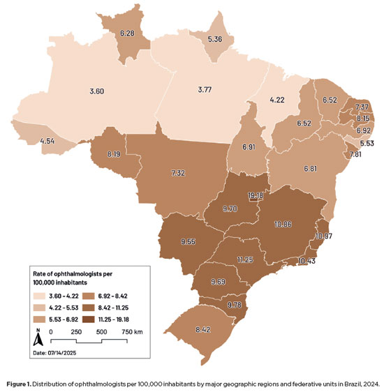
Abstract
PURPOSE: This study aimed to evaluate disparities in the distribution of ophthalmologists and the volume of cataract surgeries across Brazil, considering public and private health sectors and the country's federative units.
METHODS: Data on ophthalmologists were obtained from the National Medical Residency Commission and the Associação Múdica Brasileira. Information on cataract surgeries performed through the Unified Health System was collected from the DATASUS database, while data on procedures covered by private health plans were retrieved from the National Supplementary Health Agency. Population estimates from the 2024 Demographic Census of the Brazilian Institute of Geography and Statistics were used to calculate physician density and surgery rates per 100,000 inhabitants. Associations between the number of ophthalmologists and cataract surgery volume were analyzed using Spearman's correlation coefficient.
RESULTS: Brazil has 16,784 ophthalmologists, representing 8.96 specialists per 100,000 inhabitants. Marked disparities were observed: large cities (>500,000 inhabitants) had 18.75 ophthalmologists per 100,000 residents, whereas municipalities with <50,000 inhabitants had fewer than one. Across federative units, physician density ranged from 19.18 per 100,000 in the Federal District to 4.22 in Maranhão. In 2024, cataract surgery rates varied widely, from 1,012.61 per 100,000 inhabitants in the Southeast to 435.00 in the North. Nationally, Unified Health System performed 736.30 surgeries per 100,000 inhabitants, compared with 1,276.79 in the private sector. On average, each ophthalmologist performed 96.92 cataract surgeries annually.
CONCLUSION: Significant inequalities persist in the geographic distribution of ophthalmologists and in cataract surgery provision, with higher surgical volumes concentrated in the private sector. Targeted policies are required to address regional disparities and improve the equity and efficiency of cataract care delivery in Brazil.
Keywords: Ophthalmologists/supply & distribution; Ophthalmologists/statistics & numerical data; Cataract extraction; Health services accessibility/statistics & numerical data; Healthcare disparities; Health policy; Public health systems; Insurance, Heal

Abstract
The advantages and disadvantages of using perioperative subconjunctival steroid injections in dropless cataract surgery continue to be debated. A systematic review of PubMed, EMBASE, and the Cochrane Central database identified five studies—two randomized controlled trials and three non-randomized studies—encompassing 70,751 eyes. Among these, 12,319 eyes (17.4%) received subconjunctival steroid injections, while 58,432 eyes (82.6%) were managed with topical steroids. The Cochrane Collaboration’s RoB 2 tool was applied for bias assessments in randomized controlled trials, and heterogeneity was assessed using the I² statistics. No statistically significant differences were found between the two groups regarding macular edema (p=0.249), visual acuity (p=0.73), or laser flare count (p=0.45). Both subconjunctival injections and topical steroids demonstrated comparable efficacy and safety in controlling postoperative inflammation after cataract surgery. Additional research is warranted to validate these conclusions.
Keywords: Cataract extraction; Phacoemulsification; Lens implantation, intraocular; Postoperative care; Intravitreal injections; Anti-inflammatory agents, non-steroidal/administration & dosage; Glucocorticoids; Triamcinolone acetonide; Research design; Randomiz

Abstract
PURPOSE: Posterior capsule rupture is defined as an intraoperative posterior capsule tear resulting in vitreous loss. This study aimed to analyze the clinical characteristics, preoperative risk factors, intraoperative management strategies, and postoperative complications associated with posterior capsule rupture during phacoemulsification surgery.
METHODS: This was a retrospective observational cohort study of the medical records for 25,224 phacoemulsification surgeries performed at our tertiary eye care center between 2017 and 2022. We collected and collated the demographic characteristics and clinical findings of the patients in our cohort. Intraoperative management strategies and postoperative outcomes over a 1-year followup period were also recorded.
RESULTS: Posterior capsule rupture occurred in 351 eyes (351 patients), giving an overall posterior capsule rupture rate of 1.3%. The mean patient age was 68.6 ± 10.8 years. Pseudoexfoliation syndrome, mature cataracts, brown cataracts, and surgery performed by a resident were identified as risk factors for posterior capsule rupture (p<0.05 for each; the risk ratios were 2.70, 2.15, 2.44, 1.34, respectively). The most common intraoperative complications were dislocated lens fragments in the vitreous (8%) and iris damage (7.1%). The mean best-corrected visual acuity improved from 1.31 ± 0.84 (logMAR) postoperatively to 0.51 ± 0.56 at the end of the 1-year follow-up period (p<0.001). Corneal edema (55.6%) and elevated intraocular pressure (33.3%) were the most common early postoperative complications. Persistently elevated intraocular pressure (11.1%) and cystoid macular edema (5.1%) were the most common late postoperative complications.
CONCLUSION: Posterior capsule rupture is a common complication of phacoemulsification surgery that requires prolonged postoperative follow-up and a multidisciplinary approach. Despite the increased incidence of complications when rupture occurs, appropriate intraoperative and postoperative management can lead to satisfactory visual outcomes.
Keywords: Cataract extraction; Phacoemulsification; Posterior capsule rupture; Corneal edema; Risk factors; Postoperative complications; Intraoperative complications

Abstract
PURPOSE: To compare endothelial corneal cell changes following cataract surgery performed by phacoemulsification with intraocular lens implantation, conducted by surgeons with varying levels of experience.
METHODS: Two hundred and eighty-three eyes diagnosed with cataract were included. Lens opacity was classified into three categories (I, II, and III). Surgeons were categorized into four experience levels (1, 2, 3, and 4), based on years of practice and lifetime surgeries performed. Corneal endothelial characteristics were assessed using non-contact specular microscopy, with measurements taken before surgery and 30-60 days post-surgery.
RESULTS: Pre- and postoperative endothelial analysis showed no significant differences between surgeon levels regarding visual acuity achieved, corneal thickness, and endothelial hexagonality. However, the central endothelial cell density index showed a significantly greater reduction among level 1 surgeons (p=0.026). Grade II cataracts exhibited significant variations in the central endothelial cell density (p=0.011) and average cell size, with level 1 surgeons showing the largest increases (p=0.024).
CONCLUSIONS: The analysis revealed significant differences in visual acuity and endothelial indices between surgeon experience levels, with less experienced surgeons showing greater variations and poorer performance. Clinical protocols should consider these data to establish safer training protocols.
Keywords: Cataract extraction; Phacoemulsification; Endothelium; corneal; Lens implantation, intraocular; Visual acuity; Internship and residency; Surgeons
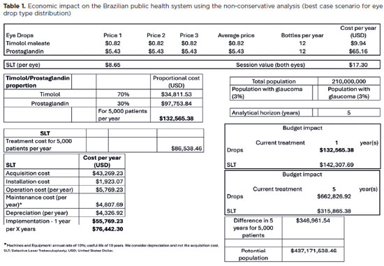
Abstract
PURPOSE: To evaluate the economic impact of the following initial treatment scenarios for glaucoma on the Brazilian Public Health System (SUS): (1) traditional continuous instillation of hypotensive eye drops and (2) single session of selective laser trabeculoplasty.
METHODS: Economic impact was analyzed in three scenarios, from the least to the most conservative, for a hypothetical cohort of 5,000 individuals with open-angle glaucoma. Thereafter, projections were made on the basis of a glaucoma prevalence of 3% in the 2021 Brazilian population size.
RESULTS: All three scenarios demonstrated that selective laser trabeculoplasty exhibited a significantly lower economic impact than the eye drops on SUS over one and five years. Furthermore, the difference was more than United States Dollar 8 billion at five years when considering 3% of the Brazilian population aged >40 years in 2021.
CONCLUSION: As the initial treatment for primary open-angle glaucoma, selective laser trabeculoplasty exhibited a lower economic impact on SUS than latanoprost and timolol maleate eye drop instillation in all the studied scenarios over one and five-year periods.
Keywords: Glaucoma; Trabeculotomy; Laser therapy; Cost analysis; Health care cost Unified Health System; Brazil
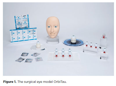
Abstract
PURPOSE: The OrbiTau surgical simulator is a synthetic eye model developed to enhance cataract surgical training. Herein, we aimed to describe the perspectives of Harvard’s Ophthalmology faculty and residents regarding the effectiveness of OrbiTau.
METHODS: A cross-sectional study was conducted in which 11 surgeons from the Massachusetts Eye and Ear Infirmary, with prior experience utilizing simulated phacoemulsification platforms, conducted cataract surgery with the OrbiTau. Subsequently, they completed a satisfaction questionnaire using the Likert scale.
RESULTS: Regarding the various OrbiTau components, 90.90% of the participants reported that the OrbiTau lens capsule was comparable to that of the human lens during capsulotomy. Furthermore, 72.72% of the participants found that the OrbiTau lens consistency was analogous to that of the human lens nucleus. Approximately 63.63% of the participants reported that the model’s posterior lens capsule resembled the native posterior capsule, and 72.72% of the participants noted that the model’s red reflex was similar to that of the dilated human pupil. Most participants believed that the OrbiTau was easier to use and more realistic than other commercially available simulators.
CONCLUSION: Our single-institution survey of the Orbitau demonstrated that this model realistically replicates ocular structures and may be a viable option for cataract surgery training.
Keywords: Cataract extraction/education; Simulation training/methods; Ophthalmology/education; Phacoemulsification/education; Ophthalmologists/education; Surgeons/education; High fidelity simulation training
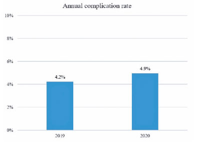
Abstract
PURPOSE: To assess the effect of the coronavirus disease 2019 (COVID-19) pandemic on cataract surgery by residents who had mandatory surgical simulator training during residency.
METHODS: In this retrospective, observational analytical study, the total number of cataract surgeries and surgical complications by all senior residents of 2019 (2019 class; prepandemic) and 2020 (2020 class; affected by the reduced number of elective surgeries due to the COVID-19 pandemic) were collected and compared. All residents had routine mandatory cataract surgery training on a virtual surgical simulator during residency. The total score obtained by these residents on cataract challenges of the surgical simulator was also evaluated.
RESULTS: The 2020 and 2019 classes performed 1275 and 2561 cataract surgeries, respectively. This revealed a reduction of 50.2% in the total number of procedures performed by the 2020 class because of the pandemic. The incidence of surgical complications was not statistically different between the two groups (4.2% in the 2019 class and 4.9% in the 2020 class; p=0.314). Both groups also did not differ in their mean scores on the simulator’s cataract challenges (p<0.696).
CONCLUSION: Despite the reduction of 50.2% in the total number of cataract surgeries performed by senior residents of 2020 during the COVID-19 pandemic, the incidence of surgical complications did not increase. This suggests that surgical simulator training during residency mitigated the negative effects of the reduced surgical volume during the pandemic.
Keywords: COVID-19; Pandemics; Cataract extraction/education; Internship and residency/methods; Simulation training/methods; Phacoemulsification/education; Surgery, computer-assisted; Computer simulation; Clinical competence; Ophthalmology/education
06-fig01.jpg)
Abstract
O presente relato de caso identificou a maculopatia média aguda paracentral como a causa de baixa de acuidade visual severa e irreversível após cirurgia de catarata. Existem fatores de risco bem estabelecidos para o desenvolvimento da maculopatia média aguda paracentral que devem ser conhecidos pelos cirurgiões de catarata. Nesse contexto cirúrgico, precauções extras no tocante a procedimentos anestésicos, pressão intraocular e alguns outros aspectos da cirurgia devem ser consideradas. A maculopatia média aguda paracentral é descrita como um sinal clínico observado no exame de tomografia de coerência óptica por domínio espectral e se trata, provavelmente, da evidência de um evento isquêmico no tecido vascular retiniano. Esse diagnóstico deve ser cogitado nos casos de perda de acuidade visual súbita no pós-operatório imediato associada com exame fundoscópico normal, como evidenciado no caso apresentado.
Keywords: Tomografia, coerência óptica; Procedimentos cirúrgicos oftalmológicos; Complicações pós-operatórias; Fatores de risco; Catarata; Extração de catarata; Baixa visão; Saúde oc
 ABO is licensed under a Creative Commons Attribution-NonComercial 4.0 Internacional.
About
Issues
Editorial Board
Submission
Arquivos Brasileiros de Oftalmologia
Official publication of Brazilian Council of Ophthalmology - Conselho Brasileiro de Oftalmologia (CBO)
Rua Casa do Ator, 1.117 - 2nd floor - Zip Code: 04546-004
São Paulo - SP, Brazil
TEL: +55 11 3266-4000
E-mail: [email protected]