Arq. Bras. Oftalmol. 2021;84 (5 )
:436-441
| DOI: 10.5935/0004-2749.20210068
Abstract
Objetivo: A Escala Bayley de Desenvolvimento Infantil (Bayley-III) é uma ferramenta que avalia o desenvolvimento de crianças nos 3 primeiros anos de vida, incluindo os domínios cognitivo e motor. Este estudo tem como objetivo correlacionar a acuidade visual de grades e a funcionalidade visual em crianças saudáveis usando a Bayley-III.
Métodos: A acuidade visual binocular de grades foi medida usando o teste dos Cartões de Acuidade de Teller seguido pela Bayley-III em crianças saudáveis com idade entre 1-42 meses. Os escores da acuidade visual (logMAR) e da Bayley-III para habilidades cognitivas e motoras (grossa e fina) foram comparados.
Resultados: Um grupo de 40 crianças (20 meninos) com idades entre 1,2-42,1 meses foi testado e a média da acuidade visual foi de 0,39 ± 0,27 logMAR, sendo que todas estavam dentro dos limites normais para a idade. Houve uma forte correlação negativa e significante entre acuidade visual e idade (r=-0,83; p<0,001). A média do escore cognitivo foi de 49,92 ± 18,93 pontos, com forte correlação positiva e significante entre o escore cognitivo e a idade (r=0,81; p<0,001). A média do escore motor grosso foi de 41,72 ± 16,23 pontos, com forte correlação positiva e significante entre o escore motor grosso e a idade (r=0,75; p<0,001). A média do escore motor fino foi de 39,75 ± 14,63 pontos, com uma forte correlação positiva e significante entre o escore motor fino e a idade (r=0,77; p<0,001). A regressão linear múltipla mostrou que maior idade e melhor acuidade visual foram significantemente associadas à escores cognitivo e motor mais altos.
Conclusões: Neste estudo foi encontrada alta correlação entre a acuidade visual de grades medida pelos cartões de acuidade de Teller e os escores cogninitivo e motor medidos pela Bayley-III em crianças saudáveis. A Bayley-III pode ser uma ferramenta útil para avaliar a repercussão da deficiência visual no desenvolvimento cognitivo e motor de crianças.
Keywords: Desenvolvimento infantil; Acuidade visual; Cognição; Destreza motora; Transtornos da visão; Testes neuropsicológicos; Criança
Arq. Bras. Oftalmol. 2021;84 (5 )
:442-448
| DOI: 10.5935/0004-2749.20210069
Abstract
Objetivo: Verificar se pacientes com dislexia do desenvolvimento (DD) apresentam déficits coerentes com uma disfunção magnocelular visual.
Métodos: Participantes com diagnóstico confirmado de dislexia do desenvolvimento (n=62; faixa etária=8 a 25 anos; Média da idade=13.8 anos, desvio padrão=3.9; 77% homens) foram comparados a um grupo controle com desenvolvimento típico, pareado por idade, sexo, dominância ocular, acuidade visual e compreensão de texto. A perimetria Frequency-Doubling Technology avaliou o limiar de sensibilidade ao contraste do campo visual periférico. O rastreador ocular Visagraph-III registrou os movimentos dos olhos durante leitura de texto.
Resultados: O grupo com dislexia do desenvolvimento apresentou piores limiares de sensibilidade no Frequency-Doubling Technology, com tamanho de efeito forte, do que o grupo controle. O grupo com dislexia do desenvolvimento apresentou mais olhos classificados com déficits na sensibilidade à ilusão de frequência duplicada do que o grupo controle. O grupo com dislexia do desenvolvimento apresentou pior habilidade motora ocular e no desempenho de leitura, revelado pela diferença entre os grupos em relação às fixações oculares, regressões, alcance de reconhecimento, taxa de leitura e eficiência relativa. Foi encontrada correlação significativa entre a sensibilidade ao contraste e as habilidades motoras oculares. Os participantes com boa eficiência relativa apresentaram uma sensibilidade ao contraste significativamente melhor do que os participantes com baixa eficiência relativa.
Conclusões: O grupo com dislexia do desenvolvimento apresentou desempenho inferior nas variáveis visuais relacionadas à função visual magnocelular (i.e., perimetria de frequência duplicada e habilidades motoras oculares), quando comparado ao grupo controle pareado. Os profissionais precisam estar cientes da importância de investigar a visão dos pacientes com dislexia do desenvolvimento além da acuidade visual e incluir nos seus procedimentos diagnósticos instrumentos para avaliar o processamento temporal, com limiar de sensibilidade ao contraste.
Keywords: Dislexia; Leitura; Percepção visual; Transtornos da visão; Músculos oculomotores; Movimentos oculares
Arq. Bras. Oftalmol. 2023;86 (6 )
:1-7
| DOI: 10.5935/0004-2749.2021-0130
Abstract
Objetivo: Avaliar a acuidade visual através de potenciais evocados visuais de varredura em crianças saudáveis e ambliópicas, comparando-a com a acuidade visual pelo teste de Snellen.
Métodos: Foram incluídas no estudo 160 crianças com idades entre 6 e 17 anos. Desse total, 104 crianças (65%) estavam entre 7 e 17 anos de idade, eram capazes de comunicação verbal e não tinham nenhuma patologia ocular ou sistêmica (Grupo 1). O grupo 2 incluiu 56 crianças verbais (35%) com idades entre 6 e 17 anos e portadoras de estrabismo ou ambliopia anisometrópica, com a melhor acuidade visual corrigida entre 0,1 e 0,8. Todos os pacientes foram submetidos a um exame oftalmológico detalhado e a uma medição do potencial evocado visual por varredura. Registraram-se as características demográficas, os achados oculares, a melhor acuidade visual corrigida e os resultados do potencial evocado visual por varredura.
Resultados: No Grupo 1, os valores médios e máximos da acuidade visual pelo potencial evocado visual por varredura mostraram-se menores que a melhor acuidade visual corrigida medida através do teste de Snellen (p<0,001 para ambas as medições). Uma análise de Bland-Altman revelou que no grupo 1, a distribuição das diferenças entre a melhor acuidade visual corrigida pelo teste de Snellen e a média do potencial evocado visual por varredura foi de ± 0,11 logMAR, enquanto a distribuição das diferenças entre a melhor acuidade visual corrigida pelo teste de Snellen e o valor máximo do potencial evocado visual por varredura foi de ± 0,023 logMAR. No Grupo 2, os valores médio e máximo do potencial evocado visual por varredura mostraram-se menores que a melhor acuidade visual corrigida pelo teste de Snellen (respectivamente, p<0,001 e p=0,009). A análise de Bland-Altman revelou que a distribuição das diferenças entre a melhor acuidade visual corrigida pelo teste de Snellen e a média do potencial evocado visual por varredura foi de ± 0,16 logMAR, enquanto a distribuição das diferenças entre a melhor acuidade visual corrigida pelo teste de Snellen e o valor máximo do potencial evocado visual por varredura foi de ± 0,19 logMAR.
Conclusões: As medidas da acuidade visual através do potencial evocado visual por varredura mostram resultados comparáveis às medidas da acuidade visual pelo teste de Snellen. Essa técnica é um método objetivo e confiável de se avaliar a acuidade visual em crianças.
Keywords: Ambliopia; Acuidade visual; Potenciais evocados visuais; Testes visuais; Humanos; Criança; Adolescente.
Arq. Bras. Oftalmol. 2023;86 (4 )
:330-336
| DOI: 10.5935/0004-2749.20230051
Abstract
Objetivo: Avaliar a eficácia das lentes de contato gelatinosas HydroCone, de hidrogel com silicone, em pacientes com microftalmia posterior.
Métodos: Foram revisados retrospectivamente 26 olhos com microftalmia posterior, a partir dos prontuários de 13 pacientes que receberam lentes de contato gelatinosas HydroCone, de hidrogel com silicone. Todos os pacientes foram submetidos ao exame de acuidade visual não corrigida e com melhor correção por óculos e com refração cicloplégica. Todos os pacientes receberam lentes de contato de acordo com os parâmetros obtidos na análise topográfica e foi obtida a melhor acuidade visual corrigida com lentes de contato.
Resultados: O equivalente esférico do olho direito variou de 10,00 a 19,25 dioptrias, e o do olho esquerdo de 11,00 a 21,5 dioptrias. Os comprimentos médios axiais e das câmaras posteriores foram menores do que para a população de mesma idade. No entanto, os valores médios dos parâmetros do segmento anterior, como o diâmetro horizontal visível da íris, a profundidade da câmara anterior central, a espessura da lente e a espessura central da córnea estavam dentro da faixa normal. Os valores médios da ceratometria revelaram curvatura corneana aumentada em relação à população normal. A média da melhor acuidade visual corrigida com lentes de contato foi significativamente maior que a média da melhor acuidade visual corrigida com óculos em ambos os olhos (p=0,045).
Conclusão: As lentes de contato gelatinosas de silicone HydroCone proporcionam melhor acuidade visual que óculos em pacientes com microftalmia posterior.
Keywords: Microftalmia; Lentes de contato hidrofílicas; Silicones; Transtornos da visão/reabilitação; Acuidade visual.
Arq. Bras. Oftalmol. 2025;88 (1 )
:1-4
| DOI: 10.5935/0004-2749.2022-0375
Abstract
PURPOSE: This study aimed to assess grating visual acuity and functional vision in children with congenital Zika syndrome.
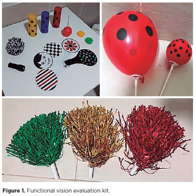
METHODS: Initial and final grating visual acuity was measured using Teller acuity cards. Cerebral vision impairment standardized tests were used to assess functional vision. Patients were referred to the early visual intervention program for visually disabled children. Neuroimaging was performed.
RESULTS: In this study, 10 children were included with an age range of 1–37 months. Eight patients presented with macular atrophic scars. Neuroimaging revealed microcephaly and cerebral abnormalities in all patients. Low vision and cerebral vision impairment characteristics were observed in all children. The final grating visual acuity in this group varied from 3.00 to 0.81 logMAR.
CONCLUSIONS: The grating visual acuity test revealed low vision in all children with congenital Zika syndrome. Functional vision evaluation revealed cerebral vision impairment characteristics in all patients, who were referred to the early visual intervention program. Visual acuity improved in six children.
Keywords: Zika virus infection/congenital; Low vision; Vision disorders; Atrophy, Microcephaly; Visual acuity; Child
Arq. Bras. Oftalmol. 2021;84 (1 )
:51-57
| DOI: 10.5935/0004-2749.20210009
Abstract
Objetivo: Determinar o impacto do uso de unidade móvel no acesso à saúde ocular e avaliar o perfil da população que necessita de cuidados oftalmológicos, as doenças oculares mais frequentes e o tratamento.
Métodos: Estudo transversal realizado em 14 municípios da região sudoeste do Estado de São Paulo utilizando uma unidade móvel oftalmológica. Os participantes eram usuários do Sistema Único de Saúde que procuraram atendimento oftalmológico, sem restrição quanto a idade, gênero ou condição socioeconômica. Os dados foram transferidos para a tabela Excel para análise estatística.
Resultados: Participaram do estudo 6.878 pessoas, com média de idade de 44 anos (variação de 4 meses a 96 anos) e 65,5% eram mulheres. Erros refrativos estavam presentes em 78,6% dos participantes, catarata em 9,6% e pterígio em 8,3%. Para 60% foram prescritos óculos, para 10% foi mantida a correção óptica em uso e para 28% foram necessárias apenas orientações. Exames especializados ou procedimentos cirúrgicos foram indicados para 18,1% dos casos que foram encaminhados para tratamento em serviço terciário. Dentre os pacientes referenciados, 36,4% necessitavam de cirurgia oculoplástica ou para tratar afecções externas do olho e 31,8%, de cirurgia de catarata.
Conclusão: A grande maioria dos pacientes que procurou atendimento na unidade móvel necessitava de prescrição de óculos. A unidade móvel oftalmológica possui alto grau de resolutividade para os problemas oculares, com oportunidade de tratar os erros refrativos e referenciar os pacientes que necessitam de atendimento especializado, geralmente relacionado a condições cirúrgicas. Unidades móveis podem ser uma alternativa aos cuidados oftalmológicos básicos, melhorando o acesso, atuando na promoção da saúde ocular e prevenindo a cegueira.
Keywords: Unidades móveis de saúde; Saúde ocular; Transtornos da visão; Erros de refração; Óculos; Cegueira/prevenção & controle
Arq. Bras. Oftalmol. 2025;88 (4 )
:1-12
| DOI: 10.5935/0004-2749.2023-0263
Abstract
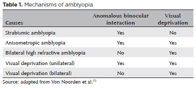
PURPOSE: Amblyopia is a cortical neurological disorder caused by abnormal visual experiences during the critical period for visual development. Recent works have shown that, in addition to the well-known visual alterations, such as changes in visual acuity, several perceptual aspects of vision are affected. This study aims to analyze and compare the effects of different types of amblyopia on visual color processing and determine whether these effects are correlated with visual acuity.
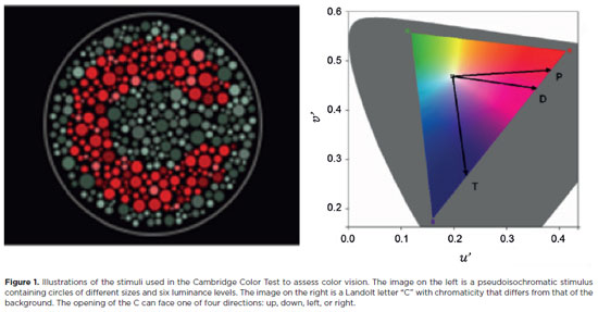
METHODS: Our study sample comprised 42 amblyopic individuals, aged 7-40 years, (strabismus, n=16; anisometropia, n=18; and mixed-cause, n=8) and 33 age-matched controls. Color vision was tested by measuring the chromaticity threshold of each patient on the protan, deutan, and tritan axes using version 02 of the Cambridge Color Test. Spatial stimulation cues were eliminated using spatial noise and luminance.
RESULTS: The color discrimination thresholds on the protan, deutan, and tritan axes were similar between control participants and amblyopic patients (p>0.05). There was no correlation between VA values and color thresholds (p>0.05).
CONCLUSION: Patients with amblyopia have normal color vision in contexts that include luminance and spatial noise. Our results may be indicative of independent neural pathways for spatial and chromatic visual processing.
Keywords: Amblyopia; Anisometropia; Color vision; Strabismus; Vision disorders; Visual acuity
Arq. Bras. Oftalmol. 2026;89 (1 )
:1-6
| DOI: 10.5935/0004-2749.2025-0071
Abstract
PURPOSE: This study aimed to evaluate the outcomes of strabismus surgical correction in patients with Down syndrome.
METHODS: We conducted a retrospective chart review of patients with Down syndrome who underwent strabismus surgery between January 1997 and May 2024 at an Ophthalmology Outpatient Clinic in São Paulo, Brazil. The data collected included age, sex, medical and ocular history, surgical details, and follow-up outcomes. The patients were categorized by strabismus type into esotropia, fourth nerve palsy, and mixed groups. Surgical success was defined as final alignment within 10Δ of orthotropia and, where applicable, whether there was resolution of abnormal head posture of ocular origin. Patients with postoperative follow-up <6 months were excluded.
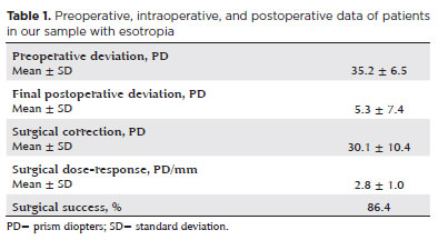
RESULTS: A total of 37 patients (21 females) were included. Of these, 22 (59.5%) were in the esotropia group, 10 (27.0%) in the fourth nerve palsy group, and 5 (13.5%) in the mixed group. The surgical success rate in the esotropia group was 86.4%, with a mean preoperative deviation of 35.2 (± 6.5)Δ, and mean surgical correction of 30.1 (± 10.4)Δ. The success rate in the fourth nerve palsy group was 40.0%, with a mean preoperative deviation of 10.4 (± 4.3)Δ. Overall, success was achieved with a single surgical procedure in 73.0% of the sample. No significant associations were found between surgical success and the clinical and demographic variables, including sex, age at surgery, oblique muscle overaction, pattern strabismus, visual acuity, amblyopia, preoperative deviation, or postoperative follow-up duration (p>0.05).
CONCLUSIONS: When standard surgical tables are applied, strabismus surgery in patients with Down syndrome appears to be safe and effective. We found high success rates, particularly among patients with esotropia. We observed no tendencies toward over- or under-correction. These findings support the use of conventional surgical protocols with this patient population.
Keywords: Down Syndrome/complications; Strabismus/surgery; Esotropia/surgery; Oculomotor nerve diseases/physiopathology; Vision disorders; Humans; Brazil.
Arq. Bras. Oftalmol. 2025;88 (2 )
:1-8
| DOI: 10.5935/0004-2749.2023-0268
Abstract
PURPOSE: This prospective, randomized, unmasked, clinical trial aimed to report the visual outcomes of cataract surgery on both eyes versus cataract surgery on one eye in Brazilian patients.
METHODS: This study included patients with bilateral cataracts and binocular visual acuity worse than or equal to 0.3 logarithm of the minimum angle of resolution. The patients were randomly assigned to undergo surgery on one (Control Group) or both eyes (one eye at a time; Intervention Group). Postoperatively, self-reported visual function using Catquest-9SF (primary outcome measure), binocular visual acuity, stereopsis, and ocular dominance (secondary outcome measures) were compared.
RESULTS: A total of 151 patients (77 and 148 eyes in the Control and Intervention Groups, respectively) completed the follow-up. Patients who underwent surgery on both eyes exhibited significantly better self-reported visual function (p=0.036) and stereopsis (p=0.026) than those who underwent surgery on one eye. Binocular visual acuity and ocular dominance did not affect the group comparisons.
CONCLUSIONS: Surgery on both eyes resulted in significantly better self-reported visual function and stereopsis than surgery on one eye.
Keywords: Cataract; Cataract extraction; Quality of life; Treatment outcome; Visual acuity; Binocular vision; Stereopsis
Arq. Bras. Oftalmol. 2024;87 (4 )
:1-6
| DOI: 10.5935/0004-2749.2022-0035
Abstract
Objetivo: Comparar as diferenças entre a chord aparente µ e o chord real µ.
Métodos: Estudo prospectivo, comparativo, não randomizado e não intervencionista. Os exames de imagem (Pentacam e HD Analyzer) foram realizados na mesma sala e nas mesmas condições escotópicas. Os critérios de inclusão foram idade de 21 a 71 anos; compreensão do termo de consentimento; miopia até 4D e astigmatismo topográfico anterior até 1D. Os critérios de exclusão foram usuários de lentes de contato; pacientes com doenças oculares prévias ou cirurgias; opacidades da córnea; a presença de alterações tomográficas da córnea ou suspeita de ceratocone.
Resultados: Em nosso estudo foram analisados 116 olhos de 58 pacientes. A média de idade foi de 30,69 anos (± 7,85). Análises de correlação foram desenvolvidas e o coeficiente de correlação de Pearson (0,647) indica uma relação linear positiva moderada entre as variáveis. A média do chord µ real foi 226,21± 128,53 µm e a média do chord µ média foi 278,66 ± 123,90 µm, com diferença média de 52,45 µm (p=0,01).
A análise do diâmetro pupilar médio apresentou: 5,76mm no HD Analyzer e 3,31mm no Pentacam.
Conclusões: Entendemos a existência de uma diferença significativa entre os métodos e assim a medida de ambos os dispositivos com base em princípios diferentes devemos respeitar suas peculiaridades. Como encontramos correlação entre as duas medidas, acreditamos que ambas podem ser utilizadas na prática diária.
Keywords: Imagem óptica; Percepção visual; Pupila; Segmento anterior do olho; Córnea; Técnicas de diagnóstico oftalmológico
Arq. Bras. Oftalmol. 2025;88 (3 )
:1-11
| DOI: 10.5935/0004-2749.2024-0049
Abstract
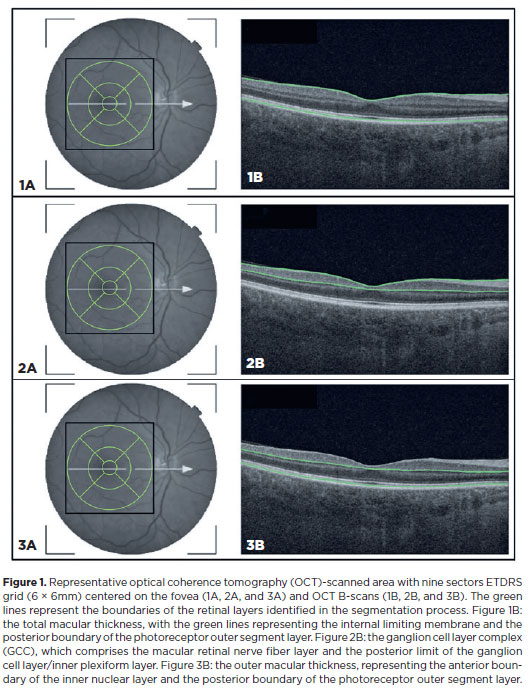
PURPOSE: This study aimed to evaluate the total macular thickness as well as the thickness of the inner and outer retinal layers in patients with Parkinson's disease. It also aimed to verify the correlation of these parameters with motor symptoms and cognitive function.
METHODS: A total of 46 eyes of 23 patients with Parkinson's disease and 40 eyes of 20 healthy controls were included in the study. The patients' cognitive, functional, and nonmotor symptoms were evaluated using the Katz Index of Independence and Pfeffer's Activities of Daily Living, Mini-Mental State Examination, Frontal Assessment Battery, Schwab and England Staging Scales, and Movement Disorders Society Nonmotor Symptoms Scale. The macular thickness measurements obtained via total, inner, and outer optical coherence tomography were recorded. Furthermore, the correlation of the parameters of optical coherence tomography with cognitive, functional, and nonmotor symptoms was assessed.
RESULTS: The scores of the Katz Index of Independence and Pfeffer's Activities of Daily Living as well as the Movement Disorders Society Nonmotor Symptoms Scale were significantly lower in patients with Parkinson's disease than in healthy controls. Moreover, the former had greater total macular thickness. The temporal and inferior outer sectors were significantly greater for the ganglion cell complex thickness in patients. A significant correlation was observed between the total macular thickness and the Movement Disorder Society-Unified Parkinson's Disease Rating Scale, Parte III (MDS-UPDRS-III) values. Contrarily, there was a negative correlation between the outer macular thickness and the MDS-UPDRS-III values. Meanwhile, the total macular thickness and ganglion cell complex thickness were significantly correlated with the scores of the Mini-Mental State Examination, Schwab and England Staging Scale, Frontal Assessment Battery, and Katz Index of Independence and Pfeffer's Activities of Daily Living. In addition, the Schwab and England scale was correlated with the outer macular thickness.
CONCLUSION: The total and inner macular thicknesses at the temporal and inferior outer sectors were greater in patients with Parkinson's disease than in the control group. These findings indicate that macular thickness may be greater in those with Parkinson's disease, particularly when associated with mild motor symptoms. In addition, the parameters of the total, inner, and outer optical coherence tomography were significantly associated with motor and nonmotor symptoms as well as cognitive function impairment.
Keywords: Parkinson's disease; Tomography, optical coherence; Neurodegenerative diseases; Cognitive dysfunction; Cognition; Motor perception; Visual acuity; Retina
Arq. Bras. Oftalmol. 2024;87 (3 )
:1-6
| DOI: 10.5935/0004-2749.2022-0366
Abstract
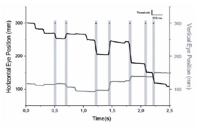
PURPOSE: To evaluate the saccadic movements of patients with visual field loss due to primary open-angle glaucoma.
METHODS: Thirteen patients with good visual acuity (0.2 logMAR or better) (seven patients with primary open-angle glaucoma 65 ± 13 years) and six controls (51 ± 6 years) yielded a comprehensive ophthalmological examination, including Humphrey Visual Field tests (SITA-Standard 24-2), and performed a monocular, exploratory digital visual search task that quantifies the duration for finding the number “4” on a random array of digits distributed on the screen. After individual adjustments of the angle and distance positioning, the screen was spatially matched with the 24-2 visual field, and divided into five areas for analysis. During the task, saccades were simultaneously recorded in the same eye with a video-based eye tracker.
RESULTS: The patients with primary open-angle glaucoma showed a significantly higher number of saccades/screen (median ± interquartile range, 59.00 ± 29.00 vs. 32.50 ± 19.75 saccades (p=0.027) and visual search time per screen (38.50 ± 60.14 vs. 23.75 ± 8.90 seconds (p=0.035) than the controls did. Although the univariate analysis indicated a significant correlation with visual field mean deviation (coefficient=26.19 (p=0.02), only the visual search time/screen was significantly associated with the number of saccades/screen in the multivariate regression model (coefficient=0.55 (p<0.001). Overall, no significant correlation was observed between the sectorial number of saccades and the sensitivity of the five visual field areas.
CONCLUSIONS: The patients with primary open-angle glaucoma show impaired search performance and showed a higher number of saccades needed to find stimuli when performing the exploratory visual task.
Keywords: Glaucoma, open angle; Saccades; Eye movements; Visual fields; Vision disorders
 ABO is licensed under a Creative Commons Attribution-NonComercial 4.0 Internacional.
03-tab01.jpg)
04-tab01tb.jpg)
07-fig01.jpg)
07-tab01.jpg)
08-fig01.jpg)

07-fig01tb.jpg)
13-fig01.jpg)