Arq. Bras. Oftalmol. 2022;85 (5 )
:498-505
| DOI: 10.5935/0004-2749.20220066
Abstract
Objetivo: Avaliar alterações da coroide através de imagens de tomografia de coerência óptica (OCT) com profundidade realçada na doença por coronavírus de 2019 (COVID-19).
Métodos: Foram incluídos no estudo 32 pacientes com COVID-19 moderada e 34 indivíduos saudáveis. A espessura da coroide foi medida em 3 pontos: subfoveal, a 1500 mm da fóvea na direção nasal e a 1500 mm da fóvea na direção temporal. A área total da coroide, a área luminal, a área estromal e o índice vascular da coroide foram medidos com o programa ImageJ. Todas as medições foram feitas durante a doença ativa e 4 meses após a remissão.
Resultados: No grupo de pacientes, as espessuras subfoveal, nasal e temporal da coroide mostraram-se reduzidas em comparação com os controles. A diferença não foi estatisticamente significativa (respectivamente, p=0,534, p=0,437 e p=0,077). As médias das áreas total da coroide, estromal e luminal, bem como o índice vascular da coroide, mostraram-se diminuídos com significância estatística no grupo de pacientes (respectivamente, p<0,001, p=0,001, p=0,001 e p=0,003). Aos 4 meses após a remissão, os parâmetros estruturais e o índice vascular da coroide revelaram um aumento estatisticamente significativo em pacientes com COVID-19, em comparação com as medidas iniciais (todos os valores de p<0,001 para os parâmetros estruturais e p=0,047 para o índice vascular da coroide).
Conclusão: Os parâmetros vasculares da coroide e do estroma mostraram uma diminuição transitória, mas significativa em pacientes com COVID-19 durante a doença.
Keywords: Coróide; COVID-19; Infecções por coronavirus; Tomografia de coerência óptica
Arq. Bras. Oftalmol. 2023;86 (2 )
:150-151
| DOI: 10.5935/0004-2749.20230021
Abstract
Objetivo: Avaliar o efeito da doença por coronavírus de 2019 (COVID19) na espessura da coroide usando tomografia de coerência óptica com profundidade de imagem aprimorada.
Métodos: Este estudo consistiu em 41 casos pós-COVID19 (Grupo 1) e 41 indivíduos saudáveis (Grupo 2). Apenas os olhos direitos dos participantes foram incluídos no estudo. A espessura da coroide foi medida usando tomografia de coerência óptica com profundidade de imagem aprimorada. Nos casos pós-COVID19, as medições foram realizadas dentro de 1 mês da doença. A espessura da coroide foi medida por dois oftalmologistas experientes nos quadrantes subfoveal, temporal e nasal, em sete pontos diferentes, a intervalos de 500 a 1500 μm da fóvea. Além disso, a espessura macular central e a espessura da camada de células ganglionares foram medidas com OCT e os dois Grupos foram comparados.
Resultados: As espessuras coroidais foram estatisticamente mais espessas no Grupo 1 que no Grupo 2, com 500 μm no quadrante subfoveal, 500 Symbol (OTF)m no temporal e 1000 μm no nasal (p=0,011, p=0,043, p=0,009 e p=0,019, respectivamente). Embora outras medidas tenham se mostrado mais espessas no Grupo 1, elas não foram estatisticamente significativas (p>0,05). Também não houve diferenças significativas entre os Grupos quanto à espessura macular central e à espessura da camada de células ganglionares (p>0,05).
Conclusão: A espessura da coroide mostrou-se aumentada em pacientes pós-COVID19. Isso pode estar relacionado à inflamação que faz parte da patogênese do COVID19.
Keywords: COVID-19; Infecções por coronavirus; Tomografia de coerência óptica; Coróide/patologia; Manifestações oculares
Arq. Bras. Oftalmol. 2021;84 (4 )
:367-373
| DOI: 10.5935/0004-2749.20210053
Abstract
OBJETIVO: A doença de Stargardt é a forma mais comum de distrofia macular de início juvenil. É bilateral e simétrica em aparência, afeta a mácula e sua característica principal é a diminuição da visão central que geralmente inicia-se na primeira ou segunda década de vida. O objetivo do estudo é descrever o perfil clínico dos pacientes avaliados no Complexo Hospital de Clínicas da Universidade Federal do Paraná, bem como descrever os achados eletrorretinográficos destes pacientes com o eletrorretinograma de campo total.
MÉTODOS: Foi realizado um estudo observacional retrospectivo, baseado na análise de prontuários e eletrorretinograma de 27 pacientes com Doença de Stargardt e Fundus Flavimaculatus, atendidos em consulta oftalmológica no ambulatório de Eletrofisiologia Ocular e Neuro-Oftalmologia do Complexo Hospital de Clínicas da Universidade Federal do Paraná, entre 1997 e 2014. Os pacientes incluídos no estudo apresentavam quadro clínico, fundoscopia e/ou achados eletrorretinográficos compatíveis com a doença.
RESULTADOS: A acuidade visual no melhor olho variou de 0 a 1,6 logMAR (20/20 a 20/800), com média de 0,89 ± 0,42 logMAR. A idade de aparecimento dos sintomas variou desde o nascimento a 36 anos (19,2 ± 9,2), sendo a maioria nas 1ª e 2ª década de vida. Em relação ao tempo entre o início dos sintomas e o diagnóstico, a média foi de 7,3 anos. Na fundoscopia, todos os pacientes apresentaram alguma alteração. Na análise do eletrorretinograma, a maioria dos pacientes demonstrou resultados que diferem da amostra de pacientes controles, ou seja, amplitudes reduzidas e tempos de culminação aumentados nas fases fotópicas e escotópicas.
CONCLUSÕES: A acuidade visual e idade de início de aparecimento dos sintomas encontrados neste estudo são compatíveis com a evolução desta distrofia. Achados fundoscópicos típicos da doença de Stargardt e eletrorretinograma alterados foram mais frequentes em decorrência do atraso no diagnóstico. Novos estudos prospectivos são necessários para avaliar estes pacientes, fundamentando-se em novas tecnologias.
Keywords: Eletrorretinografia; Doenças retinianas; Epitélio pigmentado da retina; Degeneração macular; Lipofuscina
Arq. Bras. Oftalmol. 2020;83 (5 )
:410-416
| DOI: 10.5935/0004-2749.20200080
Abstract
Objetivo: Avaliar as espessuras internas da retina e da coroide em pacientes com retinite pigmentosa precoce.
Métodos: Foram analisadas imagens de tomografia de coerência óptica de domínio espectral de 35 pacientes com retinite pigmentosa e 40 indivíduos saudáveis. Medimos a espessura do complexo de células maculares e ganglionares. Realizamos medições da espessura da coroide na região subfoveal e a 500 µm, 1000 µm e 1500 µm do centro da fóvea.
Resultados: Pacientes com retinite pigmentosa apresentaram espessuras maculares e da coroide significativamente mais finas em todas as medições e suas medidas individuais da espessura do complexo de células ganglionares foram inferiores às de indivíduos saudáveis. A espessura média do complexo de células ganglionares foi significativamente menor nos pacientes com retinite pigmentosa do que nos controles. A espessura macular média foi significativamente correlacionada com as espessuras médias do complexo das células de coroide e das células ganglionares médias. Não encontramoscorrelação entre a espessura media da coroide e a espessura media do complexo de células ganglionares.
Conclusões: A coroide foi levemente afetada em nossos pacientes com retinite pigmentosa precoce. A tendência à significância na retina interna foi possivelmente causada por uma boa acuidade visual.
Keywords: Coroide/anatomia & histologia; Retina/anatomia & histologia; Células ganglionares da retina; Retinite pigmentosa; Tomografia de coerência óptica
Arq. Bras. Oftalmol. 2026;89 (1 )
:1-10
| DOI: 10.5935/0004-2749.2025-0184
Abstract
PURPOSE: To investigate choroidal structural and vascular changes in patients with mild autonomous cortisol secretion using enhanced depth imaging optical coherence tomography and optical coherence tomography angiography.
METHODS: This cross-sectional study included 60 eyes of 30 patients with mild autonomous cortisol secretion and 60 eyes of 30 subjects with nonfunctional adenoma (controls) between February 2023 and January 2024. Subfoveal choroidal thickness, pachychoroid spectrum disease and choroidal vascularity index were evaluated using spectral-domain optical coherence tomography. Group comparisons were performed, and correlations between subfoveal choroidal thickness and clinical features were analyzed.
RESULTS: Pachyvessels were more common in patients with mild autonomous cortisol secretion than in controls (71.4% vs. 42.9%, p=0.002). The frequency of pachychoroidal spectrum disease was significantly higher in the mild autonomous cortisol secretion Group (68.3% vs. 31.7%; p<0.001). Median subfoveal choroidal thickness was 355 μm (range, 150–535) in the mild autonomous cortisol secretion Group and 297 μm (range, 162–597) in controls (p=0.014). Choroidal vascularity index was comparable between groups (p=0.072). Subfoveal choroidal thickness correlated significantly with axial length, spherical equivalent, post-1-mg dexamethasone suppression test cortisol level, and disease duration.
CONCLUSION: Patients with mild autonomous cortisol secretion exhibited greater subfoveal choroidal thickness and a higher frequency of pachychoroidal spectrum disease compared with controls, whereas stromal and vascular structural alterations were proportionally similar between groups.
Keywords: Adrenal gland neoplasms; Central serous chorioretinopathy; Choroid; Cushing syndrome; Hydrocortisone; Optical coherence tomography
Arq. Bras. Oftalmol. 2025;88 (3 )
:1-8
| DOI: 10.5935/0004-2749.2023-0115
Abstract
PURPOSE: To evaluate the presence of congenital hypertrophy of the retinal pigment epithelium in a large family affected by familial adenomatous polyposis and identify the causative mutation in the adenomatous polyposis coli gene. Thus, we aimed to determine the significance of congenital hypertrophy of the retinal pigment epithelium as a phenotypic marker of the disease.
METHODS: A family consisting of 95 individuals was evaluated. Among these, 45 individuals were randomly selected by convenience sampling method to undergo ophthalmological evaluation. A funduscopic exam, including slit lamp and indirect ophthalmoscopy, were performed in the selected patients. In those with retinal lesions, a retinography was obtained. The adenomatous polyposis coli gene was sequenced in one affected family member to identify the pathogenic mutation. Once the variant was identified, six undiagnosed family members were tested for the mutation via capillary electrophoresis sequencing.
RESULTS: Congenital hypertrophy of the retinal pigment epithelium was observed in 13 (28.9%) of the 45 individuals evaluated. Of these, nine patients were confirmed to have familial adenomatous polyposis (via colonoscopy or molecular testing). However, four patients had not been investigated. Of the 32 (71.1%) family members without the lesion, 14 did not have familial adenomatous polyposis and 18 were yet to be evaluated. The lesions were bilaterally present and exhibited a peculiar fish-tail shape in all the evaluated individuals. Adenomatous polyposis coli gene sequencing revealed a pathogenic variant c.4031del. (Ser1344*), in heterozygosity (49.27%), in exon 16.
CONCLUSIONS: The study findings confirmed the significance of congenital hypertrophy of the retinal pigment epithelium as a phenotypic marker for familial adenomatous polyposis. Furthermore, it is an effective first-line screening method for at risk family members of such patients. The novel mutation identified in our study participants, which is yet to be described in the literature, causes an aggressive form of the disease.
Keywords: Retinal diseases/congenital; Retinal pigment epithelium; Hypertrophy/congenital; Adenomatous polyposis coli / genetics; Phenotype; Optical coherence tomography
Arq. Bras. Oftalmol. 2025;88 (3 )
:1-8
| DOI: 10.5935/0004-2749.2024-0104
Abstract
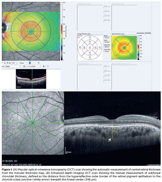
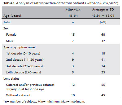
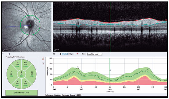
PURPOSE: This study aimed to characterize retinitis pigmentosa associated with the eyes shut homolog gene, which causes hereditary retinal degeneration.
METHODS: The anatomical and functional findings of retinitis pigmentosa in patients with variants of the eyes shut homolog gene were characterized and compared using multimodal imaging and genetic analysis of the variants. Clinical data such as visual acuity, lens status, and refraction were obtained from medical records. Patients underwent an ophthalmic examination, including static visual field, microperimetry, optical coherence tomography, fundus autofluorescence, and fundus photography.
RESULTS: Twenty-two patients were included in the study. Several anatomical and functional characteristics of retinitis pigmentosa-eyes shut homolog were identified, including the presence of cataracts, cystoid macular edema, epiretinal membrane, and a tubular visual field. Genetic results revealed 26 distinct variants in the cohort, with 7 novel variants not previously documented or reported in the scientific literature or databases.
CONCLUSION: The findings demonstrate that eyes shut homolog-retinitis pigmentosa manifests in specific patterns, starting in adolescence with mild progression and advancing with age. The integration of multimodal imaging and genetic analysis has provided a detailed understanding of the anatomical and functional features of retinitis pigmentosa-eyes shut homolog. Seven novel variants of the eyes shut homolog gene have been identified. These findings enhance the understanding of eyes shut homolog-related retinitis pigmentosa characteristics of by detailing the spectrum of mutations in this gene within the Brazilian population.
Keywords: Retinal diseases/diagnostic imaging; Retinitis pigmentosa/genetics; Retinal degeneration; Eye proteins/genetics; Eye diseases, hereditary/genetics; Genes, recessive; Phenotype; Multimodal imaging; Tomography, optical coherence/methods; Fluorescein angiogr
Arq. Bras. Oftalmol. 2024;87 (4 )
:1-7
| DOI: 10.5935/0004-2749.2023-0047
Abstract
PURPOSE: We aimed to evaluate retinal nerve fiber and choroidal layer alterations in adolescents with anorexia nervosa using spectral-domain optical coherence tomography.
METHODS: Thirty patients with anorexia nervosa and 30 healthy adolescents aged 12-18 years were included in this study. Their age, sex, body mass index, anorexia nervosa type, disease duration, and spectral-domain optical coherence tomography data were recorded.
RESULTS: Central macular thickness and retinal nerve fiber layer thickness in the temporal and inferior regions were significantly lesser in patients with anorexia than in healthy controls (p<0.05). Moreover, significant choroidal thinning around the foveal and subfoveal regions in patients with anorexia was observed (p<0.05). In addition, a statistically significant relation between the increase in disease duration and the thinning of the inferior retinal nerve fiber layer was detected (p<0.05).
CONCLUSION: The retinal nerve fiber layer and choroidal layer thicknesses were lesser in patients with anorexia than in healthy controls. Screening for retinal indices might prevent the development of irreversible retinal pathologies in adolescents with anorexia nervosa. In addition, thinning of the retinal nerve fiber and choroidal layers could reflect structural or functional changes in the brain of adolescents with anorexia nervosa.
Keywords: Tomography; Optical coherence; Nerve fibers; Choroid; Adolescents
Arq. Bras. Oftalmol. 2024;87 (3 )
:1-6
| DOI: 10.5935/0004-2749.2022-0369
Abstract
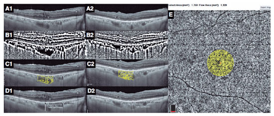
PURPOSE: To evaluate the choroidal vascular alterations and effect of surgical treatment in the setting of idiopathic epiretinal membranes.
METHODS: The structure of the choroid was studied in 33 patients with unilateral idiopathic epiretinal membrane using optical coherence tomography with enhanced depth imaging and optical coherence tomography angiography. Eyes with epiretinal membrane underwent 25-gauge vitrectomy with epiretinal membrane and internal limiting membrane peeling. The choroidal vascularity index, Haller layer/choroidal thickness ratio, and choriocapillaris flow density were used to evaluate changes in choroidal structure after surgery and compare with the healthy fellow eyes.
RESULTS: The choroidal vascularity index and Haller layer/choroidal thickness ratio of the eyes with epiretinal membrane were higher than those of the fellow eyes at baseline (p=0.009 and p=0.04, respectively) and decreased postoperatively compared with preoperative values (p=0.009 and p=0.001, respectively). The choriocapillaris flow of eyes with epiretinal membrane was lower than that of the fellow eyes at baseline (p=0.001) and increased after surgery compared with the preoperative value (p=0.04). The choroidal vascularity index, Haller layer/choroidal thickness ratio, and choriocapillaris flow values of the healthy fellow eyes were comparable at baseline and final visit. In eyes with epiretinal membrane, the final choroidal vascularity index correlated with the final choriocapillaris flow (r=-0.749, p=0.008) in the multivariate analysis.
CONCLUSION: Idiopathic epiretinal membrane appears to affect the choroidal structure with increased choroidal vascularity index and Haller layer/choroidal thickness ratio and decreased choriocapillaris flow. These macrovascular (choroidal vascularity index and Haller layer/choroidal thickness) and microvascular (choriocapillaris flow) alterations appear to be relieved by surgical treatment of the epiretinal membranes.
Keywords: Epiretinal membrane/surgery; Vitrectomy; Choroid/pathology; Choroid/blood supply; Tomography, optical coherence/methods; Optical coherence tomography angiography; Humans
 ABO is licensed under a Creative Commons Attribution-NonComercial 4.0 Internacional.
12-fig01tb.jpg)
07-fig01.jpg)
10-tab01tb.jpg)
09-fig01tb.jpg)

13-fig01.jpg)
10-fig01tb.jpg)
13-fig01.jpg)
06-fig01.jpg)