Showing of 1 until 7 from 7 result(s)
Search for: Pterygium/epidemiology; pathogenesis; Biomarkers; Review
Abstract
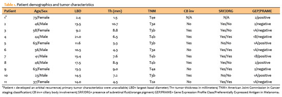
Objetivo: O melanoma da conjuntiva é um tumor raro e agressivo, com propensão à disseminação metastática regional e distante. Este estudo tem como objetivo analisar os marcadores BRAF e NRAS no melanoma da conjuntiva e sua relação com recidivas tumorais e com o prognóstico do paciente.
Métodos: Este foi um estudo retrospectivo, observacional e unicêntrico de pacientes consecutivos com diagnóstico anatomopatológico de melanoma da conjuntiva feito entre janeiro de 1992 e dezembro de 2019. As mutações BRAF e NRAS foram analisadas com o kit cobas® 4800 (Roche®) em amostras obtidas através de biópsia excisional ou por mapa. Além disso, foi avaliada a presença de lesões pré-cancerosas ou tumorais associadas.
Resultados: Foram incluídos 12 pacientes com amostras histológicas positivas para melanoma da conjuntiva (7 mulheres e 5 homens), com idade média ao diagnóstico de 60 anos e tempo médio de evolução de 6,38 ± 3,4 anos. A mutação BRAF V600E foi encontrada em 3 biópsias (25%), bem como a NRAS Q61X (25%). Ocorreram recidivas em todos os pacientes positivos para mutações de BRAF ou NRAS e 5 desses pacientes desenvolveram disseminação sistêmica (83,33%). Além disso, 4 dos 6 pacientes com BRAF ou NRAS mutante (66,66%) apresentaram achados histopatológicos de lesões tumorais ou pré-cancerosas.
Conclusões: As mutações BRAF e NRAS podem ser fatores de risco para recorrência e menor sobrevida no melanoma da conjuntiva, o que tornaria esses pacientes candidatos a terapias direcionadas e a um acompanhamento mais abrangente e individualizado. Todos esses dados justificam mais estudos prospectivos padronizados.
Keywords: Neoplasias da túnica conjuntiva; Melanoma; Biomarcadores tumorais; Proteínas proto-oncogênicas B-raf; Genes ras.
Abstract
Objetivo: Este estudo avaliou os níveis de calprotectina fecal em uma série de pacientes com uveíte anterior na tentativa de determinar se pacientes com uveíte associada com espondiloartrites apresentam níveis mais elevados desta proteína do que pacientes com uveíte anterior de outras etiologias. Um terceiro grupo com espondiloartrites sem uveíte também foi incluído na avaliação para entendimento do papel da uveíte anterior no aumento da calprotectina fecal.
Métodos: Estudo transversal de 28 pacientes divididos em três grupos: (a) com espondiloartrites e uveíte (n=9); (b) com espondiloartrites sem uveíte (n=10) e (c) com uveíte sem espondiloartrites (n=9). A dosagem de calprotectina fecal foi avaliada.
Resultados: Pacientes com uveíte anterior associada a espondiloartrites apresentaram valores medianos maiores de calprotectina fecal (101 µg/g) que os valores dos pacientes com uveíte sem espondiloartrites (9 µg/g), pacientes com espondiloartrites sem uveíte que também demonstraram valores maiores (93.0 µg/g) que os dos pacientes com uveíte sem espondiloartrites (p=0,02).
Conclusão: Pacientes com espondiloartrites com e sem uveíte anterior aguda demonstraram níveis significativamente elevados de calprotectina fecal. Este teste pode ser útil na diferenciação entre uveítes associadas com espondiloartrites de uveítes de outras etiologias. Entretanto, não foi possível demonstrar associação entre o aumento dos níveis de calprotectina fecal e a presença da uveíte em espondiloartrites.
Keywords: Calprotectin; Uveitis; Spondyloarthritis; Inflammatory bowel diseases; Biomarkers
Abstract
PURPOSE: This pilot study was conducted to investigate the presence of various bioactive compounds (copeptin, asprosin, and salusins) in the blood and tears of patients with glaucoma.
METHODS: A total of 83 subjects, including 28 patients with open-angle glaucoma, 28 patients with ocular hypertension, and 27 control volunteers, were enrolled in this study. The levels of salusin-α, salusin-β, copeptin, and asprosin in tears and venous blood samples were measured by enzyme linked immunosorbent assay (ELISA).
RESULTS: Patients with open-angle glaucoma and those with ocular hypertension showed statistically significantly decreased levels of salusin-α and salusin-β in their blood and tears compared with those of control subjects (p<0.05), with the decrease being the most pronounced in patients with ocular hypertension (p<0.05). In contrast, the levels of copeptin and asprosin showed a statistically significant increase in both these patient groups compared with those of control subjects (p<0.05). There was a negative correlation between intraocular pressure and blood and tear salusins.
CONCLUSIONS: Fluids from patients with open-angle glaucoma and ocular hypertension showed lower salusin levels. Patients with ocular hypertension had higher levels of copeptin and asprosin, but not those with open-angle glaucoma (except for asprosin, whose levels showed a slight but remarkable increase in plasma in patients with open-angle glaucoma). The pathogenesis of ocular hypertension and open-angle glaucoma may be significantly impacted by these biomarkers.
Keywords: Glaucoma, open-angle/physiopathology; Intraocular pressure/physiopathology; Retinal ganglion cells/pathology; Biomarkers/blood; Glycopeptides; Fibrillin-1; Tears/chemistry; Intercellular signaling peptides and proteins/blood; Enzyme-linked immunosorbent a
Abstract
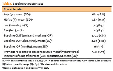
PURPOSE: To evaluate the accuracy of a short-term intravitreal dexamethasone sodium phosphate challenge in predicting the anatomical response to a sustained-release dexamethasone implant (Ozurdex) in patients with refractory diabetic macular edema.
METHODS: This prospective, non-randomized, Phase 2 pilot study enrolled 12 pseudophakic eyes with diabetic macular edema refractory to anti-vascular endothelial growth factor (anti-VEGF) therapy. Participants underwent a challenge phase (Day 0) consisting of a single intravitreal injection of 0.08-mg dexamethasone sodium phosphate, followed by a maintenance phase (Day 7), during which all subjects received a 0.7-mg dexamethasone implant. The primary outcome was concordance between the anatomical response at Day 3 (post-dexamethasone sodium phosphate) and Day 60 (post-implant), defined as a ≥10% reduction in central macular thickness. Secondary outcomes included achieving a clinically "dry" macula (central macular thickness ≤300 μm) at Day 60, changes in best-corrected visual acuity, safety outcomes (intraocular pressure), and spectral-domain optical coherence tomography biomarker analysis.
RESULTS: The dexamethasone sodium phosphate challenge induced significant macular drying by Day 3 (median central macular thickness reduction, −21 μm; p=0.002). A positive response to dexamethasone sodium phosphate strongly predicted response to the dexamethasone implant, with a positive predictive value and specificity of 100%. The negative predictive
value was 80%. Irreversible biomarkers, including disorganization of retinal inner layers and ellipsoid zone disruption, were more prevalent among nonresponders (60% vs. 28.6%). Safety outcomes were acceptable; 16.7% of patients developed ocular hypertension, which was successfully managed with topical therapy.
CONCLUSION: A short-term dexamethasone sodium phosphate challenge is a safe,
low-cost, and highly specific predictor of dexamethasone implant efficacy. This "test-and-treat" strategy may optimize resource allocation in resource-constrained settings by identifying responders before high-cost implantation.
Keywords: Diabetic retinopathy; Macular edema; Dexamethasone; Drug implants; Biomarkers, pharmacological
Abstract
OBJETIVO: Os níveis séricos de N-óxido de trimetilamina têm sido associados ao diabetes mellitus tipo 2 e suas complicações. O presente estudo tem como objetivo responder a duas questões, entre elas: O nível plasmático de N-óxido de trimetilamina poderia ser um novo marcador no diagnóstico de retinopatia diabética? e Ele poderia ser utilizado no diagnóstico diferencial de retinopatia diabética e não diabética?
MÉTODOS: Trinta pacientes com retinopatia diabética, 30 pacientes com retinopatia não diabética, 30 pacientes com diabetes mellitus tipo 2 sem retinopatia e 30 participantes saudáveis do grupo controle foram incluídos no estudo. Parâmetros bioquímicos, níveis séricos de IL-6, de TNF-α e de N-óxido de trimetilamina foram medidos em todos os participantes.
RESULTADOS: O nível de N-óxido de trimetilamina foi significativamente maior na retinopatia diabética do que nos outros grupos (p<0,001). Não houve diferença significativa no nível de N-óxido de trimetilamina entre o grupo de retinopatia não diabética, do grupo controle ou do grupo de diabetes mellitus tipo 2. Houve uma correlação positiva significativa entre o nível de N-óxido de trimetilamina e os níveis elevados de FPG, IMC, HOMA-IR, BUN, IL-6 e TNF-α.
CONCLUSÃO: O estudo atual mostrou que o nível de N-óxido de trimetilamina encontra-se elevado na retinopatia diabética. Esses achados sugerem que o nível sérico de N-óxido de trimetilamina pode ser um novo marcador na retinopatia diabética, podendo ser usado no diagnóstico diferencial de retinopatia diabética e não diabética.
Keywords: Biomarcadores; Retinopatia diabética; Diagnóstico diferencial; Óxidos; Diabetes mellitus tipo 2; N-óxido de trimetilamina; Trimetilamina
Abstract
PURPOSE: We aimed to study reported cases of nasopharyngeal carcinoma presenting with ophthalmic manifestations with and without a prior diagnosis of nasopharyngeal carcinoma.
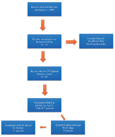
METHODS: We conducted a systematic review following the Preferred Reporting Items for Systematic Reviews and Meta-Analyses (PRISMA). A literature search was conducted using the MEDLINE database in PubMed and Google Scholar. We included patients with a previous diagnosis of nasopharyngeal carcinoma in Group I and those without a prior diagnosis of nasopharyngeal carcinoma in Group II. Data included demographics, clinical presentation, history of nasopharyngeal carcinoma, treatment, histopathological description, World Health Organization classification, and outcome.
RESULTS: Fifty-eight patients (26 in Group I and 32 in Group II) were included. The male-to-female ratio was 3:1. The mean age of the patients (53.3 ± 11.7 years and 54.8 ± 16.2 years, respectively) and gender did not differ significantly between the two groups. The most common ocular presentations were diplopia and proptosis in the first group (each in 34.6%), whereas visual disturbance was most common in the second group (46.9%). Treatment options and World Health Organization grading were comparable. The outcome in 38 patients (after a comparable follow-up period) was significantly better in group II (p=0.003). There was no statistically significant difference in the outcome of 23 patients in correlation with World Health Organization grades II versus III irrespective of group (p=0.094).
CONCLUSIONS: The demographics of patients with nasopharyngeal carcinoma presenting with ophthalmic manifestations were similar between the two study groups, with a wide age range and male predominance. Patients presenting initially to ophthalmologists with no history of nasopharyngeal carcinoma have a more favorable outcome. World Health Organization grading may have less value as a prognostic indicator.
Keywords: Nasopharyngeal carcinoma; Carcinoma; Eye manifestations; Exophthalmos; Diplopia; Systematic review
Abstract
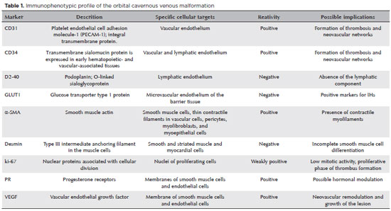
Vascular anomalies comprise a wide spectrum of clinical manifestations related to disturbances in the blood or lymph vessels. They correspond to mainly tumors (especially hemangiomas), characterized by high mitotic activity and proliferation of the vascular endothelium, and malformations, endowed with normal mitotic activity and no hypercellularity or changes in the rate of cell turnover. However, the classifications of these lesions go beyond this dichotomy and consist various systems adapted for and by different clinical subgroups. Thus, the classifications have not reached a consensus and have historically caused confusion regarding the nomenclatures and definitions. Cavernous venous malformations of the orbit, previously called cavernous hemangiomas, are the most common benign vascular orbital lesions in adults. Herein, we have compiled and discussed the various evidences, including clinical, radiological, morphological, and molecular evidence that indicate the non-neoplastic nature of these lesions.
Keywords: Orbital neoplasms; Vascular malformations; Vascular system injuries; Cavernous hemangioma; Cavernous venous malformations; Biomakers, tumor; Endothelium, vascular; Cell proliferation

ABO is licensed under a Creative Commons Attribution-NonComercial 4.0 Internacional.

About
Issues
Editorial Board
Submission
Official publication of Conselho Brasileiro de Oftalmologia
Rua Casa do Ator, 1.117 - 2º andar - CEP: 04546-004
São Paulo - SP, Brazil
Phone: +55 11 3266-4000