Arq. Bras. Oftalmol. 2023;86 (5 )
:1-6
| DOI: 10.5935/0004-2749.20230064
Abstract
Objetivo: Avaliar a resposta tecidual e clínica a um implante orbitário de polimetilmetacrilato, oco e multiperfurado em sua porção posterior em modelo animal após evisceração.
Métodos: Dezesseis coelhos da raça Nova Zelândia foram submetidos à evisceração do globo ocular direito. Todos receberam implante oco de polimetilmetacrilato de 12 mm de diâmetro, multiperfurado em sua semiesfera posterior. O estudo foi dividido em avaliação clínica e histopatológica. A avaliação clínica foi diária até 14 dias pós-evisceração e, a cada sete dias, até completar 180 dias. Os animais foram divididos em grupos de quatro animais e cada um foi submetido à exenteração com 07, 30, 90 e 180 dias e depois à eutanásia. A análise histopatológica teve por fim caracterizar o padrão inflamatório, a estrutura do colágeno e o grau de neovascularização. Para isso, além da tradicional coloração pela hematoxilina-eosina, utilizou-se o corante Picrosirius Red (PSR) e imuno-histoquímica com o marcador CD 34.
Resultados: Não houve sinais de infecção, afinamento conjuntival ou escleral, exposição ou extrusão do implante em nenhum animal durante o estudo. Já no sétimo dia, o tecido neoformado migrou para dentro do implante formando uma rede fibrovascular através dos canais posteriores. A resposta inflamatória diminuiu ao longo do tempo avaliado e não foram encontradas células gigantes multinucleadas.
Conclusão: O implante analisado permite a sua integração aos tecidos orbitários pelo crescimento fibrovascular em seu interior. Os autores acreditam que este modelo de implante orbital pode fazer parte de testes com humanos.
Keywords: Implantes orbitários; Polimetilmetacrilato; Evisceração ocular; Anoftalmia; Procedimentos cirúrgicos oftalmológicos; Coelhos.
Arq. Bras. Oftalmol. 2025;88 (4 )
:1-6
| DOI: 10.5935/0004-2749.2024-0278
Abstract
PURPOSE: This study aimed to evaluate the prevalence of orbital conditions in a tertiary ophthalmic outpatient hospital in Sao Paulo, Brazil, with a focus on the main diagnoses and their distribution.
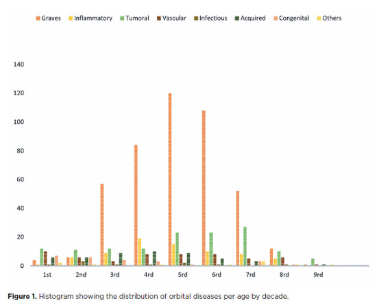
METHODS: A retrospective chart review was conducted involving patients registered and admitted to the orbital disease unit at the Department of Ophthalmology, University of São Paulo Medical School, from January 2004 to March 2018. A total of 838 medical charts were analyzed, of which 37 were excluded due to incomplete data. The remaining charts were categorized into eight diagnostic groups: Graves’ orbitopathy , inflammatory disorders, tumors, vascular lesions, acquired structural abnormalities, congenital structural abnormalities, infectious diseases, and others.
RESULTS: Of the 837,300 ophthalmological appointments, 3,372 (0.4%) were related to orbital diseases. The study included 801 patients, of whom 63.45% were women. The patients’ mean age was 42.86 years. Graves’ orbitopathy was the most common (55%), followed by tumor (17%), inflammatory disorders (9%), vascular lesions (7%), acquired structural abnormalities (5%), congenital structural abnormalities (4%), others (2%), and infectious diseases (1%). The study found significant differences in the incidence and types of orbital diseases, indicating the specialized nature of tertiary care and referral biases.
CONCLUSION: Published data on epidemiological orbital diseases is scarce. Therefore, this study focused on the diverse nature of orbital diseases and their low incidence among ophthalmology appointments. The major trends align with other epidemiological studies, demonstrating a preponderance of Graves’ orbitopathy in middle-aged adults and a bimodal distribution of tumors. These findings are essential in shaping resident training programs and healthcare policies, particularly in tertiary settings. Understanding the epidemiology of orbital diseases can improve diagnostic accuracy, treatment approaches, and patient outcomes as well as support future systemic prospective studies.
Keywords: Orbital diseases; Orbital tumors; Neoplasms; Inflammation; Graves’ ophthalmopathy; Outpatients
Arq. Bras. Oftalmol. 2025;88 (6 )
:1-4
| DOI: 10.5935/0004-2749.2025-0033
Abstract
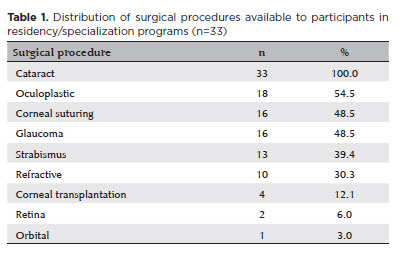
PURPOSE: In Brazil, it has traditionally been standard practice to teach a wide range of surgical techniques to all ophthalmology residents, with the aim of equipping them to manage most ocular conditions. However, with modern developments, access to subspecialists has expanded to nearly the entire country. This raises the question of whether it is still necessary to teach numerous surgical techniques to every resident. This study evaluates the effectiveness of surgical training in Brazilian ophthalmology residency programs to determine if comprehensive surgical training for all residents is truly effective, thereby providing evidence to inform educational policy decisions.
METHODS: A cross-sectional study using a questionnaire distributed to physicians engaged in eye care.
RESULTS: A total of 137 physicians responded to the survey, with 104 (76.0%) having already completed their specialization. The findings indicate that most practicing ophthalmologists received surgical training during residency in cataract, glaucoma, oculoplastic, and strabismus surgeries. Nonetheless, many of these specialists no longer perform most of these surgeries in practice, except for cataract surgery. While 53.8% of those who completed residency reported satisfaction with their training, 35.6% indicated that they wished they had received better surgical preparation.
CONCLUSION: The training of ophthalmology specialists must be made more efficient. Training efficiency is reduced when time and resources are devoted to surgical procedures that many specialists will not perform in their careers.
Keywords: Opthalmologists; Teaching; Education, medical; Ophthalmological surgical procedures; Simulation training; Wet lab; Surveys and questionnaires
Arq. Bras. Oftalmol. 2025;88 (3 )
:1-5
| DOI: 10.5935/0004-2749.2023-0174
Abstract
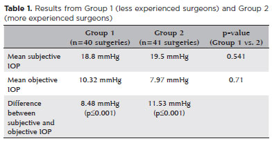
PURPOSE: To compare objective and subjective intraocular pressure measurements immediately after cataract surgery and intraocular pressure measurements between less experienced surgeons (Group 1) and experienced surgeons (Group 2).
METHODS: Surgeons were asked to estimate the IOP after corneal sealing after surgery based on their tactile perception of eye tension (subjective intraocular pressure) Objective intraocular pressure was measured using a Perkins tonometer while patients were still in the surgical field. Objective intraocular pressure was compared to subjective intraocular pressure. Results from less experienced surgeons were compared to more experienced surgeons.
RESULTS: The study comprised 81 surgeries (81 eyes) performed by 27 surgeons. The mean objective intraocular pressure (9.14 mmHg; SD=5.86) was statistically significantly lower (p<0.001) than the mean subjective intraocular pressure (19.21 mmHg; SD=4.82). Hypotony (intraocular pressure <6mmHg) was observed in 25 eyes (30.86%). The mean subjective intraocular pressure was 18.8 mmHg (SD=5.19) for less experienced surgeons and 19.5 mmHg (SD=4.46) for more experienced, without statistically significant difference (p=0.541). No statistically significant difference (p=0.71) was observed when comparing objective intraocular pressure in Group 1 (10.32 mmHg; SD=6.65) and Group 2 (7.97 mmHg; SD=4.7).
CONCLUSION: Objective intraocular pressure was significantly lower than subjective intraocular pressure, regardless of surgeons' experience. This study showed that the subjective method is unreliable compared to the gold standard (Perkins tonometer) and does not improve with surgeons' experience. Establishing standard training methods is paramount to developing surgeons' skills.
Keywords: Cataract; Intraocular pressure; Hypotony, Tonometry; Eye diseases; Training
Arq. Bras. Oftalmol. 2026;89 (5 )
:1-8
| DOI: 10.5935/0004-2749.2026-0041
Abstract
PURPOSE: To evaluate the efficacy of autologous fat grafting for treating enophthalmos in anophthalmic sockets.
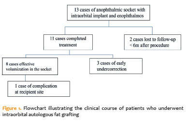
METHODS: In this prospective interventional case series, consecutive patients with anophthalmic sockets and enophthalmos were enrolled between August 2013 and July 2025. Clinical ophthalmologic examination, Hertel exophthalmometry, and orbital computed tomography scans were performed. Enophthalmos was assessed clinically and radiologically before and after orbital lipofilling with autologous fat. Fat was harvested from the inner thigh and injected into the inferolateral orbital compartment. The volume of injected fat was determined according to the degree of enophthalmos measured by Hertel exophthalmometry. Follow-up ranged from 2 to 60 months (median, 7 months).
RESULTS: Thirteen patients (13 sockets) underwent orbital autologous fat grafting using Coleman's technique. The injected fat volume ranged from 3.7 to 6 mL per procedure (median, 4.2 mL). Clinically, a reduction in enophthalmos of 1–5 mm was observed in 12 cases (median, 2 mm), which was associated with a decrease in prosthesis thickness. One patient developed a fat cyst due to anterior fat migration, and three patients required repeat grafting because of undercorrection. Computed tomography scans, available for nine patients, demonstrated a 6.8%–10.8% increase in intraorbital volume, particularly in the inferolateral quadrant. Conclusions: Clinical and radiological assessments demonstrated that lipofilling is an effective technique for restoring orbital volume in anophthalmic sockets with enophthalmos.
Keywords: Anophthalmos; Enophthalmos; Orbital volumetry; Fat grafting; Orbital reconstruction
Arq. Bras. Oftalmol. 2025;88 (3 )
:1-5
| DOI: 10.5935/0004-2749.2024-0084
Abstract
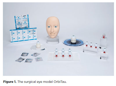
PURPOSE: The OrbiTau surgical simulator is a synthetic eye model developed to enhance cataract surgical training. Herein, we aimed to describe the perspectives of Harvard’s Ophthalmology faculty and residents regarding the effectiveness of OrbiTau.
METHODS: A cross-sectional study was conducted in which 11 surgeons from the Massachusetts Eye and Ear Infirmary, with prior experience utilizing simulated phacoemulsification platforms, conducted cataract surgery with the OrbiTau. Subsequently, they completed a satisfaction questionnaire using the Likert scale.
RESULTS: Regarding the various OrbiTau components, 90.90% of the participants reported that the OrbiTau lens capsule was comparable to that of the human lens during capsulotomy. Furthermore, 72.72% of the participants found that the OrbiTau lens consistency was analogous to that of the human lens nucleus. Approximately 63.63% of the participants reported that the model’s posterior lens capsule resembled the native posterior capsule, and 72.72% of the participants noted that the model’s red reflex was similar to that of the dilated human pupil. Most participants believed that the OrbiTau was easier to use and more realistic than other commercially available simulators.
CONCLUSION: Our single-institution survey of the Orbitau demonstrated that this model realistically replicates ocular structures and may be a viable option for cataract surgery training.
Keywords: Cataract extraction/education; Simulation training/methods; Ophthalmology/education; Phacoemulsification/education; Ophthalmologists/education; Surgeons/education; High fidelity simulation training


 ABO is licensed under a Creative Commons Attribution-NonComercial 4.0 Internacional.


06-fig01.jpg)
14-fig01.jpg)
01-fig01.jpg)
11-fig01.jpg)
01-fig01tb.jpg)
02-fig01.jpg)