Arq. Bras. Oftalmol. 2023;86 (3 )
:1-5
| DOI: 10.5935/0004-2749.20230031
Abstract
Objetivo: Descrever os resultados clínicos do tratamento do crescimento epitelial através da técnica de remoção manual seguido da utilização de um compressor de ar comprimido aquecido após a cirurgia de laser in situ keratomileusis (LASIK).
Métodos: Vinte olhos de 17 pacientes foram incluídos no estudo. Cada paciente havia sido submetido a cirurgia de LASIK com presença de crescimento epitelial e foi submetido a tratamento cirúrgico para sua retirada. O objetivo primário foi identificar a presença de crescimento epitelial recorrente ao final de 3 meses de seguimento. Os objetivos secundários foram as medidas de acuidade visual sem correção, acuidade visual com correção, e complicações pós-operatórias.
Resultados: Dez pacientes (58,8%) eram homens e 7 mulheres. Oito olhos de sete (41,2%) pacientes apresentavam cirurgia de LASIK primária e 12 olhos de 10 pacientes tinham cirurgia de LASIK com retratamento; dezesseis olhos (80%) utilizaram microcerátomo manual e quatro (20%) laser de femtosegundo. A média de idade no momento da cirurgia de remoção do epitélio era de 37,0 anos ± 9,3 (DP) (variando de 24 a 55 anos). Ocorreu recidiva do crescimento epithelial em dois olhos (10%) após 3 meses de seguimento. A acuidade visual sem correção antes da cirurgia era de 0,07 ± 0,09 logMAR, e após a cirurgia passou para 0,02 ± 0,04 logMAR (p=0,06). A chance (odds ration) de aparecimento do crescimento epithelial após uma reoperação de LASIK é 29,41 vezes maior do que no LASIK primário.
Conclusão: A técnica de remoção epitelial manual seguida da utilização de ar comprimido aquecido é segura e efetiva no tratamento do crescimento epitelial após LASIK. Ao final do último acompanhamento, nenhum olho apresentou perda de linhas de visão.
Keywords: Epitélio/crescimento & desenvolvimento; Endotélio corneano; Doenças da córnea; Ceratomileuse assistida por excimer laser in situ; Ceratectomia fotorrefrativa; Procedimentos cirúrgicos refrativos; Acuidade visual
Arq. Bras. Oftalmol. 2025;88 (4 )
:1-8
| DOI: 10.5935/0004-2749.2024-0214
Abstract
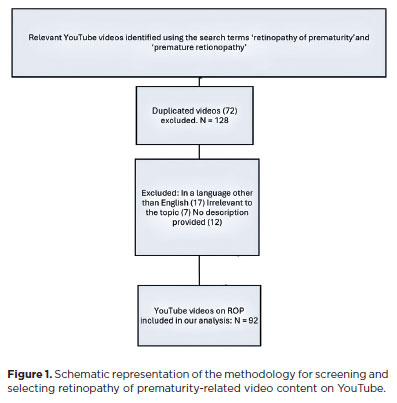
PURPOSE: This study aimed to evaluate the quality and reliability of YouTube videos as an educational resource about retinopathy of prematurity.
METHODS: Videos were sourced from YouTube using the search terms "retinopathy of prematurity" and "premature retinopathy" with the default settings. Each video was assessed on the following metrics: views, likes, dislikes, comments, upload source, country of origin, view ratio, like ratio, and video power index. The quality and reliability of the videos were evaluated by two independent researchers using the DISCERN questionnaire, the JAMA benchmarks, the Global Quality Score scale, the Health on the Net Code of Conduct, and the Ensuring Quality Information for Patients scale.
RESULTS: The study assessed 92 videos, the majority of which (42 videos, 45.7%) originated from the United States. Most of the videos focused on screening, pathophysiology, and diagnosis of retinopathy of prematurity (61.9%). The primary contributors were medical organizations (19 videos, 20.6%), nonacademic health channels (19 videos, 20.6%), and physicians (15 videos, 16.3%). Significant differences were found between the DISCERN (p=0.003), JAMA (p=0.001), Global Quality Score (p=0.003), Health on the Net Code of Conduct (p=0.006), and Ensuring Quality Information for Patients (p=0.001) scores among different video sources. However, the key video metrics did not differ. Using the DISCERN and Global Quality Score scales, the overall YouTube video content on retinopathy of prematurity was rated as moderate in quality. Using the Health On the Net Code of Conduct and Ensuring Quality Information for Patients scales, it was rated as high quality. Strong correlations were observed between the scores on all of the scales (p<0.001).
CONCLUSION: Videos from medical organizations and healthcare centers were of a higher quality than those from nonmedical sources. Despite the varied foci of each evaluation scale, the strong correlation between them indicates that they provide reliable and comprehensive assessments of the quality of informational content.
Keywords: Retinopathy of prematurity; YouTube; Information dissemination/methods; Online education; Internet access; Social media/instrumentation; Information seeking behavior; Internet/statistics & numerical data; Consumer health information; Social networking
Arq. Bras. Oftalmol. 2022;85 (6 )
:590-598
| DOI: 10.5935/0004-2749.20220081
Abstract
Objetivo: Identificar tendências no campo de pesquisa da orbitopatia de Graves nas últimas duas décadas e analisar os ramos de maior concentração de pesquisas nessa área.
Métodos: O banco de dados Web of Science foi usado para extrair artigos com “orbitopatia de Graves” ou seus sinônimos no título. Dados completos e referências foram exportados para o programa VOSviewer para serem analisados. Mapas e gráficos de visualização foram construídos a partir desses dados.
Resultados: Foram obtidos 1067 artigos sobre a orbitopatia de Graves a partir do banco de dados Web of Science. Os EUA ficaram em primeiro lugar em termos de número de publicações, seguidos pela Itália e pela República Popular da China. Dentre os autores, os artigos de Wiersinga WM tiveram o maior número de citações. Quanto às instituições, os artigos da Universidade de Amsterdã tiveram o maior número de citações, mas a Universidade de Pisa publicou o maior número de artigos. Dentre os periódicos, a revista Thyroid publicou o maior número de artigos. A análise de coautoria mostrou quatro agrupamentos de colaboração entre países. O primeiro agrupamento engloba países europeus; o segundo engloba os EUA, Brasil, Canadá, Coreia do Sul e Taiwan. A República Popular da China compreende um agrupamento por si só. O quarto agrupamento inclui Japão, Austrália e Polônia. A análise das palavras-chave revelou cinco agrupamentos de tópicos de palavras-chave: patogênese, gerenciamento, associação, qualidade de vida e cirurgia. A análise das referências citadas em conjunto revelou cinco agrupamentos: patogênese, manejo, fatores de risco, avaliação clínica e manejo cirúrgico.
Conclusão: A pesquisa no campo da orbitopatia de Graves cresceu nos últimos vinte anos. Os tópicos com a maior concentração de pesquisas são: patogênese, gerenciamento, fatores de risco, qualidade de vida e complicações. As tendências de pesquisa mudaram nas últimas duas décadas. Ficou evidente um aumento do interesse em explorar os mecanismos e associações da orbitopatia de Graves. Observou-se uma cooperação entre países europeus neste campo de pesquisa. Os EUA estabeleceram uma cooperação internacional mais ampla que outros países. Acreditamos que mais colaboração internacional envolvendo países em desenvolvimento seria recomendável.
Keywords: Oftalmopatia de Graves; Bibliometria; Oftalmopatia de Graves; Pesquisa
Arq. Bras. Oftalmol. 2025;88 (6 )
:1-6
| DOI: 10.5935/0004-2749.2024-0333
Abstract
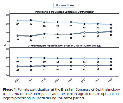
PURPOSE: To assess female participation in the Brazilian Congress of Ophthalmology.
METHODS: This retrospective, descriptive-analytical study examined the profiles of individuals involved in the scientific program of the Brazilian Congress of Ophthalmology from 2016 to 2023. Data were provided by the Brazilian Council of Ophthalmology and were categorized by ophthalmology subspecialty, participant role, and geographic region of origin within Brazil. Roles were grouped into three main categories: coordinator, speaker, and moderator/discussant.
RESULTS: Female participation at the congress increased from 33% in 2016 to 42% in 2023, showing an annual upward trend of 1.33 (p<0.001). Around 64% of female participants were from the Southeast region, while 16% were from the Northeast. The coordinator role showed the largest increase in female participation, rising from 22% in 2016 to 40% in 2023 (Slope: 2; p<0.001), followed by the speaker role, which increased from 34% to 44% (Slope: 1.5; p<0.001), and the moderator/discussant role, which rose from 32% to 38% (Slope: 1.24; p=0.0586). Changes in female representation across ophthalmology subspecialties were not statistically significant.
CONCLUSION: From 2016–2023, female participation in the Brazilian Congress of Ophthalmology increased across most subspecialties and conference roles. Although gender disparity has narrowed, continuous efforts are needed to achieve greater gender equity and equality in ophthalmology conferences.
Keywords: Ophthalmology; Gender equity; Ophthalmologists/statistics & numerical data; Physicians, women/statistics & numerical data; Leadership; Congresses as topic/statistics & numerical data.
Arq. Bras. Oftalmol. 2025;88 (4 )
:1-7
| DOI: 10.5935/0004-2749.2024-0229
Abstract
PURPOSE: The volume of the vitreous chamber varies with the size of the eye. The space created in the vitreous cavity by a vitrectomy is called the vitrectomized space. The volume of the vitrectomized space is strongly correlated with the axial length of the eye. This study aims to present guidelines for estimating the using participants stratified by axial length, sex, and history of cataract surgery.
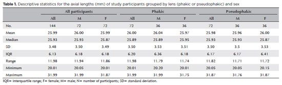
METHODS: This retrospective, observational, cross-sectional study included 144 randomly selected participants who underwent vitrectomies between 2013 and 2023. Before surgery, the axial lengths of participants' eyes were measured using optical biometrics. The axial lengths of the eyes in our sample were between 20-32 mm. In all cases, a complete vitrectomy was performed, followed by complete fluid-air exchange and injection of a balanced saline solution. The volume infused was recorded.
RESULTS: The median (interquartile range; range) volume of the vitrectomized space was 6.1 (3.8; 3.1-11.3) mL in men and 6.1 (3.3; 3.2-11.2) mL in women (p=0.811). The median volume of the vitrectomized space was 5.9 (3.6; 3.1-11.2) mL in patients with phakic lenses and 6.25 (3.6; 3.3-11.3) mL in those with pseudophakic lenses (p=0.533). A positive correlation was found between the axial length and the volume of the vitrectomized space in this sample (r=0.968; p<0.001). In a cubic polynomial regression, the coefficient of determination was 0.948. Similar results were observed in both sexes and in both phakic and pseudophakic patients. The estimated cubic polynomial regression equation for this sample was VVS = 0.000589052857847605 × AL3 - 0.025114926401582700 × AL2 + 0.685961117595624000 × AL - 5.088226672620790000.
CONCLUSION: We developed this axial length estimation of the volume of vitrectomized space as a guideline for the determination of vitrectomized space volume using axial length.
Keywords: Cataract extraction; Retinal perforations/surgery; Epiretinal membrane/surgery; Vitreous body; Axial length, eye; Vitrectomy; Biometry/methods; Diagnostic techniques, ophthalmological; Guidelines as topic.
Arq. Bras. Oftalmol. 2026;89 (1 )
:1-7
| DOI: 10.5935/0004-2749.2025-0218
Abstract
PURPOSE: This study aimed to evaluate disparities in the distribution of ophthalmologists and the volume of cataract surgeries across Brazil, considering public and private health sectors and the country's federative units.
METHODS: Data on ophthalmologists were obtained from the National Medical Residency Commission and the Associação Múdica Brasileira. Information on cataract surgeries performed through the Unified Health System was collected from the DATASUS database, while data on procedures covered by private health plans were retrieved from the National Supplementary Health Agency. Population estimates from the 2024 Demographic Census of the Brazilian Institute of Geography and Statistics were used to calculate physician density and surgery rates per 100,000 inhabitants. Associations between the number of ophthalmologists and cataract surgery volume were analyzed using Spearman's correlation coefficient.
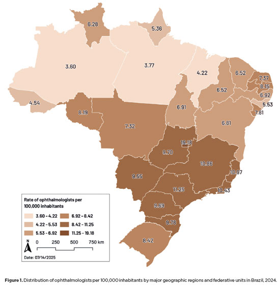
RESULTS: Brazil has 16,784 ophthalmologists, representing 8.96 specialists per 100,000 inhabitants. Marked disparities were observed: large cities (>500,000 inhabitants) had 18.75 ophthalmologists per 100,000 residents, whereas municipalities with <50,000 inhabitants had fewer than one. Across federative units, physician density ranged from 19.18 per 100,000 in the Federal District to 4.22 in Maranhão. In 2024, cataract surgery rates varied widely, from 1,012.61 per 100,000 inhabitants in the Southeast to 435.00 in the North. Nationally, Unified Health System performed 736.30 surgeries per 100,000 inhabitants, compared with 1,276.79 in the private sector. On average, each ophthalmologist performed 96.92 cataract surgeries annually.
CONCLUSION: Significant inequalities persist in the geographic distribution of ophthalmologists and in cataract surgery provision, with higher surgical volumes concentrated in the private sector. Targeted policies are required to address regional disparities and improve the equity and efficiency of cataract care delivery in Brazil.
Keywords: Ophthalmologists/supply & distribution; Ophthalmologists/statistics & numerical data; Cataract extraction; Health services accessibility/statistics & numerical data; Healthcare disparities; Health policy; Public health systems; Insurance, Heal
Arq. Bras. Oftalmol. 2025;88 (5 )
:1-7
| DOI: 10.5935/0004-2749.2024-0202
Abstract
PURPOSE: This study aimed to evaluate the relationship between the objective severity of dry eye disease subjective symptoms, and corneal sensitivity.
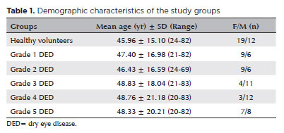
METHODS: The study included 62 eyes from 31 healthy volunteers and 150 eyes from 75 patients diagnosed with dry eye disease . Participants underwent the Schirmer I test, tear break-up time assessment, and corneal staining evaluation using the Oxford Scale. Subjective symptoms were assessed through the Ocular Surface Disease Index questionnaire and a modified Ocular Surface Pain Score questionnaire. Corneal sensitivity was measured in five corneal regions using a Cochet-Bonnet esthesiometer. Dry eye disease severity was graded from 1 to 5 based on the Oxford Scale. Comparative analyses were performed.
RESULTS: Schirmer I and tear break-up time values were significantly lower in the DED group, while Ocular Surface Disease Index and Ocular Surface Pain Score were significantly higher (p<0.001 for all). Corneal sensitivity in all quadrants was significantly lower in DED patients (p<0.001 for all). A strong correlation was observed between the Ocular Surface Pain Score and the Ocular Surface Disease Index (r=0.983, p<0.001). Central corneal sensitivity exhibited a moderate positive correlation with Schirmer I and tear break-up time (p<0.001, r=0.583 and 0.657, respectively) and a moderate negative correlation with Ocular Surface Disease Index and Ocular Surface Pain Score (p<0.001, r=0.625 and −0.631, respectively). Disease severity progression was associated with increased Ocular Surface Disease Index and Ocular Surface Pain Score, but no statistically significant difference was found between Grades 3 and 5. Similarly, corneal sensitivity decreased with advancing disease severity, yet no significant difference was observed between Grades 4 and 5.
CONCLUSION: Corneal sensitivity decreases in dry eye disease and is negatively correlated with disease severity. Subjective symptoms increase with disease progression and show a positive correlation with severity. The absence of significant differences between the advanced stages suggests that neuropathic mechanisms and subbasal nerve plexus deterioration play a role in chronic and late-stage dry eye disease.
Keywords: dry eye disease; signs and symptoms; cornea; neuralgia; Cochet-Bonnet esthesiometer; sensory thresholds; surveys and questionnaires
Arq. Bras. Oftalmol. 2025;88 (5 )
:1-7
| DOI: 10.5935/0004-2749.2024-0217
Abstract
PURPOSE: This study aimed to evaluate the influence of intrastromal corneal ring segment implants on the intraocular pressure measurements using Goldmann applanation tonometry, rebound tonometry, and noncontact tonometry in keratoconic corneas and analyze the intertonometer agreement.
METHODS: We enrolled 74 eyes in this observational and prospective study. Each participant had a complete eye examination, corneal analysis with Scheimpflug Tomography (Pentacam®), and intraocular pressure evaluation with Goldmann applanation tonometry, rebound tonometry, and noncontact tonometry, before and after intrastromal corneal ring segment implantation (on postoperative days 1, 7, 45, and 90). Intertonometer agreement was assessed using Bland-Altman analysis.
RESULTS: The mean age was 29.9 ± 10.2 years, and 47 (63.5%) eyes had keratoconus grade II. Intraocular pressures were higher for noncontact tonometry preoperatively and on 90 postoperative day (mean ± SD: 12.4 ± and 12.1 ± 2.2 mmHg, respectively), followed by Goldmann applanation tonometry (11.1 ± 3.0 and 11.2 ± 2.7 mmHg, respectively), and were lower for rebound tonometry (9.7 ± and 9.4 ± 3.2 mmHg, respectively). The variation from the Goldmann tonometry on 7 postoperative day to the baseline (p=0.022) and that of noncontact tonometry on 90 postoperative day to the baseline (p=0.021) were statistically significant. The rebound tonometry underestimated intraocular pressure when compared with the Goldmann applanation tonometry by a mean of 1.47 ± 5.19 mmHg. Noncontact tonometry, when compared with Goldmann applanation tonometry, overesti-mated intraocular pressure by a mean of 1.23 ± 4.15 mmHg.
CONCLUSION: Despite statistically significant differences between some postoperative periods, the intraocular pressure measurement differences may not be clinically relevant.
Keywords: Keratoconus; Intraocular pressure; Cornea; Corneal stroma; Postoperative period; Tonometry ocular; Prostheses and implants
Arq. Bras. Oftalmol. 2026;89 (3 )
:1-6
| DOI: 10.5935/0004-2749.2025-0272
Abstract
PURPOSE: To assess musculoskeletal symptoms, identify the most affected body areas, and investigate factors associated with the development of musculoskeletal disorders among ophthalmologists in Brazil.
METHODS: A survey was conducted using an online questionnaire and snowball sampling. Statistical analyses were performed using Jamovi version 2.3.28, and graphs were generated using RStudio version 2023.06.2 + 561.
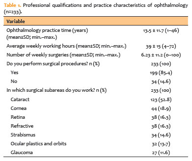
RESULTS: A total of 233 participants (42 ophthalmology residents and 191 ophthalmologists) were included, with a mean age of 40.4 years (standard deviation 11.3; range 25–73 years). Musculoskeletal symptoms were reported by 83% of participants. The cervical region (57.1%), upper back (54.5%), and lumbar region (53.6%) were the most frequently reported sites of pain. A high body mass index was identified in 54.9% of the sample, and 50.2% of participants reported using painkillers in the previous year for musculoskeletal symptoms. The mean duration of professional activity in ophthalmology was 13.5 years, and the mean weekly workload was 39 hours. A significant association was observed between weekly workload and the presence of musculoskeletal disorders (p=0.045).
CONCLUSION: This study demonstrated a high prevalence of musculoskeletal disorders among ophthalmologists in Brazil, particularly involving the cervical, lumbar, and upper back regions, consistent with findings reported in international studies. Important contributing factors include long working hours, a high patient volume, and repetitive or awkward postures during examinations and procedures. Preventive strategies and improvements in working conditions are needed to protect the health and well-being of ophthalmologists.
Keywords: Musculoskeletal Diseases/epidemiology; Back pain; Lumbar Vertebrae; Occupational diseases/epidemiology; Ergonomics; Ophthalmic practice; Ophthalmologists/statistics & numerical data; Brazil/epidemiology
Arq. Bras. Oftalmol. 2024;87 (4 )
:1-6
| DOI: 10.5935/0004-2749.2022-0128
Abstract
Objetivo: Relatar um experimento projetado para determinar alterações anatômicas em córneas porcinas após a colocação de um novo implante depolímero na córnea.
Métodos: Foi utilizado olho de porco ex vivo. Um novo agente modelador biocompatível, de colágeno tipo 1, com 6mm de diâmetro foi moldado com excimer laser em sua face posterior, para criar três formatos planocôncavos. Os implantes foram inseridos dentro de um bolsão, dissecado manualmente, a 200 micrômetros (µm). Foram definidos três grupos de tratamento: grupo A (n=3), teve a profundidade máxima de ablação de70 µm; o grupo B (n=3), profundidade máxima de ablação de 64 µm; e o grupo C (n=3), profundidade máxima de ablação de 104 µm, com buraco central. O grupo controle, D (n=3), foi incluído, com a criação do bolsão estromal, porém sem inserir o material. A avaliação desses olhos foi realizada por tomografia de coerência óptica (OCT) e por tomografia corneana.
Resultados: A tomografia corneana mostrou uma tendência para diminuição da ceratometria média em todos os 4 grupos. A tomografia de coerência óptica mostrou córneas com implantes localizados no estroma anterior e aplanamento visível, enquanto as córneas não mudaram qualitativamente o formato no grupo controle.
Conclusões: O novo implante de biomaterial planocôncavo descrito aqui foi capaz de remodelar a córnea em modelo de animal ex vivo, resultando no aplanamento corneano. Novos estudos são necessários usando modelos animais in vivo para confirmar tais achados.
Keywords: Córnea; Cirurgia da córnea a laser; Substância própria; Proteses e implantes; Lasers de excimer; Materiais biocompatíveis; Animais; Suínos


 ABO is licensed under a Creative Commons Attribution-NonComercial 4.0 Internacional.


08-fig01.jpg)
06-fig01.jpg)

09-fig01.jpg)
14-fig01.jpg)
03-fig01.jpg)