Arq. Bras. Oftalmol. 2022;85 (4 )
:344-350
| DOI: 10.5935/0004-2749.20220052
Abstract
Objetivo: Investigar a redução na densidade celular endotelial corneana associada à trabeculotomia transluminal assistida por gonioscopia (GATT) em curto prazo.
Métodos: Análise retrospectiva de prontuários médicos de pacientes com glaucoma de ângulo aberto que foram submetidos à trabeculotomia transluminal assistida por gonioscopia isolada ou combinada com facoemulsificação. Pacientes que foram submetidos à facoemulsificação isolada foram incluídos como controles. Dados da densidade celular endotelial corneana (avaliada através de microscópio especular) pré-operatória e ao primeiro mês pós-operatório foram coletados e comparados.
Resultados: Sessenta e dois olhos que foram submetidos à trabeculotomia transluminal assistida por gonioscopia (trabeculotomia transluminal assistida por gonioscopia=39 olhos; faco com trabeculotomia transluminal assistida por gonioscopia=23 olhos) passaram pelos critérios de inclusão. A idade média dos pacientes estudados era 61,3 ± 18,4 anos no grupo trabeculotomia transluminal assistida por gonioscopia isolada e 60,4 ± 11,9 anos no grupo faco com trabeculotomia transluminal assistida por gonioscopia. Homens eram 66,6% do grupo trabeculotomia transluminal assistida por gonioscopia isolada e 56,5% do grupo faco com trabeculotomia transluminal assistida por gonioscopia. O defeito perimétrico médio (Mean Deviation) era -13,9 ± 9,2 dB e -10,3 ± 7,7 dB nos grupos trabeculotomia transluminal assistida por gonioscopia isolada e faco com trabeculotomia transluminal assistida por gonioscopia respectivamente. O grupo que fora submetido à trabeculotomia transluminal assistida por gonioscopia isolada apresentou redução média da densidade celular endotelial corneana de 28,8 células/mm2 (1,31%; p=0,467). No grupo faco com trabeculotomia transluminal assistida por gonioscopia, a redução média da densidade celular endotelial corneana foi de 89,4 células/mm2 (4,36%; p=0,028). Olhos controle (23 olhos) apresentaram redução média da densidade celular endotelial corneana de 114,1 ± 159,8 células/mm2 (4,41%; p=0,505). A redução na densidade celular endotelial corneana no grupo faco com trabeculotomia transluminal assistida por gonioscopia não foi significativamente diferente do grupo controle (p=0,81).
Conclusões: A trabeculotomia transluminal assistida por gonioscopia parece ser segura para a camada endotelial corneana em um curto prazo quando realizada de forma isolada ou combinada com cirurgia de catarata.
Keywords: Glaucoma de ângulo aberto; Perda de células endoteliais da córnea; Extração de catarata; Malha trabecular; Gonioscopia/métodos; Trabeculectomia/métodos
Arq. Bras. Oftalmol. 2020;83 (4 )
:283-288
| DOI: 10.5935/0004-2749.20200040
Abstract
Objetivo: Comparar as alterações nos parâmetros do segmento anterior após a cirurgia ExPRESS Mini Glaucoma Shunt vs. trabeculectomia usando a câmera Scheimpflug Pentacam rotativa.
Métodos: Neste estudo comparativo prospectivo, 27 pacientes com glaucoma tratados no Centro Médico Rabin de 2009 a 2013 foram incluídos neste estudo comparativo prospectivo: 19 participantes (19 olhos) foram submetidos ao implante de derivação ExPRESS e 12 (13 olhos) foram submetidos à trabeculectomia. Alterações nos parâmetros da câmara anterior no dia 1 e em 3 meses de pós-operatório foram avaliadas pelas imagens de Scheimpflug.
Resultados: A pressão intraocular diminuiu significativamente em relação aos valores iniciais nos dois grupos. A diminuição nos dois grupos foi semelhante no 3º mês pós-operatório (p=0,82). A cirurgia com ExPRESS causou um aumento temporário do astigmatismo posterior da córnea (p=0,008) e uma diminuição temporária da profundidade da câmara anterior (p=0,016) e do volume (p=0,006) no primeiro dia do pós-operatório. Ao final de três meses, esses parâmetros não foram mais estatisticamente significativos (p=0,065, p=0,51 e p=0,57, respectivamente). A trabeculectomia causou um aumento temporário do astigmatismo anterior e posterior da córnea no primeiro dia do pós-operatório (p=0,003 e p=0,005, respectivamente), mas isso não foi observado ao final de 3 meses (p=1,0 e p=1,0, respectivamente). Após 3 meses, tanto o EXPRESS quanto a trabeculectomia mostraram alterações semelhantes nos parâmetros da câmara anterior.
Conclusões: O implante ExPRESS Mini para glaucoma e a trabeculectomia diminuíram significativamente a pressão intraocular e tiveram efeitos temporários nos parâmetros do segmento anterior, com pequenas diferenças entre os métodos.
Keywords: Glaucoma/cirurgia; Implantes para drenagem de glaucoma; Trabeculectomia/métodos; Pressão intraocular
Arq. Bras. Oftalmol. 2022;85 (5 )
:465-471
| DOI: 10.5935/0004-2749.20220067
Abstract
Objetivos: Relatar a distribuição dos motivos de encaminhamento de crianças para ambulatório de glaucoma infantil em um serviço oftalmológico terciário.
Métodos: Dados médicos de pacientes menores que 18 anos encaminhados para ambulatório de glaucoma infantil na cidade de São Paulo, Brasil, de 2012 a 2018 foram retrospectivamente analisados. Os dados incluíram os motivos de encaminhamento, a idade, a origem e quem detectou a alteração ocular. Para definição diagnóstica, a classificação do Childhood Glaucoma Research Network foi usada.
Resultados: 563 olhos de 328 pacientes foram incluídos no estudo. O diagnóstico de glaucoma foi confirmado em 162 crianças (49%). 83 (25%) pacientes tiveram diagnóstico de glaucoma descartado, e 83 (25%) continuaram em acompanhamento como glaucoma suspeito. Os principais motivos de encaminhamento foram relação escavação-disco >0,5 ou assimetria ≥0,2 (24%), pressão intraocular >21 mmHg (21%) e opacidade corneana (15%). No período neonatal, os motivos de encaminhamento foram opacidade corneana, buftalmo, lacrimejamento e fotofobia. Após o período neonatal, além desses sinais externos, outros sinais foram também motivos de encaminhamento, como escavação-disco >0,5 ou assimetria ≥0,2, pressão intraocular >21 mmHg e miopização. Os encaminhamentos ocorreram por profissionais de saúde em 69% e preocupação dos pais em 30%. Os pais foram os primeiros a identificar as alterações e procurar cuidado médico em 97% dos casos de glaucoma congênito primário.
Conclusões: Os motivos de encaminhamento de crianças para um serviço de glaucoma de glaucoma terciário foram determinados e diferem em diferentes faixas etárias e grupos. Os autores reforçam a necessidade de alertar diferentes grupos para os sinais mais comuns, a fim de evitar, não só o adiamento do diagnóstico, o que impacta no prognóstico, mas também despender recursos importantes direcionados ao enfrentamento dessas doenças, com encaminhamentos imprecisos.
Keywords: Glaucoma/congênito; Glaucoma/fisiopatologia; Opacidade da córnea; Criança; Acuidade visual; Encaminhamento e consulta; Serviços de saúde ocular
Arq. Bras. Oftalmol. 2025;88 (6 )
:1-6
| DOI: 10.5935/0004-2749.2024-0309
Abstract
PURPOSE: To report the surgical outcomes of patients with primary congenital glaucoma who underwent gonioscopy-assisted transluminal trabeculotomy.
METHODS: This retrospective, noncomparative, interventional study included consecutive patients with primary congenital glaucoma with uncontrolled intraocular pressure undergoing gonioscopy-assisted transluminal trabeculotomy between January 2017 and January 2020. The included participants were followed up for at least 24 months, and only one surgeon performed all the procedures. The number of glaucoma medications, pre- and postoperative intraocular pressure, treatment extension (in quadrants), surgical complications, and any associated events or interventions were documented.
RESULTS: This study included 13 eyes from 10 patients (mean age, 4.5 ± 3.2 years; range, 3 months to 10 years). After a 24-month follow-up, the mean intraocular pressure significantly decreased from 26.1 ± 3.7 to 11.8 ± 2.5 mmHg (p<0.001). The mean number of glaucoma medications was reduced from 3.3 ± 0.5 to 0.85 ± 1.0 (p<0.001). At the end of the follow-up interval, all eyes (13 out of 13) had an intraocular pressure between 7 and 15 mmHg. In 11 of 13 eyes (84.6%), gonioscopy-assisted transluminal trabeculotomy was performed in all quadrants (360º). The most frequent postoperative complication was transitory (self-limited) hyphema (7 out of 13 eyes [53.8%]). No sight-threatening adverse events occurred during the entire follow-up period.
CONCLUSIONS: The 2-year follow-up results indicated gonioscopy-assisted transluminal trabeculotomy as an efficient and safe option for primary congenital glaucoma treatment with minimal postoperative complications.
Keywords: Glaucoma, Open-angle/surgery; Gonioscopy; Trabeculectomy/methods; Intraocular pressure; Antihypertensive agents/therapeutic use.
Arq. Bras. Oftalmol. 2023;86 (3 )
:1-8
| DOI: 10.5935/0004-2749.20230034
Abstract
Purpose: To assess the outcomes of the trabecular bypass as replacement therapy for medications in pharmacologically controlled vs. pharmacologically uncontrolled open-angle glaucoma patients.
Methods: This was a retrospective study of eyes treated with first- (iStent) or second-generation (iStent inject) trabecular bypass. Group 1 consisted of eyes with pharmacologically controlled intraocular pressure <18 mmHg and Group 2 consisted of eyes with pharmacologically controlled intraocular pressure ≥18 mmHg. The main outcomes measured were qualified (with or without medications) and unqualified or complete (without medications) success rates at different target intraocular pressures, mean reduction (%) in medication use, and proportion of medication-free eyes.
Results: The mean age was 70.4 years in Group 1 (n=105) and 68.1 years in Group 2 (n=65). Qualified success rates for intraocular pressure <18 mmHg, intraocular pressure <15 mmHg, and intraocular pressure <12 mmHg were similar between the groups (Group 1: 96.2%, 88.6%, and 32.4%, respectively; Group 2: 93.8%, 78.5%, and 21.5%, respectively; all p>0.05). Complete success rates were significantly higher in Group 1 than in Group 2: for intraocular pressure <18 mmHg (76.2% vs. 47.7%), intraocular pressure <15 mmHg (73.3% vs. 40.0%), and intraocular pressure <12 mmHg (14.3% vs. 4.6%). The mean reduction in medication use was higher in Group 1 than in Group 2. At the end of follow-up, 79.0% of eyes in Group 1 and 47.7% of eyes in Group 2 became medication-free.
Conclusions: Both groups showed high qualified success rates, but eyes with baseline pharmacologically controlled intraocular pressure <18 mmHg showed higher complete success rates and greater chances of achieving no need for medications.
Keywords: Procedimentos cirúrgicos oftalmológicos; Extração de catarata; Glaucoma, ângulo aberto; Glaucoma/terapia; Glaucoma/cirurgia
Arq. Bras. Oftalmol. 2023;86 (2 )
:137-144
| DOI: 10.5935/0004-2749.20230026
Abstract
Objetivo: Descrever a frequência, as características clínicas, as complicações e o manejo do glaucoma em olhos submetidos a implantes de ceratoprótese.
Métodos: Pacientes submetidos à cirurgia de ceratoprótese entre junho de 2010 e janeiro de 2020 foram avaliados retrospectivamente em termos de glaucoma associado e prognóstico.
Resultados: Dos 17 pacientes submetidos à cirurgia de ceratoprótese, em 9 (52,9%) foi constatado glaucoma subjacente ou induzido por ceratoprótese. Cinco olhos (29,4%) tinham glaucoma subjacente e receberam a implantação de um dispositivo de drenagem de glaucoma pelo menos 6 meses antes da cirurgia de ceratoprótese. Um olho (5,9%) com pressão intraocular normal teve implantado um dispositivo de drenagem de glaucoma na mesma sessão da cirurgia de ceratoprótese, devido às características de “alto risco” das estruturas do segmento anterior. Quatro dos olhos com glaucoma preexistente apresentaram progressão após a cirurgia de ceratoprótese. Foi iniciado um tratamento antiglaucomatoso adicional em 2 olhos, enquanto outros 2 olhos receberam o implante de um segundo dispositivo de drenagem de glaucoma. Foram observadas complicações pós-operatórias em 3 olhos (100%) com dispositivo de drenagem de glaucoma implantado 6 meses antes ou na mesma sessão da cirurgia de ceratoprótese tipo afácica com vitrectomia parcial, incluindo descolamento de retina regmatogênico em 2 olhos e endoftalmite bacteriana em 1 olho. Em 1 olho observou-se migração do óleo de silicone para a área subconjuntival através do tubo após vitrectomia via pars plana. Nenhum dos 3 olhos (0%) implantados com dispositivo de drenagem de glaucoma anos antes da cirurgia de ceratoprótese apresentou complicações do segmento posterior, exceto progressão glaucomatosa. Dos 11 olhos sem história prévia de glaucoma, 3 (27,3%) apresentaram alta pressão intraocular e alterações do disco glaucomatoso após cirurgia de ceratoprótese, condições que podem ser controladas clinicamente.
Conclusões: Nesta coorte, os olhos com glaucoma pré-existente foram mais difíceis de manejar, comparados àqueles que desenvolveram glaucoma após a cirurgia de ceratoprótese. Apareceram mais complicações retinianas quando o implante do dispositivo de drenagem de glaucoma foi realizado no máximo 6 meses antes da cirurgia de ceratoprótese do tipo afácico com vitrectomia parcial.
Keywords: Glaucoma/cirurgia; Pressão intraocular; Complicação pós-operatória; Implantação de prótese; Implante para drenagem de glaucoma
Arq. Bras. Oftalmol. 2021;84 (6 )
:587-593
| DOI: 10.5935/0004-2749.20210083
Abstract
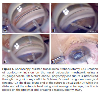
Objetivo: Reportar a curva de aprendizado dos 2 anos iniciais da trabeculotomia transluminal assistida por gonioscopia, usando a técnica de sutura termicamente atenuada e revisar os fatores que podem afetar o resultado.
Métodos: Este estudo retrospectivo incluiu 100 olhos de 89 participantes com glaucoma resistente ao tratamento clínico máximo, definido como tendo pressão intraocular superior a 21mmHg, além de três ou quatro drogas hipotensoras diferentes. Pressão intraocular inicial, 1 semana, primeiro, segundo, terceiro, sexto, 12 e 24 meses de acompanhamento; necessidade de medicação antiglaucoma; necessidade de mais cirurgias anti-glaucomatosas foram registradas. Olhos que necessitaram de intervenção cirúrgica adicional para o controle da pressão intraocular foram considerados como insucesso.
Resultados: Cinquenta e um olhos foram submetidos à trabeculotomia transluminal assistida por gonioscopia isolado e 49 olhos à trabeculotomia transluminal assistida por gonioscopia associado à extração de catarata no mesmo tempo cirúrgico. Houve diferença estatisticamente significativa entre a pressão intraocular média global no acompanhamento e a pressão intraocular média pré-operatória (p<0,001) em todas as visitas do acompanhamento. Ao avaliar a extensão do tratamento, os pacientes com extensão de 360 graus não apresentaram pressão intraocular média menor estatisticamente significativa em comparação com outras extensões. O hifema foi a única complicação presente em 50 olhos (50%), contudo todos tiveram resolução espontânea em quatro semanas. Um total de 26 olhos (26%) teve que ser submetido a trabeculectomia convencional adicional devido à pressão intraocular descontrolada, principalmente aqueles previamente submetidos à cirurgia vitreorretiniana.
Conclusões: A trabeculotomia transluminal assistida por gonioscopia, além de ser um procedimento aparentemente seguro, apresenta taxas de sucesso satisfatórias, mesmo durante a curva de aprendizado inicial do cirurgião. A técnica foi efetiva em reduzir a pressão intraocular e uso de medicamentos.
Keywords: Trabeculotomia/métdos; Glaucoma de ângulo aberto/cirurgia; Gonioscopia/métodos; Resultado de tratamento
Arq. Bras. Oftalmol. 2025;88 (5 )
:1-5
| DOI: 10.5935/0004-2749.2024-0197
Abstract
PURPOSE: This study aims to describe the technique, feasibility, efficacy, and safety of 360° trabeculotomy ab externo with double access for the treatment of congenital glaucoma.
METHODS: This paper provides a detailed description of the 360° trabeculotomy ab externo with double access used to treat pediatric glaucoma. The postoperative outcomes of six eyes from six patients who underwent this procedure for primary and secondary congenital glaucoma are also reported.
RESULTS: Six eyes from six patients were included in this study. The median age of the patients at the time of surgery was 1.25 yr (range: 0.27-5.41 yr). The mean preoperative intraocular pressure was 25 ± 5.87 mmHg (range: 18-35 mmHg). At baseline, the mean number of hypotensive eye drop medications used was 2 ± 0.63. Postoperatively, the mean intraocular pressure decreased to 10 ± 2.20 mmHg (range: 9-14 mmHg), and none of the patients required hypotensive eye drops. The most common postoperative complication was hyphema, observed in one case on the first postoperative day; however, it resolved within 7 days.
CONCLUSIONS: The 360° trabeculotomy ab externo with double access is a valuable addition to the surgical options for pediatric glaucoma. This technique facilitates a complete 360° ab externo opening of the trabecular meshwork while enhancing surgical safety.
Keywords: Glaucoma; Glaucoma/congenital; Trabeculectomy; Intraocular pressure; Ophthalmic solutions; Trabecular meshwork; Child
Arq. Bras. Oftalmol. 2026;89 (3 )
:1-8
| DOI: 10.5935/0004-2749.2025-0243
Abstract
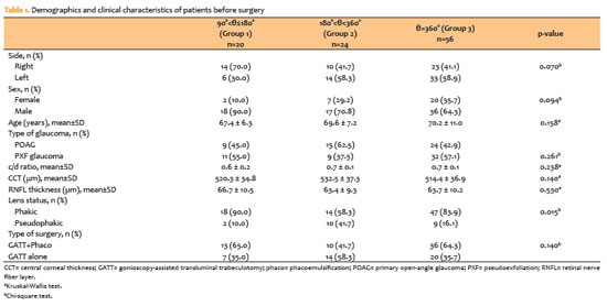
PURPOSE: To evaluate the impact of varying degrees of trabeculotomy during gonioscopy-assisted transluminal trabeculotomy surgery on postoperative intraocular pressure reduction and surgical success.
METHODS: Patients who underwent gonioscopy-assisted transluminal trabeculotomy (at least 90°) for open-angle glaucoma and had a follow-up period of at least 12 months were included. Patients were grouped according to trabeculotomy degree (Group 1: 90°<θ≤180°; Group 2: 180°<θ<360°; Group 3: θ=360°). Ophthalmic examination findings, intraocular pressure measurements, number of antiglaucoma medications, and complications were recorded. Surgical success was defined as intraocular pressure <15 mmHg with at least a 20% reduction; surgical failure was defined as failure to meet this criterion or the need for additional surgery.
RESULTS: A total of 100 patients were included: 20 in Group 1, 24 in Group 2, and 56 in Group 3. Intraocular pressure levels differed significantly only in the first postoperative month (p=0.013). At 12 months, intraocular pressure levels, percentage reduction in intraocular pressure, and mean number of antiglaucoma medications did not differ significantly (p>0.05). No correlation was found between trabeculotomy degree and percentage intraocular pressure reduction (p=0.173). At 12 months, surgical success rates were similar (60.0%, 58.3%, and 64.3% for Groups 1, 2, and 3, respectively). Complication rates were also comparable among the groups.
CONCLUSION: The degree of trabeculotomy did not affect surgical success over a 12-month follow-up period. Although early intraocular pressure reduction may differ with 360° trabeculotomy, a complete 360° incision may not be necessary to achieve optimal pressure reduction.
Keywords: Gonioscopy; Trabeculotomy; Glaucoma, open-angle; segmental GATT antiglaucoma agents; Intraocular pressure
Arq. Bras. Oftalmol. 2025;88 (3 )
:1-6
| DOI: 10.5935/0004-2749.2024-0215
Abstract
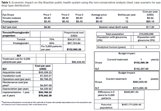
PURPOSE: To evaluate the economic impact of the following initial treatment scenarios for glaucoma on the Brazilian Public Health System (SUS): (1) traditional continuous instillation of hypotensive eye drops and (2) single session of selective laser trabeculoplasty.
METHODS: Economic impact was analyzed in three scenarios, from the least to the most conservative, for a hypothetical cohort of 5,000 individuals with open-angle glaucoma. Thereafter, projections were made on the basis of a glaucoma prevalence of 3% in the 2021 Brazilian population size.
RESULTS: All three scenarios demonstrated that selective laser trabeculoplasty exhibited a significantly lower economic impact than the eye drops on SUS over one and five years. Furthermore, the difference was more than United States Dollar 8 billion at five years when considering 3% of the Brazilian population aged >40 years in 2021.
CONCLUSION: As the initial treatment for primary open-angle glaucoma, selective laser trabeculoplasty exhibited a lower economic impact on SUS than latanoprost and timolol maleate eye drop instillation in all the studied scenarios over one and five-year periods.
Keywords: Glaucoma; Trabeculotomy; Laser therapy; Cost analysis; Health care cost Unified Health System; Brazil
Arq. Bras. Oftalmol. 2024;87 (3 )
:1-5
| DOI: 10.5935/0004-2749.2023-0033
Abstract
PURPOSE: This study aims to compare the initial ocular discomfort symptoms resulting from trabeculectomy and Ahmed glaucoma valve implantation surgeries.
METHODS: A prospective comparative study was conducted. The evaluation of ocular discomfort employed a questionnaire designed to identify the frequency and severity of distinct symptoms: ocular pain, general discomfort, tearing, foreign body sensation, and burning. This questionnaire was administered prior to surgery as a baseline, and subsequently at 7, 30, and 90 days post-surgery. Simultaneously, the Ocular Surface Disease Index (OSDI) was applied at these same time intervals.
RESULTS: The study encompassed a total of 17 patients (9 undergoing trabeculectomy and 8 undergoing Ahmed glaucoma valve implantation). The Ahmed glaucoma valve implantation group exhibited higher tearing levels at baseline (p=0.038). However, no statistically significant differences in symptoms were observed between the two surgeries at 7 and 30 days post-surgery. At the 90-day mark following surgery, patients who had undergone trabeculectomy reported a significantly higher foreign body sensation (p=0.004). Although OSDI scores did not differ between groups at baseline, the trabeculectomy group showed significantly higher OSDI scores than the Ahmed glaucoma valve implantation group at 7, 30, and 90 days after surgery (p<0.05).
CONCLUSION: Post-surgery, patients who had undergone trabeculectomy experienced increased foreign body sensation. Trabeculectomy appears to cause greater early postoperative ocular discomfort compared to the Ahmed glaucoma valve implantation group.
Keywords: Glaucoma/surgery; Paresthesia; Trabeculectomy; Glaucoma drainage implants; Postoperative care
Arq. Bras. Oftalmol. 2024;87 (2 )
:1-8
| DOI: 10.5935/0004-2749.2022-0306
Abstract
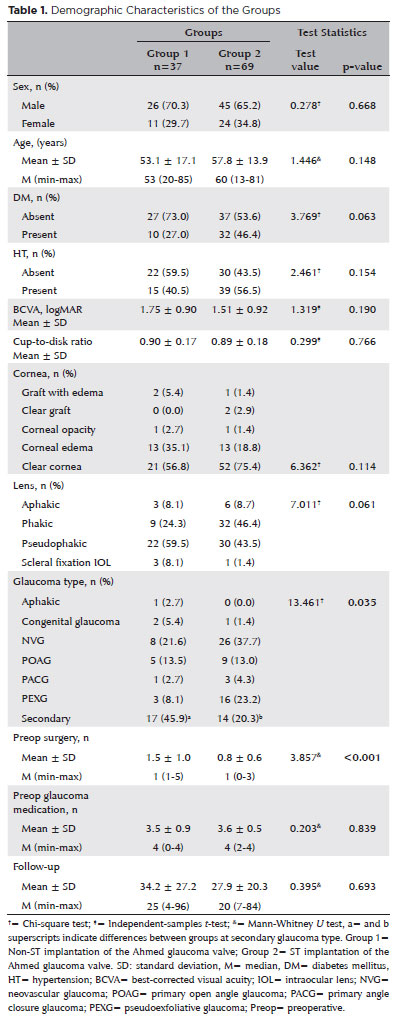
PURPOSE: As superotemporal implantation of the Ahmed glaucoma valve is not always feasible in cases of refractory glaucoma, this study examined the characteristics and surgical outcomes of cases in which the valve was implanted in a nonsuperotemporal quadrant using a modified long scleral tunnel technique.
METHODS: This retrospective case-control study included 37 eyes with nonsuperotemporal quadrant-Ahmed glaucoma valve implantation in Group 1 and 69 eyes with superotemporal Ahmed glaucoma valve implantation in Group 2. The demographic characteristics of these groups, surgical outcomes, including complications, further surgical interventions, and surgical success rates were compared. Surgical success was defined as an intraocular pressure not exceeding 21 mmHg, accompanied by a minimum reduction of 20% in intraocular pressure from the baseline without any additional intraocular pressure-lowering procedures, and the absence of light perception loss or phthisis bulbi.
RESULTS: Group 1 had significantly higher numbers of eyes with secondary glaucoma and preoperative surgical procedures than Group 2 (p<0.05). Both groups had mean preoperative intraocular pressure values, and mean intraocular pressure values at the last visit of 34.2 and 27.9 months, 35.5 ± 1.5 and 35.8 ± 1.2 mmHg, and 14.5 ± 5 and 14.9 mmHg, respectively. Although both groups had 70.2% and 75.8% as their five-year cumulative probability of success, respectively, the rates of complications, revisional surgery, and additional surgical procedures did not differ significantly (p>0.05).
CONCLUSION: The modified long scleral tunnel technique for Ahmed glaucoma valve implantation in nonsuperotemporal quadrants achieves intraocular pressure control and complication rates comparable to superotemporal implantation.
Keywords: Glaucoma/surgery; Sclera/surgery; Glaucoma drainage implant; Intraocular pressure; Tenon capsule
 ABO is licensed under a Creative Commons Attribution-NonComercial 4.0 Internacional.
09-tab01.jpg)
03-tab01.jpg)
14-fig01tb.jpg)
14-tab01.jpg)
12-fig01tb.jpg)
11-fig01.jpg)