Arq. Bras. Oftalmol. 2022;85 (2 )
:109-114
| DOI: 10.5935/0004-2749.20220018
Abstract
Objetivo: O objetivo deste estudo é determinar se o tamanho inicial do tumor se correlaciona com o tempo de regressão após terapia tópica com interferon alfa- 2b (1 milhão de UI/ml) no tratamento da neoplasia escamosa da superfície ocular.
Métodos: Foi realizado um estudo retrospectivo em 15 doentes clinicamente diagnosticados com neoplasia escamosa da superfície ocular e tratados com interferon tópico alfa- 2b (1 milhão de UI/ml, quatro vezes ao dia). Todos os casos de neoplasia escamosa da superfície ocular incluídos no estudo apresentaram envolvimento da córnea e do limbo. A extensão inicial da neoplasia escamosa da superfície ocular foi medida em milímetros quadrados usando o programa ImageJ (LOCI, University of Wisconsin, Madison, EUA) em imagens feitas dos olhos de cada paciente imediatamente antes do início do tratamento. O tempo até a resolução do tumor foi observado em cada caso.
Resultados: A resolução tumoral completa foi alcançada em todos os casos, com média da extensão inicial de 26,71 mm2 (desvio padrão ± 17,21 mm2) e tempo médio até a resolução de 77 dias (desvio padrão ± 32 dias). Um aumento do volume tumoral após 15 dias de tratamento foi observado em 2 pacientes com resolução completa do tumor. Não houve correlação significativa entre o tempo até a resolução e a extensão inicial do tumor medida em milímetros quadrados (teste de Spearman, p=0,347).
Conclusões Nosso estudo sugere que a duração do tratamento necessário com interferon alfa- 2b tópico não depende do tamanho inicial do tumor de neoplasia escamosa da superfície ocular geralmente encontrada na prática clínica.
Keywords: Neoplasia da túnica conjuntiva; Doença da córnea/tratamento farmacológico; Carcinoma de célula escamosa; Interferon alfa-2/ uso terapêutico
Arq. Bras. Oftalmol. 2021;84 (3 )
:220-224
| DOI: 10.5935/0004-2749.20210030
Abstract
OBJETIVO: Avaliar a morfologia da córnea e da câmara anterior em olhos fácicos com inflamação intraocular não infecciosa.
MÉTODOS: Esse estudo incluiu 59 olhos com uveíte ativa, 62 olhos com uveíte inativa e 95 olhos saudáveis. A densidade de células endoteliais da córnea, a proporção de células hexagonais, o coeficiente de variação, o volume e a espessura da córnea, a ceratometria máxima e o volume e profundidade da câmara anterior foram medidos com um microscópio especular e uma Pentacam HR.
RESULTADOS: A duração média da uveíte foi de 24,6 ± 40,5 (0-180) meses. O número médio de crises de uveíte foi de 2,8 ± 3,0 (1-20). O coeficiente de variação foi significativamente maior no grupo com uveíte ativa do que no grupo com uveíte inativa (p=0,017, Tukey post-hoc). Não houve diferença significativa nos demais parâmetros do segmento anterior entre os grupos com uveíte ativa, com uveíte inativa e controle (p>0,05). A análise de regressão linear múltipla demonstrou que o coeficiente de variação foi maior na uveíte ativa do que na uveíte inativa, após ajustes para a duração e tipo de uveíte e a presença ou não de doença reumática e de tratamento imunossupressor (p=0,003). A duração da uveíte e o número de crises não demonstraram correlação significativa com os parâmetros oculares (p>0,05, correlação de Spearman). A diferença nos parâmetros não demonstrou correlação significativa com o tipo de uveíte (p>0,05).
CONCLUSÕES: O coeficiente de variação foi maior nos olhos com uveíte ativa do que naqueles com uveíte inativa, ao passo que a densidade de células endoteliais e a morfologia da câmara anterior não mostraram diferenças significativas entre os grupos com uveíte ativa, com uveíte inativa e controle.
Keywords: Câmara anterior; Inflamação; Epitélio posterior; Contagem de células; Uveites
Arq. Bras. Oftalmol. 2021;84 (5 )
:454-461
| DOI: 10.5935/0004-2749.20210071
Abstract
Objetivo: Comparar a estrutura da córnea e as alterações morfológicas endoteliais após cirurgia de catarata por facoemulsificação sem intercorrências entre pacientes com diabetes mellitus tipo 2 e não diabéticos; e determinar quais fatores pré e intra-operatórios relacionados com a maior redução da densidade celular endotelial.
Métodos: Quarenta e cinco diabéticos (45 olhos) e 43 (43 olhos) controlos com catarata relacionada à idade foram incluídos neste estudo observacional prospectivo. Os parâmetros da córnea (espessura e volume) e do segmento anterior foram medidos pela tomografia Scheimpflug; a densidade e morfologia celular endotelial (coeficiente de variação do tamanho das células, células hexagonais) foram registrados usando microscopia especular não contato. Os pacientes foram avaliados no pré-operatório, 1 e 6 meses após a cirurgia. Foi realizada uma análise de regressão linear uni e multivariada para avaliar a relação entre os parâmetros demográficos, clínicos, oculares e intra-operatórios com a redução da densidade celular endotelial aos 6 meses.
Resultados: Nos dois grupos houve uma perda significativa de células endoteliais ao 1º mês pós-operatório (p<0,001), que permaneceu estável até ao 6º mês; sem diferenças estatisticas entre os grupos diabetes mellitus e não diabetes mellitus em qualquer avaliação. A espessura média da córnea no pós-operatório central aos 1 e 6 meses não mudou significativamente em relação ao valor médio pré-operatório nos dois grupos (p>0.05). A análise de regressão multivariada linear mostrou que a idade avançada (p=0.042) e os graus mais elevados de catarata (p=0.001) foram significativamente associados à maior redução densidade celular endotelial aos 6 meses de seguimento.
Conclusão: Este estudo mostrou que a idade avançada e as cataratas mais densas podem predispor a uma maior redução densidade celular endotelial após a cirurgia de catarata. Outros fatores, como diabetes mellitus e parâmetros pré-operatórios do segmento anterior, não influenciaram significativamente as alterações pós-operatórias da densidade celular endotelial.
Keywords: Catarata; Facoemulsificação; Diabetes mellitus tipo 2; Retinopatia diabética; Epitélio posterior; Paquimetria corneana; Perda de células endoteliais da córnea
Arq. Bras. Oftalmol. 2025;88 (5 )
:1-9
| DOI: 10.5935/0004-2749.2024-0326
Abstract
PURPOSE: This study was conducted to investigate the effect of injectable platelet-rich fibrin on the recovery of compromised epithelium due to crosslinking treatment.
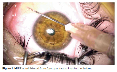
METHODS: In this comparative study, the epithelial closure rates and in vivo confocal biomicroscopy results of 26 patients with keratoconus who underwent subconjunctival injection of injectable platelet-rich fibrin near the limbus after epithelium-off corneal crosslinking treatment were compared with those of 25 patients who did not receive the injection of injectable platelet-rich fibrin.
RESULTS: The average time to epithelial defect closure in the injectable platelet-rich fibrin group was 2.76 ± 0.90 days compared to 3.56 ± 0.86 days in the non-injectable platelet-rich fibrin group (p=0.003). At the end of the 1st month, the mean subbasal nerve plexus density was 1.26 ± 1.61 nerves/mm2 in the injectable platelet-rich fibrin group, whereas it was 0.72 ± 0.89 nerves/mm2 in the non-injectable platelet-rich fibrin group (p=0.016). By the 3rd month, the density increased to 3.42 ± 1.13 nerves/mm2 in the injectable platelet-rich fibrin group and 2.36 ± 1.15 nerves/mm2 in the non-injectable platelet-rich fibrin group (p=0.002). Similarly, the anterior stromal keratocyte density at the end of the 1st month was 93.6 ± 33.5 cells/mm2 in the injectable platelet-rich fibrin group compared to 67.3 ± 26.4 cells/mm2 in the non-injectable platelet-rich fibrin group (p=0.001). By the end of the 3rd month, the density increased to 255.2 ± 45.7 cells/mm2 in the injectable platelet-rich fibrin group and 222.1 ± 43.6 cells/mm2 in the non-injectable platelet-rich fibrin group (p=0.011). In the non-injectable platelet-rich fibrin group, one patient developed a sterile infiltrate at the end of the 1st week, whereas no complications were observed in the injectable platelet-rich fibrin group.
CONCLUSION: Subconjunctival injectable platelet-rich fibrin application is an effective and safe method for corneal epithelial healing after crosslinking treatment.
Keywords: Keratoconus; Platelet-rich fibrin; Epithelium; corneal; Corneal crosslinking; Wound healing
Arq. Bras. Oftalmol. 2024;87 (3 )
:1-7
| DOI: 10.5935/0004-2749.2023-0049
Abstract
PURPOSE: To investigate the association of pre-photorefractive keratectomy Schirmer-1 test value with post-photorefractive keratectomy central corneal epithelial thickness, ocular surface disease index score, and uncorrected distance visual acuity.
METHODS: Patients were categorized according to preoperative Schirmer-1 value: the normal Schirmer Group (n=54; Schirmer-1 test value, >10 mm) and the low Schirmer Group (n=52; Schirmer-1 test value, between 6 and 10 mm). We analyzed ablation depth, visual acuity, result of Schirmer-1 test (with anesthesia), tear film break-up time, ocular surface disease index score, central corneal epithelial thickness, and spherical equivalent refraction.
RESULTS: We found significant differences between the groups in Schirmer-1 test value, tear film break-up time, and ocular surface disease index score, both preoperatively and postoperatively (p<0.001). The preoperative central corneal epithelial thicknesses of the two groups were similar (p>0.05). After photorefractive keratectomy, the Schirmer-1 test value and spherical equivalent refraction decreased in both groups (p<0.05), and ocular surface disease index scores and central corneal epithelial thickness values increased in the low Schirmer Group (p<0.001) but not in the normal Schirmer Group (p>0.05). The postoperative central corneal epithelial thicknesses of the low Schirmer Group were significantly higher than those of the normal Schirmer Group (p<0.001). Postoperative uncorrected distance visual acuity did not differ significantly between the two groups (p>0.05).
CONCLUSIONS: In patients with low Schirmer-1 test values before photorefractive keratectomy, the corneal epithelium thickened and ocular surface complaints increased during the postoperative period. However, changes in the corneal epithelium did not affect the postoperative uncorrected distance visual acuity. To reduce postoperative problems on the ocular surface in these patients, we recommend that dry eye be treated before photorefractive keratectomy.
Keywords: Epithelium, corneal; Cornea; Photorefractive keratectomy; Schirmer test; Visual acuity
Arq. Bras. Oftalmol. 2024;87 (4 )
:1-8
| DOI: 10.5935/0004-2749.2023-0144
Abstract
PURPOSE: To assess the outcomes of deep anterior lamellar keratoplasty or penetrating keratoplasty at the scar and the edema stages.
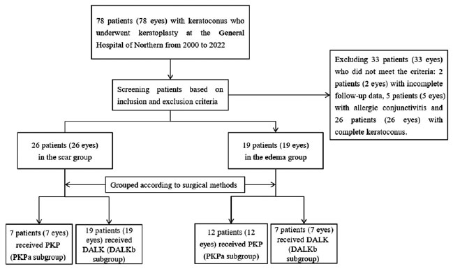
METHODS: Forty-five patients (45 eyes) with keratoconus scar stage (scar group, n=26; penetrating keratoplasty a subgroup, n=7; deep anterior lamellar keratoplasty b subgroup, n=19) and keratoconus edema stage (edema group, n=19; penetrating keratoplasty c subgroup, n=12; deep anterior lamellar keratoplasty d group, n=7) who received penetrating keratoplasty or deep anterior lamellar keratoplasty from 2000 to 2022 were retrospectively studied. At 1, 6, and 12 months after surgery, the best-corrected visual acuity, astigmatism, spherical equivalent, corneal endothelial cell density, and complications were analyzed.
RESULTS: The best-corrected visual acuity and average corneal endothelial cell loss rate were not significantly different between the scar and edema groups (p>0.05). At 6 and 12 months after surgery, the astigmatism and spherical equivalent in the scar group were significantly lower than those in the edema group (p<0.05). The spherical equivalent of the deep anterior lamellar keratoplasty b subgroup was lower than that of the penetrating keratoplasty a subgroup in the scar group 6 months after surgery (p<0.05). In the edema group, there was no significant difference in spherical equivalent between subgroups (p>0.05). There were no significant differences in best-corrected visual acuity and astigmatism between subgroups within the two groups (p>0.05). In comparison to the scar group, the edema group experienced more complications. According to a survival analysis, there was no statistically significant difference between the scar group and the edema group regarding the progression of vision.
CONCLUSIONS: In terms of the outcomes and prognosis for vision after keratoplasty with edema stage and scar stage, deep anterior lamellar keratoplasty may be as effective as penetrating keratoplasty.
Keywords: keratoconus; Edema; Cicatrix; keratoplasty, penetrating; Corneal transplantation; Astigmatism; Corneal endothelial cell loss; Endothelial cells
Arq. Bras. Oftalmol. 2024;87 (2 )
:1-7
| DOI: 10.5935/0004-2749.2022-0084
Abstract
OBJETIVOS: Foram estudadas cinco técnicas de cultivo primário de células epiteliais de córnea humana para se determinar o melhor protocolo para a obtenção do maior rendimento de meio de cultivo condicionado para ser utilizado na diferenciação de células tronco mesenquimais para células epiteliais de córnea.
MÉTODOS: As técnicas de cultivo estudadas foram: explantes em frascos de cultivo com e sem tratamento hidrofílico de superfície, sobre membrana amniótica, com digestão enzimática e por raspado de córnea. O meio de cultivo condicionado foi coletado e as células tronco mesenquimais induzidas a se diferenciarem em células epiteliais da córnea utilizando o meio de cultivo condicionado. As células foram caracterizadas por citometria de fluxo com pan-citoqueratina e com os marcadores específicos da córnea, citoqueratina 3 e citoqueratina 12.
RESULTADOS: A técnica utilizando frascos com o tratamento de superfície apresentou o maior rendimento de meio de cultivo condicionado. Os frascos sem tratamento de superfície levaram a uma taxa de sucesso muito baixa. A digestão enzimática e a raspagem da córnea mostraram contaminação das culturas com fibroblastos de córnea. A cultura sobre membranas amnióticas só permitiu a coleta do meio de cultivo condicionado durante a 1ª confluência celular. A análise de citometria de fluxo confirmou o sucesso da diferenciação celular utilizando o meio de cultivo condicionado coletado, demonstrada pela expressão de citoqueratina 3 (95,3%), citoqueratina 12 (93,4%) e pan-citoqueratina (95,3%).
CONCLUSÃO: O cultivo de explantes de células tronco mesenquimais em frascos com tratamento hidrofílico de superfície é a melhor técnica para a obtenção de um alto rendimento de meio de cultivo condicionado.
Keywords: Cultivo de células; Células tronco mesenquimais; Diferenciação celular; Células epiteliais; Córnea; Meio de cultivo condicionado; Técnicas de cultivo


 ABO is licensed under a Creative Commons Attribution-NonComercial 4.0 Internacional.


03-fig01.jpg)
04-tab01.jpg)
06-tab01tb.jpg)
02-fig01.jpg)
12-fig01.jpg)
17-equ01.jpg)
15-fig01.jpg)
14-fig01.jpg)