Arq. Bras. Oftalmol. 2023;86 (1 )
:79-82
| DOI: 10.5935/0004-2749.20220077
Abstract
Um homem de 53 anos, com história de 3 dias de edema periorbital e perda de visão no olho esquerdo, apresentou trombose séptica do seio cavernoso com envolvimento bilateral das veias orbitais, causando uma orbitopatia congestiva. O paciente foi tratado com uma cantotomia e cantólise de emergência, colírios para redução da pressão intraocular, antibióticos, anticoagulantes e exames seriados. A tomografia de coerência óptica finalmente demonstrou destruição isquêmica difusa de ambas as camadas da retina, sugerindo uma oclusão da artéria oftálmica ou das artérias ciliares posteriores curtas e da artéria retiniana central, com ausência de envolvimento do segmento intracavernoso da artéria carótida interna. O paciente permaneceu sem percepção luminosa no olho esquerdo.
Keywords: Trombose do corpo cavernoso; Doenças orbitárias; Tomografia de coerência óptica; Humanos; Relato de casos
Arq. Bras. Oftalmol. 2024;87 (4 )
:1-4
| DOI: 10.5935/0004-2749.2023-0219
Abstract
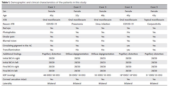
Bilateral acute depigmentation of the iris and bilateral acute iris transillumination (BAIT) are similar clinical entities. The former causes acute-onset depigmentation of the iris stroma without transillumination, whereas the latter causes depigmentation of the iris pigment epithelium with transillumination. The etiopathogenesis of these conditions is not yet fully understood, but the proposed causes include the use of systemic antibiotics (especially moxifloxacin) and viral triggers. We present a case series of five female patients with a mean age of 41 (32-45) years, all of whom suffered acute onset of bilateral pain and redness of the eyes after moxifloxacin use (oral or topical). It is important for ophthalmologists to be aware of the two forms of iris depigmentation since this case series suggests that SARS-CoV-2 or its empirical treatment with moxifloxacin may trigger iris depigmentation. If this is the case, clinicians will likely see increased incidences of bilateral acute depigmentation of the iris and bilateral acute iris transillumination during and after the COVID-19 pandemic.
Keywords: SARS-CoV-2; Pigment epithelium of eye; Iris diseases/pathology; Transillumination; Ocular hypertension; Intraocular pressure; Drug related side effect and adverse reactions; Iris diseases/drug therapy; Moxifloxacin/therapeutic Use; Humans; Case reports
Arq. Bras. Oftalmol. 2024;87 (3 )
:1-4
| DOI: 10.5935/0004-2749.2022-0357
Abstract
We present a rare case of primary caruncle basal cell carcinoma (BCC), a condition with limited occurrences. Our patient, an 80-year-old woman without prior ocular pathological history, presented a 2x2mm pedunculated blackish nodular lesion on the caruncle of her left eye, without local conjunctival or cutaneous involvement. Histological analysis following complete excision confirmed the presence of basal cell carcinoma within the caruncle. Over a span of 30 months, no recurrence has been observed. While scant cases are documented in the literature, we conducted a review of these instances. Despite its infrequent manifestation, this condition should be taken into account when evaluating caruncular tumors, given its tendency to invade the orbit. Complete excision with free surgical margins is the treatment of choice, and adjuvant radiotherapy or chemotherapy might be considered.
Keywords: Conjunctival diseases; Eye neoplasms; Sebaceous gland neoplasms; Conjunctival neoplasms; Carcinoma, basal cell; Diagnosis, differential; Humans; Case reports
Arq. Bras. Oftalmol. 2024;87 (2 )
:1-5
| DOI: 10.5935/0004-2749.2022-0237
Abstract
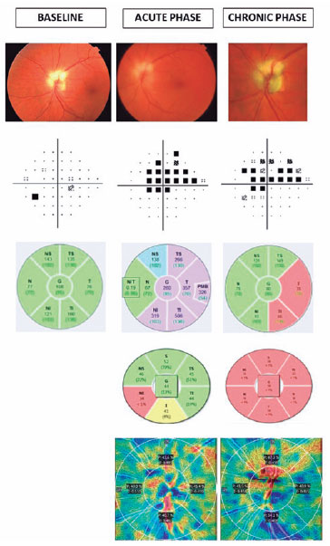
Uma mulher de 71 anos de idade apresentou neuropatia óptica isquêmica anterior não arterítica no nervo óptico com fibras nervosas peripapilares mielinizadas previamente registradas. Seu histórico médico foi significativo para hipertensão arterial sistêmica controlada, hiperlipidemia e diabetes mellitus. Em ambos os discos ópticos, a tacícula fisiológica esteve ausente. A neuropatia óptica isquêmica anterior não arterítica afetou principalmente os setores temporal e inferior da camada de fibras nervosas da retina peripapilar, como demonstrado pela tomografia de coerência óptica da camada de fibras nervosas da retina e pela angiotomografia de coerência óptica do disco óptico. Ao contrário de outros relatórios publicados, apenas uma ligeira regressão das fibras nervosas mielinizadas foi observada após um ano de acompanhamento. Isto pode ser explicado pelo fato da isquemia ter afetado principalmente os setores temporal e inferior peripapilares, enquanto as fibras nervosas de mielina eram nasal superior ao disco óptico.
Keywords: Neuropatia óptica isquêmica; Fibras nervosas mielinizadas; Doenças do nervo óptico; Tomografia de coerência óptica; Neovascularização retiniana; Acuidade visual; Humanos; Relato de casos
Arq. Bras. Oftalmol. 2024;87 (2 )
:1-3
| DOI: 10.5935/0004-2749.2022-0091
Abstract
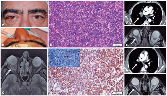
Hepatitis C virus infection may be implicated in 12.7% of ocular adnexal marginal zone lymphomas. We present the first case of an orbital-systemic mucosa-associated lymphoid tissue lymphoma that responded to hepatitis C virus medical treatment. A 62-year-old male with a right-sided orbital mass was diagnosed with stage IIA orbital marginal zone lymphoma in addition to hepatitis C virus infection based on clinical, imaging, laboratory, and histological examinations. The systemic and orbital responses were achieved 1 year after undergoing hepatitis C virus treatment with glecaprevir/pibrentasvir. The association between the hepatitis C virus infection and orbital-systemic mucosa-associated lymphoid tissue lymphoma is relevant. Accordingly, patients with orbital mucosa-associated lymphoid tissue lymphoma should be assessed for hepatitis C virus seroreactivity for therapeutic and prognostic purposes.
Keywords: Orbital disease; Orbital neoplasms; Lymphoma, B-cell marginal zone; Hepacivirus; Hepatitis C; Humans; Case reports
Arq. Bras. Oftalmol. 2025;88 (2 )
:1-4
| DOI: 10.5935/0004-2749.2024-0007
Abstract
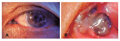
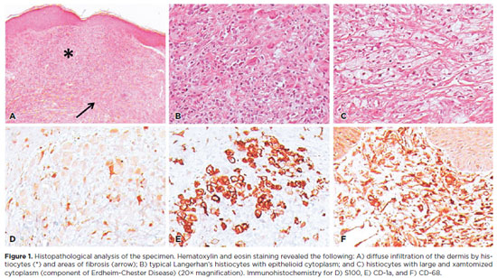
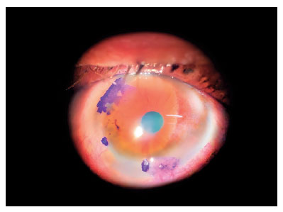
Langerhans cell histiocytosis comprises a heterogeneous range of clinical manifestations secondary to clonal proliferation of histiocytes, characterized by the accumulation of these cells in various organs and tissues. The ophthalmological component commonly involved is the orbit. Herein, we report a rare case of Langerhans cell histiocytosis with eyelid involvement, which resulted in severe ocular surface complications, which subsequently significantly impacted the patient's quality of life. This case report highlights the fact that despite being rare, Langerhans cell histiocytosis should be included in the differential diagnosis of eyelid lesions. Furthermore, a multidisciplinary approach with a systemic overview is crucial for managing the ocular complications.
Keywords: Dry eye syndromes; Erdheim-Chester disease/drug therapy; Human; Female; Case reports


 ABO is licensed under a Creative Commons Attribution-NonComercial 4.0 Internacional.


08-fig01.jpg)
11-fig01.jpg)
08-fig01.jpg)
14-fig01.jpg)
02-fig01.jpg)
01-fig01tb.jpg)
03-fig01.jpg)
14-fig01.jpg)
02-fig01.jpg)