Arq. Bras. Oftalmol. 2023;86 (3 )
:1-5
| DOI: 10.5935/0004-2749.20230031
Abstract
Objetivo: Descrever os resultados clínicos do tratamento do crescimento epitelial através da técnica de remoção manual seguido da utilização de um compressor de ar comprimido aquecido após a cirurgia de laser in situ keratomileusis (LASIK).
Métodos: Vinte olhos de 17 pacientes foram incluídos no estudo. Cada paciente havia sido submetido a cirurgia de LASIK com presença de crescimento epitelial e foi submetido a tratamento cirúrgico para sua retirada. O objetivo primário foi identificar a presença de crescimento epitelial recorrente ao final de 3 meses de seguimento. Os objetivos secundários foram as medidas de acuidade visual sem correção, acuidade visual com correção, e complicações pós-operatórias.
Resultados: Dez pacientes (58,8%) eram homens e 7 mulheres. Oito olhos de sete (41,2%) pacientes apresentavam cirurgia de LASIK primária e 12 olhos de 10 pacientes tinham cirurgia de LASIK com retratamento; dezesseis olhos (80%) utilizaram microcerátomo manual e quatro (20%) laser de femtosegundo. A média de idade no momento da cirurgia de remoção do epitélio era de 37,0 anos ± 9,3 (DP) (variando de 24 a 55 anos). Ocorreu recidiva do crescimento epithelial em dois olhos (10%) após 3 meses de seguimento. A acuidade visual sem correção antes da cirurgia era de 0,07 ± 0,09 logMAR, e após a cirurgia passou para 0,02 ± 0,04 logMAR (p=0,06). A chance (odds ration) de aparecimento do crescimento epithelial após uma reoperação de LASIK é 29,41 vezes maior do que no LASIK primário.
Conclusão: A técnica de remoção epitelial manual seguida da utilização de ar comprimido aquecido é segura e efetiva no tratamento do crescimento epitelial após LASIK. Ao final do último acompanhamento, nenhum olho apresentou perda de linhas de visão.
Keywords: Epitélio/crescimento & desenvolvimento; Endotélio corneano; Doenças da córnea; Ceratomileuse assistida por excimer laser in situ; Ceratectomia fotorrefrativa; Procedimentos cirúrgicos refrativos; Acuidade visual
Arq. Bras. Oftalmol. 2020;83 (5 )
:366-371
| DOI: 10.5935/0004-2749.20200077
Abstract
Objetivo: O comprimento da membrana de Descemet e o tamanho do enxerto doador na ceratoplastia lamelar anterior profunda não coincidem em córneas muito íngremes, o que pode levar às dobras da membrana de Descemet. O objetivo deste estudo é estabelecer um modelo teórico para cálculo do tamanho do enxerto para ceratoplastia lamelar anterior profunda e avaliar a sua eficácia na prevenção de dobras da membrana de Descemet.
Métodos: Calculamos o diâmetro do arco do leito receptor usando a fórmula do cosseno e desenvolvemos uma tabela para auxiliar os cirurgiões na seleção do tamanho da punção no doador. Para testar a utilidade dessa fórmula, avaliamos o desenvolvimento das dobras da membrana de Descemet em pacientes com ceratocone com córneas muito íngremes (K>60D). No grupo 1, foram realizadas cirurgias de ceratoplastia lamelar anterior profunda, utilizando tamanhos de enxerto que foram determinados com base em nosso modelo (n=31). No grupo 2, os tamanhos dos enxertos foram determinados com base no julgamento empírico do cirurgião sem qualquer cálculo formal (n=30).
Resultados: Nossos cálculos teóricos demonstraram que o diâmetro dos tamanhos da punção do doador necessários para evitar as dobras na membrana de Descemet aumenta quando a córnea é mais íngreme ou o tamanho da trefina é maior. Testamos a eficácia deste modelo no resultado clínico da ceratoplastia lamelar anterior profunda. A média de idade (28,9 ± 10,1 anos vs. 32,8 ± 8,3 anos, p=0,11) e K1 pré-operatório (59,2 ± 9,3 D vs. 58,1 ± 9,4 D, p=0,67), K2 (66,2 ± 6,0 D vs. 65,7 ± 7,4) D, p=0,81) e Km (62,1 ± 7,7 D vs. 61,8 ± 8,1 D, p=0,88) foram semelhantes entre os dois grupos. Três pacientes desenvolveram dobras na membrana de Descemet no grupo 2, e nenhum dos pacientes desenvolveu dobras na membrana de Descemet no grupo 1. Estes resultados apoiam nossos cálculos teóricos.
Conclusão: O ajuste do tamanho do enxerto doador com base no diâmetro do arco calculado do leito receptor reduziu o desenvolvimento das dobras na membrana de Descemet após ceratoplastia lamelar anterior profunda em córneas íngremes.
Keywords: Membrana de Descemet; Ceratocone; Ceratoplastia penetrante; Topografia da córnea; Córnea/patologia
Arq. Bras. Oftalmol. 2024;87 (1 )
:1-7
| DOI: 10.5935/0004-2749.2021-0048
Abstract
Objetivo: Identificar parâmetros tomográficos de normalidade em córneas de crianças e adolescentes sem a presença de atopias sistêmicas e alergias oculares.
Métodos: Este estudo descritivo transversal avaliou pacientes com idade entre 8 e 16 anos que foram submetidos a exame biomicroscópico completo por lâmpada de fenda e avaliação tomográfica da córnea por tomógrafo dual Scheimpflug, excluindo pacientes com doença ocular (incluindo conjuntivite alérgica) ou prick test positivo para atopias sistêmicas.
Resultados: Cento e setenta pacientes foram avaliados e após cumpridos os critérios de exclusão, 34 (68 olhos) foram analisados. A média etária da amostra foi 10,76 ± 2,31 anos; 19 (55,9%) eram meninos e 15 (44,1%) meninas. A média da ceratometria em dioptrias (D) no meridiano mais plano (Kflat), mais curvo (Ksteep) e máxima (Kmax) foram 42,37 ± 1,63D, 43,53 ± 1,65D e 43,90 ± 1,73D, respectivamente. Os valores médios da asfericidade corneana (ε2) e do ponto mais fino da córnea foram 0,28 ± 0,11 e 550,20 ± 37,90 micras (μm). A assimetria corneana inferior-superior (I-S) e coma foi em média 0,74 ± 0,59D e 0,28 ± 0,12D, respectivamente.
Conclusão: O conhecimento dos valores médios e sua variação de parâmetros tomográficos da córnea em crianças e adolescentes sem atopias sistêmicas ou alergias oculares pode ser útil para o diagnóstico precoce do ceratocone e o seu tratamento em estágio inicial.
Keywords: Córnea; Topografia corneana; Astigmatismo; Conjuntivite alérgica; Tomografia; Ceratocone/diagnóstico; Humanos; Criança; Adolescente.
Arq. Bras. Oftalmol. 2021;84 (1 )
:11-16
| DOI: 10.5935/0004-2749.20210002
Abstract
OBJETIVO: Determinar o efeito da blefaroplastia superior na topografia corneana e no cálculo do poder das lentes intraoculares usando Galilei e IOLMaster.
MÉTODOS: Trinta pacientes submetidos a blefaroplastia superior de maio de 2014 a março de 2017 no Hospital Oftalmológico de Sorocaba, São Paulo, Brasil foram incluídos neste estudo de série de casos observacional. Todos os pacientes foram submetidos a sessões de imagem com Galilei e IOLMaster antes da cirurgia (exame de base) e no 1º e 6º mês pós-operatório. Os resultados primários utilizando os dois aparelhos incluíram ceratometria, astigmatismo corenano e astigmatismo corneano induzido pela blefaroplastia. O comprimento axial e o cálculo do poder da lente intraocular foram realizados unicamente com o IOLMaster (fórmula de Holladay). Teste-t pareado e análise vetorial foram usados na análise estatística.
RESULTADOS: Sessenta olhos de 30 pacientes foram incluídos prospectivamente. A análise vectorial mostrou que após 6 meses da cirurgia, a blefaroplastia superior induziu na média 0,39 D de astigmatismo corneano medido com o Galilei e 0,31 D com IOLMaster. As medidas com o IOLMaster mostraram que a ceratometria média (44,56 vs 44,64 D, p=0,01), ceratometria máxima (45,17 vs 45,31, p=0,01) e o astigmatismo corneano (1,22 vs 1,34, p=0,03) foram maiores após 6 meses da blefaroplastia. As medidas com IOLMaster mostraram que o poder da lente intraocular foi significativamente menor 6 meses após a blefaroplastia (22,07 vs 21,93, p=0,004). Todos os outros parâmetros não mostraram mudanças entre o pré-operatório e o 6º mês da cirurgia (p>0,05 para todas as comparações).
CONCLUSÕES: A blefaroplastia superior influenciou o cálculo da lente intraocular utilizando o IOLMaster. Contudo, a influência não foi clinicamente significativa. Não foram encontradas mudanças topográficas com o Galilei.
Keywords: Blefaroplastia; Lentes intraoculares; Ceratometria; Topografia da córnea; Biometria
Arq. Bras. Oftalmol. 2022;85 (4 )
:351-358
| DOI: 10.5935/0004-2749.20220051
Abstract
Objetivo: Desenvolver um aplicativo (TopEye) na plataforma iOS para dispositivos móveis que possibilite a captação e interpretação do mapa de cores gerados por qualquer topógrafo corneano através da inteligência artificial (IA).
Métodos: A execução, acompanhamento e avaliação do projeto foi utilizada a metodologia Scrum, processo de desenvolvimento interativo e incremental para gerenciamento de projetos e desenvolvimento ágil de software. O banco de padrões de diagnóstico gerado consiste em 1172 exemplos, divididos em: 275 padrões esféricos, 302 regulares simétricos, 295 regulares assimétricos e 300 irregulares (ceratocone). Para o desenvolvimento da inteligência artificial do aplicativo, foi estabelecido o treinamento da rede com 240 imagens de cada tipo de padrão, totalizando 960 (81,91%) padrões. O restante das imagens, 212 (18,09%), foram utilizadas para testar o aplicativo e usadas para gerar os resultados. O processo é semiautomático, assim a captação da imagem topográfica é realizada com smartphone, o examinador realiza o contorno do relevo corneano manualmente para em seguida a rede neural realizar o diagnóstico.
Resultados: O aplicativo diagnosticou 201 (94,81%) imagens corretamente. De um total de 212 imagens, o algoritmo errou a classificação de apenas 11 (5,19%). A principal ocorrência de erro foi na distinção das classes simétrica e assimétrica. No rastreio do ceratocone o aplicativo alcançou 95,00% de sensibilidade e 98,68% especificidade.
Conclusão: O trabalho resultou na obtenção de um aplicativo eficiente na captura da imagem topográfica pela câmera do smartphone e na interpretação da mesma através da inteligência artificial aplicada.
Keywords: Dispositivos móveis; Inteligência artificial; Topografia corneana; Astigmatismo
Arq. Bras. Oftalmol. 2021;84 (3 )
:230-234
| DOI: 10.5935/0004-2749.20210037
Abstract
OBJETIVO: Investigar o efeito do uso de uma substância viscoelástica na ruptura da membrana de Descemet em casos de ceratoplastia lamelar anterior profunda em “bolha dupla”.
MÉTODOS: Foram avaliados retrospectivamente prontuários e vídeos de cirurgias de 40 pacientes operados entre janeiro de 2014 e julho de 2015. Os pacientes foram divididos em dois grupos: 20 pacientes nos quais a parede posterior do estroma foi puncionada sem a colocação de nenhuma substância viscoelástica (grupo 1) e 20 pacientes nos quais uma substância viscoelástica foi aplicada sobre o estroma posterior ao ser puncionada a parede posterior do estroma (grupo 2). A taxa de perfuração da membrana de Descemet foi comparada entre os grupos.
RESULTADOS: Observou-se perfuração da membrana de Descemet em 12 casos (60,0%) no grupo 1 e em apenas 3 casos (15,0%) no grupo 2. Essa diferença foi estatisticamente significativa (p=0,003). Apenas um caso (5%) no grupo 2 teve macroperfuração durante o procedimento, sendo a cirurgia então convertida em uma ceratoplastia penetrante. Onze casos (55,0%) no grupo 1 tiveram macroperfuração da membrana de Descemet e essas cirurgias foram convertidas em ceratoplastias penetrantes. Essa diferença entre os grupos foi estatisticamente significativa (p=0,001).
CONCLUSÕES: A aplicação de substância viscoelástica sobre o lado posterior do estroma logo antes da punção é um método eficaz para diminuir o risco de perfuração da membrana de Descemet na ceratoplastia lamelar anterior profunda.
Keywords: Lâmina limitante posterior/cirurgia; Substâncias viscoelásticas; Transplante de córnea; Substância propria; Ceratoplastia penetrante
Arq. Bras. Oftalmol. 2025;88 (1 )
:1-5
| DOI: 10.5935/0004-2749.2023-0160
Abstract
PURPOSE: To determine the clinical outcomes in patients after type 1 Boston keratoprosthesis surgery and the significance of ultrasound biomicroscopy imaging for postoperative follow-up.
METHODS: This retrospective analysis included 20 eyes of 19 patients who underwent corneal transplantation with type 1 Boston keratoprosthesis between April 2014 and December 2021. Data on patient demographics, preoperative diagnosis, visual acuity, and postoperative clinical findings were analyzed.
RESULTS: Type 1 Boston keratoprosthesis implantation resulted in intermediate- and long-term positive outcomes. However, blindness and other serious complications such as glaucoma, retroprosthetic membrane formation, endophthalmitis, or retinal detachment also occurred. The use of ultrasound biomicroscopy imaging allowed for better evaluation of the back of the titanium plate, anterior segment structures, and the relationship of the prosthesis with surrounding tissues, which provided valuable postoperative information.
CONCLUSION: Regular lifetime monitoring and treatment are necessary in patients who undergo Boston type 1 keratoprosthesis implantation for high-risk corneal transplantation. ultrasound biomicroscopy imaging can be a valuable imaging technique for the evaluation of patients with Boston type 1 keratoprosthesis, providing important information on anterior segment anatomy and potential complications. Further studies and consensus on postoperative follow-up protocols are required to optimize the management of patients with Boston type 1 keratoprosthesis.
Keywords: Boston Keratoprosthesis; Corneal transplantation; Ultrasound biomicroscopy; Anterior segment; Prostheses and implants
Arq. Bras. Oftalmol. 2026;89 (2 )
:1-5
| DOI: 10.5935/0004-2749.2025-0208
Abstract
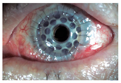
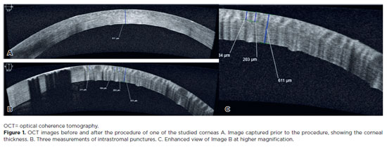
PURPOSE: To assess the reliability and penetration depth of an automated micropuncture system using a tattoo machine.
METHODS: Twenty human corneas were obtained and subjected to intrastromal micropuncture using a tattoo machine. Each cornea was divided into two halves: one received pigment, while the other received saline solution as a control. The Cheyenne tattoo machine was operated at 60 Hz, with standardized needle exposure (six passes per application). The machine used cartridges containing five microneedles. The study was registered with Agência Nacional de Vigilância Sanitária ANVISA (numbers 80281110015, 80281110016, and 80281110019). The pigment used was Electric Ink black ink, with a density of 1,271,460 μg/mL. Puncture depth was measured before and after the procedure using both anterior segment optical coherence tomography and histopathological analysis. Puncture depth measurements were analyzed using ImageJ software. Each cornea was measured thrice, and the results were subsequently compared.
RESULTS: No corneal perforations were observed with the use of the tattoo machine, and puncture depth measurements ranged from 107 to 486 µm.
CONCLUSIONS: The use of a tattoo machine represents a viable and accessible approach for keratopigmentation, with potential for both cosmetic and therapeutic applications. Its adaptation for controlled intrastromal drug delivery may enable the targeted treatment of deep infectious keratitis, corneal neovascularization, and stromal inflammatory disorders, representing a promising approach for corneal stromal diseases. Further research is needed to optimize techniques and evaluate long-term safety and efficacy, particularly for the delivery of antimicrobial, anti-inflammatory, and anti-vascular endothelial growth factor agents.
Keywords: Eye banks; Cadaver; Cornea; Corneal stroma; Drug delivery systems; Tissue donors; Tattooing/instrumentation; Punctures
Arq. Bras. Oftalmol. 2025;88 (1 )
:1-4
| DOI: 10.5935/0004-2749.2023-0056
Abstract
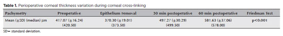
PURPOSE: This study aimed to analyze variations in intraoperative corneal thickness during corneal cross-linking in patients with keratoconus and to investigate its possible correlation with presurgical maximal keratometry (Kmax) and pachymetry.
METHODS: This was a prospective case series. We used a method similar to the Dresden protocol, with the application of hydroxypropyl methylcellulose 0.1% hypo-osmolar riboflavin in corneas between 330 and 400 µm after epithelium removal. Corneal thickness was measured using portable calipers before and immediately after epithelium removal, and 30 and 60 min after the procedure.
RESULTS: The 30 patients in this study were followed up for one year. A statistically significant difference was observed in pachymetry values during the intraoperative period (p<0.0001) and an increase of 3.05 µm (95%CI: 0.56–5.54) for each diopter was seen after epithelium removal (p0.019). We found an average Kmax difference of −2.12 D between men and women (p0.013). One year after treatment, there was a statistically significant reduction in pachymetry (p<0.0001) and Kmax (p0.0170) values.
CONCLUSIONS: A significant increase in pachymetry measurements was seen during the procedure, and most patients showed a regression in Kmax and pachymetry values one year after surgery.
Keywords: Corneal pachymetry; corneal topography; cross-linking reagents/therapeutic use; hypromellose derivatives; keratoconus/surgery; riboflavin/therapeutic use
Arq. Bras. Oftalmol. 2025;88 (6 )
:1-8
| DOI: 10.5935/0004-2749.2025-0118
Abstract
PURPOSE: Using advanced imaging techniques, this study aimed to evaluate corneal stability, epithelial remodeling, and tear film changes over a one-year period in first-time soft-contact lens wearers.
METHODS: A retrospective study was conducted on 100 eyes of 50 first-time daily soft-contact lens users aged 21–65 years with no prior rigid gas-permeable lens wear. The Sirius Scheimpflug imaging system was used to assess corneal topography, epithelial thickness, and non-invasive tear break-up time at baseline, 3, 6, and 12 months. Corneal warpage was evaluated using symmetry indices and Baiocchi Calossi Versaci indices. We performed statistical analysis using repeated-measures analyses of variance with Greenhouse-Geisser correction.
RESULTS: The mean baseline central corneal thickness was 537.83 (±7.92) µm, with no significant thinning after one year. The average simulated keratometry values remained stable, indicating no progressive corneal steepening or flattening. There were no significant changes in warpage indices over time, suggesting corneal shape preservation. Higher-order aberrations (coma, trefoil, and spherical aberrations) and non-invasive tear break-up time remained unchanged throughout the study period.
CONCLUSIONS: Modern silicone hydrogel soft-contact lenses do not induce significant corneal warpage, epithelial remodeling, or optical aberrations over a one-year period. We found that corneal morphology and tear film stability were preserved, supporting the safety of soft-contact lens use. These findings provide clinically relevant insights into the long-term impact of contact lens wear. They may facilitate improved lens fitting strategies and preoperative refractive surgery assessments.
Keywords: Contact lenses, hydrophilic; Cornea/surgery; Corneal diseases; Corneal topography; Adaptation, ocular/physiology; Endothelium, corneal/pathology; Refractive errors; Tears/metabolism.
Arq. Bras. Oftalmol. 2026;89 (4 )
:1-8
| DOI: 10.5935/0004-2749.2025-0313
Abstract
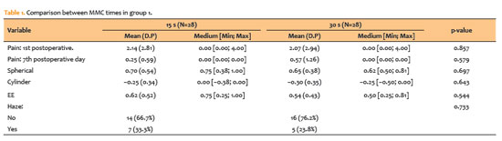
PURPOSE: To compare clinical outcomes associated with different intraoperative mitomycin C exposure times during photorefractive keratectomy for myopia and astigmatism correction.
METHODS: This prospective, comparative, contralateral-eye study included 41 patients (82 eyes), comprising 28 eyes with ablation <60µm and 13 eyes with ablation >60µm, who underwent photorefractive keratectomy with varying mitomycin C application times based on ablation depth. In eyes with ablation <60µm, mitomycin C was applied for 15 s in one eye and 30 s in the fellow eye. In eyes with ablation >60µm, mitomycin C was applied for 30 s in one eye and 60 s in the fellow eye. Outcomes included visual acuity, postoperative pain (visual analog scale), subjective tearing, corneal haze, and refractive results at 3 months.
RESULTS: No statistically significant differences were observed between mitomycin C application times within either group for postoperative pain, tearing, visual acuity, refractive outcomes (spherical, cylindrical, and spherical equivalent), or haze prevalence (p>0.05 for all comparisons). Visual acuity improved in all groups, and no eyes lost ≥2 lines of corrected distance visual acuity.
CONCLUSIONS: Shorter mitomycin C exposure times (15 or 30 s) appear to be as effective and safe as longer durations (30 or 60 s) for haze prevention after photorefractive keratectomy without compromising refractive outcomes or increasing postoperative discomfort at 3-month follow-up.
Keywords: Mitomicin/therapeutic use; Photorefractive keratectomy; Lasers, excimer; Intraoperative period; Miopia/surgery; Astigmatismo/surgery; Corneal opacity; Postoperative pain; Comparative study
Arq. Bras. Oftalmol. 2025;88 (5 )
:1-7
| DOI: 10.5935/0004-2749.2024-0217
Abstract
PURPOSE: This study aimed to evaluate the influence of intrastromal corneal ring segment implants on the intraocular pressure measurements using Goldmann applanation tonometry, rebound tonometry, and noncontact tonometry in keratoconic corneas and analyze the intertonometer agreement.
METHODS: We enrolled 74 eyes in this observational and prospective study. Each participant had a complete eye examination, corneal analysis with Scheimpflug Tomography (Pentacam®), and intraocular pressure evaluation with Goldmann applanation tonometry, rebound tonometry, and noncontact tonometry, before and after intrastromal corneal ring segment implantation (on postoperative days 1, 7, 45, and 90). Intertonometer agreement was assessed using Bland-Altman analysis.
RESULTS: The mean age was 29.9 ± 10.2 years, and 47 (63.5%) eyes had keratoconus grade II. Intraocular pressures were higher for noncontact tonometry preoperatively and on 90 postoperative day (mean ± SD: 12.4 ± and 12.1 ± 2.2 mmHg, respectively), followed by Goldmann applanation tonometry (11.1 ± 3.0 and 11.2 ± 2.7 mmHg, respectively), and were lower for rebound tonometry (9.7 ± and 9.4 ± 3.2 mmHg, respectively). The variation from the Goldmann tonometry on 7 postoperative day to the baseline (p=0.022) and that of noncontact tonometry on 90 postoperative day to the baseline (p=0.021) were statistically significant. The rebound tonometry underestimated intraocular pressure when compared with the Goldmann applanation tonometry by a mean of 1.47 ± 5.19 mmHg. Noncontact tonometry, when compared with Goldmann applanation tonometry, overesti-mated intraocular pressure by a mean of 1.23 ± 4.15 mmHg.
CONCLUSION: Despite statistically significant differences between some postoperative periods, the intraocular pressure measurement differences may not be clinically relevant.
Keywords: Keratoconus; Intraocular pressure; Cornea; Corneal stroma; Postoperative period; Tonometry ocular; Prostheses and implants
Arq. Bras. Oftalmol. 2025;88 (2 )
:1-9
| DOI: 10.5935/0004-2749.2023-0292
Abstract
PURPOSE: Myopia, or nearsightedness, is one of the most common eye conditions worldwide. However, a comparison of the effectiveness of different laser-assisted interventions is lacking. Thus, we aimed to compare the efficacy and safety of LASIK and IntraLASIK in addressing myopia.
METHODS: The study was conducted in two ophthalmology clinics in Beijing, China, in 2022. A total of 84 patients (152 eyes) with different degrees of myopia were examined and underwent LASIK (n=46, 80 eyes) or IntraLASIK (n=38, 72 eyes). Keratometry, corneal topography, pachymetry, visual acuity evaluation, and corneal biomechanical analysis were performed before and after the intervention.
RESULTS: IntraLASIK produced more precise flaps than LASIK, with deviations of <8 mm and 0.1 mm from the intended thickness and diameter, respectively. LASIK resulted in nonuniform flaps, with thickness deviations of 5-86 mm. IntraLASIK demonstrated a superior efficacy for patients with severe myopia and thin corneas, with a mean spherical equivalent of 0.9 D at 6 months compared to the 1.4 D for LASIK. Approximately 91% and 83% of the patients with mild to moderate and severe myopia, respectively, achieved results within ± 0.49 D from the refractive target with IntraLASIK.
CONCLUSIONS: Corneal hysteresis and corneal resistance factor decreased with an increase in laser intensity, and they decreased faster with thinner corneas. Thus, IntraLASIK is more useful than LASIK in patients with thin corneas and severe myopia.
Keywords: Myopia; Lasers; Cornea; Keratomileusis; Laser in situ
 ABO is licensed under a Creative Commons Attribution-NonComercial 4.0 Internacional.
08-fig01.jpg)
02-fig01tb.jpg)
06-fig01.jpg)
02-fig01.jpg)
08-fig01.jpg)
06-fig01.jpg)


09-fig01.jpg)