Arq. Bras. Oftalmol. 2020;83 (4 )
:289-293
| DOI: 10.5935/0004-2749.20200041
Abstract
Objetivo: A dilatação pupilar farmacológica é realizada em exames oftalmológicos abrangentes e antes das medições biométricas. Até o momento, não há consenso sobre seu impacto nas medições biométricas. O objetivo deste estudo foi investigar os efeitos da dilatação pupilar nas medidas biométricas oculares em crianças saudáveis.
Métodos: Estudo prospectivo, observacional e não randomizado de crianças (4-18 anos) que foram admitidas para exame oftalmológico de rotina. As medidas biométricas foram realizadas usando um dispositivo de biometria óptica sem contato, antes e após a dilatação pupilar farmacológica com cloridrato de ciclopentolato. Os cálculos de potência das lentes intraoculares foram realizados utilizando as fórmulas de Hill-RBF, Barrett, Olsen, Sanders-Retzlaff-Kraff/ Teórica, Holladay e Hoffer Q. Análises estatísticas descritivas também foram realizadas. O teste dos postos sinalizados de Wilcoxon foi usado para comparar as medidas antes e após a dilatação pupilar farmacológica. As relações entre as variáveis foram analisadas pelo coeficiente de correlação de Spearman-Brown.
Resultados: O estudo incluiu 116 olhos de 58 crianças (idade média de 8,4 ± 0,32 anos; 34 meninas). Alterações significativas foram observadas após a dilatação pupilar, em termos de profundidade da câmara anterior, profundidade do humor aquoso e espessura central da córnea e do cristalino. Nenhuma mudança significativa ocorreu no comprimento axial. Os cálculos de potência da lente intraocular não revelaram alterações significativas após a dilatação pupilar na maioria das fórmulas, com exceção da fórmula Olsen.O poder da lente intraocular foi significativamente inversa correlacionada com o comprimento axial e a profundidade da câmara anterior.
Conclusões: A dilatação pupilar farmacológica em crianças parece não ter impacto no comprimento axial e no poder da lente intraocular, mas causou um aumento significativo na profundidade da câmara anterior. A diferença nas medidas da profundidade da câmara anterior antes e após a dilatação pupilar pode estar relacionada ao modelo do dispositivo de biometria óptica utilizado. Tais resultados devem ser considerados nos cálculos de potência da lente intraocular realizados usando parâmetros de profundidade da câmara anterior.
Keywords: Dilatação; Paquimetria corneana; Lentes intraoculares; Câmara anterior; Criança
Arq. Bras. Oftalmol. 2026;89 (2 )
:1-6
| DOI: 10.5935/0004-2749.2025-0244
Abstract
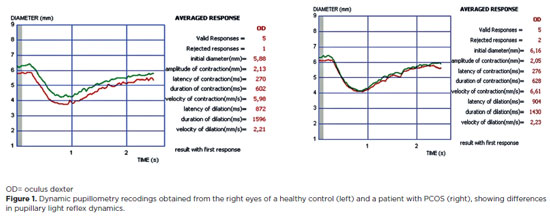
PURPOSE: Polycystic ovary syndrome is frequently associated with autonomic nervous system dysfunction, even in the absence of obesity or overt metabolic abnormalities. Alterations in pupillary responses may reflect early autonomic involvement and serve as a potential tool for early diagnosis, risk stratification, and disease monitoring. This study aimed to investigate pupillary reflex parameters using dynamic pupillometry in newly diagnosed non-obese women with polycystic ovary syndrome and to compare the findings with those of healthy controls. Methods: This prospective cross-sectional study included 48 newly diagnosed women with polycystic ovary syndrome and 44 age- and sex-matched healthy controls. Pupillary function parameters were measured using dynamic pupillometry (MonPackOne; Metrovision, France). Results: The mean age did not differ significantly between the groups (p=0.870). Initial pupil diameter, pupil contraction amplitude, and contraction velocity were significantly lower in the PCOS group than in the control group, whereas pupillary dilation duration was significantly longer (p<0.001, p<0.001, p=0.007, and p=0.032, respectively). No significant differences were observed between the groups regarding contraction latency, contraction duration, dilation latency, or dilation velocity (p=0.749, p=0.925, p=0.653, and p=0.310, respectively). Conclusion: Newly diagnosed non-obese women with polycystic ovary syndrome exhibit significant alterations in pupillary dynamics, suggesting a generalized reduction in both sympathetic and parasympathetic activity. Dynamic pupillometry may represent a practical, noninvasive tool for detecting early autonomic hypoactivity and identifying patients at risk for future metabolic or cardiovascular complications.
Keywords: Autonomic nervous system; IPupil/physiology; Reflex, Pupillary/physiology; Polycystic ovary syndrome/diagnosis; Menstruation disturbances; Ideal body weight
Arq. Bras. Oftalmol. 2025;88 (1 )
:1-10
| DOI: 10.5935/0004-2749.2023-0073
Abstract
PURPOSE: To describe the epidemiological and clinical profile of hospitalized patients with retinoblastoma in Brazil.
METHODS: Using data from the Hospital Cancer Registry of the , patients with the morphological codes of retinoblastoma who were diagnosed between 2000 to 2018, aged 0–19 years, and followed up in registered hospitals (analytical cases) were selected. The relative and absolute frequencies of demographic, clinical, diagnostic, therapeutic, and outcome variables were described. Hospital performance indicators were calculated and compared between hospitals qualified and not qualified to treat pediatric oncology cases and between hospitals with different case volumes (<20, 20–75, >75 cases).
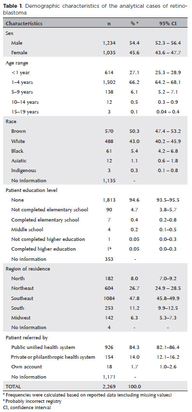
RESULTS: Of the 2,269 identified analytical cases from 86 institutions, 48% were from the Southeast, 54% were male, and 66% were aged <4 years. The proportion of missing data (NA) was too high for several variables. Approximately 84% of the patients were from the public health system, 40% had a positive family history, and 88% had unilateral involvement. The first treatment included surgery in 58.3% of the patients (NA=2), Approximately 36.6% of these patients achieved complete remission, 10.8% achieved partial remission, and 12.7% died (NA=59%). Hospital performance indicators were within the target in >90% of the patients. The median time between the first appointment and diagnosis (6 days, interquartile range [IQR] 1–14) was significantly lower and the median time to death was longer (343 days, IQR, 212-539) in high-volume hospitals (>75 cases) than in medium- and low-volume hospitals.
CONCLUSIONS: Despite the high proportion of missing data, we found that the delay in diagnosis is due to prehospital factors. Additionally, there is a need for educational programs for healthcare professionals and families that emphasize early identification and referral to specialized centers. Future studies should focus on the impact of Hospital Cancer Registry data completeness on outcomes, causes of delay in diagnosis, regional inequalities, and barriers to accessing specialized services.
Keywords: Retinoblastoma/diagnosis; Retinoblastoma/epidemiology; Patient care; Humans; Children; Adolescents; Brazil.
Arq. Bras. Oftalmol. 2026;89 (1 )
:1-6
| DOI: 10.5935/0004-2749.2025-0071
Abstract
PURPOSE: This study aimed to evaluate the outcomes of strabismus surgical correction in patients with Down syndrome.
METHODS: We conducted a retrospective chart review of patients with Down syndrome who underwent strabismus surgery between January 1997 and May 2024 at an Ophthalmology Outpatient Clinic in São Paulo, Brazil. The data collected included age, sex, medical and ocular history, surgical details, and follow-up outcomes. The patients were categorized by strabismus type into esotropia, fourth nerve palsy, and mixed groups. Surgical success was defined as final alignment within 10Δ of orthotropia and, where applicable, whether there was resolution of abnormal head posture of ocular origin. Patients with postoperative follow-up <6 months were excluded.
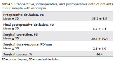
RESULTS: A total of 37 patients (21 females) were included. Of these, 22 (59.5%) were in the esotropia group, 10 (27.0%) in the fourth nerve palsy group, and 5 (13.5%) in the mixed group. The surgical success rate in the esotropia group was 86.4%, with a mean preoperative deviation of 35.2 (± 6.5)Δ, and mean surgical correction of 30.1 (± 10.4)Δ. The success rate in the fourth nerve palsy group was 40.0%, with a mean preoperative deviation of 10.4 (± 4.3)Δ. Overall, success was achieved with a single surgical procedure in 73.0% of the sample. No significant associations were found between surgical success and the clinical and demographic variables, including sex, age at surgery, oblique muscle overaction, pattern strabismus, visual acuity, amblyopia, preoperative deviation, or postoperative follow-up duration (p>0.05).
CONCLUSIONS: When standard surgical tables are applied, strabismus surgery in patients with Down syndrome appears to be safe and effective. We found high success rates, particularly among patients with esotropia. We observed no tendencies toward over- or under-correction. These findings support the use of conventional surgical protocols with this patient population.
Keywords: Down Syndrome/complications; Strabismus/surgery; Esotropia/surgery; Oculomotor nerve diseases/physiopathology; Vision disorders; Humans; Brazil.
Arq. Bras. Oftalmol. 2025;88 (4 )
:1-5
| DOI: 10.5935/0004-2749.2024-0279
Abstract
PURPOSE: Trachoma is the major infectious cause of preventable blindness in the world, and its sequelae include the presence of cicatricial entropion and trachomatous trichiasis. Trachoma can be corrected by surgical treatment of the eyelids and, if left untreated, may result in corneal opacification, low vision, and blindness. There are limited data on trachomatous trichiasis in Brazil. This study was conducted to estimate the frequency of entropion and trichiasis surgeries of trachomatous origin based on the records of procedures performed in specialized hospitals that served the Unified Health System (SUS) in the years 2016 and 2017.
METHODS: This was a retrospective study conducted in the oculoplastic sectors of the ophthalmology services of the following three hospitals in the state of São Paulo: Hospital das Clínicas da Faculdade de Medicina de Botucatu (HC Botucatu), Hospital das Clínicas da Faculdade de Medicina de Ribeirão Preto da Universidade de São Paulo (HC Ribeirão Preto), and Hospital Estadual de Bauru (HE Bauru). Medical records corresponding to the codes of interest were evaluated.
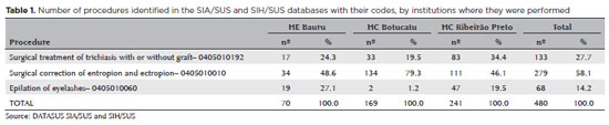
RESULTS: In total, 462 medical records were evaluated, including 170 (36.8%) at HC Botucatu, 61 (13.2%) at HE Bauru, and 231 (50.0%) at HC Ribeirão Preto. There were 39 (8.4%) cases of trachomatous trichiasis, ranging from 9 (14.8%) at HE Bauru to 15 (6.5%) at HC Ribeirão Preto.
CONCLUSIONS: The frequency of surgery due to trachoma was low in these oculoplastic services. The state of São Paulo might have reached the goal for trachoma elimination in the surgical component. The questionnaire used for data collection was successfully tested despite some difficulties in collecting data from the medical records. Studies with the same methodology are recommended in other services in the areas of endemic trachoma in the past to understand the frequency of eye lid surgeries performed for treating trachomatous sequelae.
Keywords: Trachoma; Trichiasis; Medical records; Epidemiology; Neglected diseases; Unified Health System; Brazil
Arq. Bras. Oftalmol. 2024;87 (3 )
:1-7
| DOI: 10.5935/0004-2749.2021-0514
Abstract
Objetivo: Comparar os achados oculares em longo prazo de crianças que se submeteram à cirurgia de catarata congênita antes dos dois anos de idade e receberam uma injeção intracameral de triancinolona no intraoperatório ou usaram prednisolona oral no pós-operatório para modular a inflamação ocular.
Métodos: Neste estudo prospectivo de coorte, todos os pacientes que participaram de um ensaio clínico anterior, que analisou os resultados cirúrgicos de 1 ano da cirurgia de catarata congênita usando triancinolona intracameral (Grupo de Estudo) ou prednisolona oral (Grupo Controle), eram elegíveis para participar. Os prontuários médicos dos pacientes foram revisados e as crianças foram submetidas a um exame oftalmológico completo no acompanhamento final. As principais medidas de desfecho foram: achados biomicroscópicos, pressão intraocular, espessura central da córnea, a necessidade de intervenções cirúrgicas adicionais e achados compatíveis com glaucoma.
Resultados: Vinte e seis olhos (26 pacientes) foram incluídos (Grupo de Estudo = 11 olhos; Grupo de Controle = 15 olhos). O seguimento médio foi de 8,2 ± 1,2 anos e 8,1 ± 1,7 anos nos Grupos de Estudo e Controle, respectivamente (p=0,82). Todos os olhos apresentavam lente intraocular centrada. Não houve diferença estatisticamente significativa entre os grupos com relação à presença de sinéquia posterior (p=0,56), pressão intraocular (p=0,49) ou espessura central da córnea (p=0,21). Nenhum dos olhos preencheu os critérios diagnósticos para glaucoma, apresentou opacificação secundária do eixo visual ou foi reoperado.
Conclusão: Os achados oculares em longo prazo de crianças que se submeteram à cirurgia de catarata congênita e receberam uma injeção intracameral de triancinolona no intraoperatório foram semelhantes aos que usaram prednisolona oral no pós-operatório para modular a inflamação ocular, sugerindo que a triancinolona intracameral pode substituir a prednisolona oral na cirurgia de catarata congênita, facilitando o tratamento pós-operatório e a adesão ao mesmo.
Keywords: Catarata congênita; Triancinolona; Prednisolona; Esteroides; Complicações pós-operatórias; Criança
Arq. Bras. Oftalmol. 2024;87 (3 )
:1-7
| DOI: 10.5935/0004-2749.2022-0374
Abstract
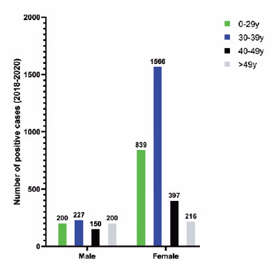
PURPOSE: To describe a 2019 acute toxoplasmosis outbreak in the city of São Paulo, Brazil, and to evaluate the laboratory serological profile for toxoplasmosis for three consecutive years. The ophthalmological manifestations of the patients involved in the outbreak were also studied.
METHODS: A cross-sectional descriptive study of a toxoplasmosis outbreak in São Paulo, Brazil, between February and May 2019. Epidemiological data were described, as were the observed ocular manifestations. As part of this study the number of patients with positive IgM toxoplasmosis serology was obtained from a large laboratory network (DASA) for three consecutive years, including the year of the outbreak (2018, 2019, 2020).
RESULTS: Eighty-three individuals were identified in the outbreak and two clusters were studied. The clinical picture of at least 77% of the patients, the epidemiological analysis, and the short incubation period (5-8 days) suggested contamination by oocysts. Serological laboratory data analysis revealed an increase of positive toxoplasmosis IgM in 2019 of 73% compared to the previous year. Ophthalmological examination revealed that at least 4.8% of the patients developed toxoplasmic retinochoroiditis, none of whom had been treated during the acute systemic disease.
CONCLUSION: Our findings indicate vegetable contamination as the possible source of this outbreak, a high prevalence of toxoplasmosis in São Paulo during the outbreak period, and a drop in the number of tests during the COVID-19 pandemic. Retinochoroiditis was observed in at least 4.8% of the cases. We confirm the need to implement effective means for the prevention, diagnosis, and treatment of the disease. This may involve raising awareness among the population of the importance of vegetable hygiene, and improved quality control of food and water.
Keywords: Toxoplasmosis/etiology; Food parasitology; Water/parasitology; Uveitis, posterior/parasitology; Chorioretinitis/parasitology; Visual acuity; Disease outbreaks; Eye manifestations; Humans.
 ABO is licensed under a Creative Commons Attribution-NonComercial 4.0 Internacional.
04-tab01.jpg)
12-fig01tb.jpg)
12-fig01.jpg)
08-fig01.jpg)
11-fig01tb.jpg)
01-fig01tb.jpg)
03-fig01.jpg)
02-fig01.jpg)