Arq. Bras. Oftalmol. 2022;85 (3 )
:223-228
| DOI: 10.5935/0004-2749.20220030
Abstract
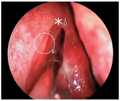
Objetivos: O objetivo deste estudo é comparar as curvas de aprendizagem dos especialistas em dois campos diferentes sem experiência prévia de dacriocistorrinostomia endonasal endoscópica e revelar as complicações com as taxas de sucesso cirúrgico.
Métodos: Foram investigados retrospectivamente 90 pacientes que receberam dacriocistorrinostomia endonasal endoscópica consecutiva com preservação da mucosa realizada por um oftalmologista (Grupo 1, n=45) e realizada por um otorrinolaringologista (Grupo 2, n=45) entre outubro de 2017 e outubro de 2019. Foram incluídos no estudo pacientes admitidos com epífora e diagnosticados com obstrução primária do ducto nasolacrimal adquirido como resultado do teste de irrigação lacrimal, com idade superior a 18 anos e com, pelo menos, 6 meses de acompanhamento. Em todos os casos, patologias adicionais, como o desvio do septo, foram avaliadas por meio da realização de imagens maxilofaciais. Os prontuários dos pacientes foram avaliados quanto à duração da cirurgia, complicações e desempenho funcional.
Resultados: A média de duração cirúrgica dos pacientes no Grupo-2 foi de 36,27 ± 11,61 minutos, enquanto no Grupo-1 foi de 43,62 ± 16,89 minutos, sendo a diferença estatisticamente significativa (p=0,018). O desempenho funcional no Grupo 1 foi de 84,4% (73,3% nos primeiros 15 casos, 93,3% nos últimos 15 casos) no Grupo 2, essa taxa foi de 88,9% (80% nos primeiros 15 casos, 93,3% nos últimos 15 casos) e a diferença não foi estatisticamente significativa (p=0,53). A intervenção do septo além da cirurgia endoscópica em ambos os grupos (p=0,03, p=0,005, respectivamente) e sangramento intenso durante a cirurgia (para ambos os grupos, p<0,0001) diminuiu significativamente o sucesso funcional.
Conclusão: A dacriocistorrinostomia endonasal endoscópica, realizada após o treinamento necessário, pode ser realizada com alto sucesso e com baixas taxas de complicações por oftalmologistas que não estão familiarizados com a cirurgia endoscópica após adquirirem experiência com trinta casos.
Keywords: Obstrução dos ductos lacrimais; Ducto nasolacrimal/cirurgia; Dacriocistorinostomia/métodos; Endoscopia; Oftalmologia/educação
Arq. Bras. Oftalmol. 2021;84 (4 )
:311-315
| DOI: 10.5935/0004-2749.20210044
Abstract
OBJETIVO: Portadores de catarata podem apresentar concomitantemente obstrução do ducto lacrimo-nasal (DLN), com risco de desenvolver endoftalmite no pós-operatório da facectomia. O objetivo do presente estudo é apresentar as percepções dos cirurgiões de catarata sobre a propedêutica e a conduta frente a pacientes com obstrução do ducto lacrimo-nasal concomitante com catarata.
MÉTODOS: Trata-se de uma pesquisa baseada em um questionário envolvendo cirurgiões brasileiros de catarata, realizado no período de março a abril de 2018. Foram levantados dados sobre o perfil dos participantes, o tempo e a experiencia da prática oftalmológica, o treinamento prévio para diagnóstico e tratamento da obstrução do ducto lacrimo-nasal e os conhecimentos de endoftalmite após cirurgia de catarata. Todos os dados foram inseridos em planilha Excel e analisados de acordo com a frequência de ocorrência.
RESULTADOS: Noventa e um oftalmologistas responderam ao questionário. A maioria (63,7%) deles realiza cirurgias de catarata há mais de 10 anos e a maioria (84,6%) recebeu treinamento para diagnóstico e tratamento da obstrução do ducto lacrimo-nasal durante o curso de residência médica. A pesquisa da obstrução crônica do ducto lacrimo-nasal no pré-operatório da catarata é feita pelo teste do refluxo de secreção pelos pontos lacrimais (53,8%) ou por irrigação das vias lacrimais (23,1%). A obstrução do ducto lacrimo-nasal é tratada com colírios antibióticos por 47,2% dos respondentes. Para os portadores de obstrução do ducto lacrimo-nasal , 78% indicam a desobstrução das vias lacrimais previamente à facectoma, aguardando de 4 a 6 semanas para tal. O procedimento de escolha para tratar a obstrução do ducto lacrimo-nasal antes da facectomia é a dacriocistorrinostomia (88,4%). A necessidade de um protocolo para auxiliar na detecção e tratamento da obstrução do ducto lacrimo-nasal em portadores de catarata é reconhecida pela maioria dos participantes deste estudo.
CONLUSÃO: É necessário melhorar a propedêutica e o manejo da catarata em portador de obstrução do ducto lacrimo-nasal porque esse é um fator de risco para endoftalmite.
Keywords: Catarata; Extração de catarata; Endoftalmite; Obstrução dos ductos lacrimais; Dacriocistite; Intervenção baseada em internet; Inquéritos e questionários
Arq. Bras. Oftalmol. 2025;88 (4 )
:1-5
| DOI: 10.5935/0004-2749.2024-0160
Abstract
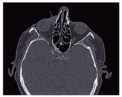
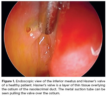
PURPOSE: Congenital epiphora can be related to anomalies of the nasolacrimal duct. This study aimed to assess the distal end of the nasolacrimal duct and the outcomes of endoscopic treatment in children older than 12 months with congenital epiphora.
METHODS: This retrospective analysis describes the clinical characteristics, management, and outcomes of symptomatic congenital lacrimal obstruction in 32 lacrimal systems of 23 children. Data was collected on the preoperative symptoms, age at the time of surgery, intraoperative findings, treatment modalities, and outcomes of the children in our cohort. All patients underwent a standard endoscopic lacrimal examination, including irrigation and diagnostic probing, viewed via the inferior meatus. Cases with complex anomalies characterized by obstructions in the canaliculi, nasolacrimal junction, or nasolacrimal duct were excluded.
RESULTS: The mean age at the time of surgery was 48.03 (±27.99) months. Four different types of distal nasolacrimal duct obstruction were diagnosed. These were obstructions by a membrane (n=12), ostium stenosis (n=15), impacted turbinate (n=3), and membranous residual flaps (n=2). They were all managed with inferior meatus microsurgery and nasal endoscopic probing without silicone intubation. After a mean follow-up period of 14.75 (±11.93) months, successful outcomes were achieved in all cases.
CONCLUSION. Microsurgery to the inferior meatus, performed under nasal endoscopy, is a safe and effective treatment for isolated anomalies of the distal end of the nasolacrimal duct in children older than 12 months. We do not recommend silicone intubation in the absence of complex lacrimal system anomalies.
Keywords: Lacrimal duct obstruction; Nasolacrimal duct; Silicone; Microsurgery; Endoscopy; Epiphora; Intubation; Child
Arq. Bras. Oftalmol. 2025;88 (1 )
:1-8
| DOI: 10.5935/0004-2749.2023-0103
Abstract
PURPOSE: This study aimed to compare the safety and effectiveness of intraocular pressure reduction between micropulse transscleral cyclophotocoagulation and “slow cook” transscleral cyclophotocoagulation in patients with refractory primary open-angle glaucoma.
METHODS: We included patients with primary open angle glaucoma with at least 12 months of follow-up. We collected and analyzed data on the preoperative characteristics and postoperative outcomes. The primary outcomes were a reduction of ≥20% of the baseline value (criterion A) and/or intraocular pressure between 6 and 21 mmHg (criterion B).
RESULTS: We included 128 eyes with primary open-angle glaucoma. The preoperative mean intraocular pressure was 25.53 ± 6.40 and 35.02 ± 12.57 mmHg in the micropulse- and “slow cook” transscleral cyclophotocoagulation groups, respectively (p<0.001). The mean intraocular pressure was reduced significantly to 14.33 ± 3.40 and 15.37 ± 5.85 mmHg in the micropulse- and “slow cook” transscleral cyclophotocoagulation groups at the last follow-up, respectively (p=0.110). The mean intraocular pressure reduction at 12 months was 11.20 ± 11.46 and 19.65 ± 13.22 mmHg in the micropulse- and “slow cook” transscleral cyclophotocoagulation groups, respectively (p<0.001). The median preoperative logMAR visual acuity was 0.52 ± 0.69 and 1.75 ± 1.04 in the micropulse- and “slow cook” transscleral cyclophotocoagulation groups, respectively (p<0.001). The mean visual acuity variation was -0.10 ± 0.35 and -0.074 ± 0.16 in the micropulse- and “slow cook” transscleral cyclophotocoagulation, respectively (p=0.510). Preoperatively, the mean eye drops were 3.44 ± 1.38 and 2.89 ± 0.68 drugs in the micropulse- and “slow cook” transscleral cyclophotocoagulation groups, respectively (p=0.017), but those were 2.06 ± 1.42 and 1.02 ± 1.46 at the end of the study in the slow cook” and micropulse transscleral cyclophotocoagulation groups, respectively (p<0.001). The success of criterion A was not significant between both groups. Compared with 11 eyes (17.74%) in the slow cook” transscleral cyclophotocoagulation group, 19 eyes (28.78%) in the micropulse transscleral cyclophotocoagulation group showed complete success (p=0.171). For criterion B, 28 (42.42%) and 2 eyes (3.22%) showed complete success after micropulse- and slow cook” transscleral cyclophotocoagulation, respectively (p<0.001).
CONCLUSION: Both techniques reduced intraocular pressure effectively.
Keywords: Sclera/surgery; Glaucoma, open-angle/surgery; Ciliary body/surgery; Intraocular pressure; Laser coagulation/methods; Lasers, semiconductor; Comparative study; Effectiveness
Arq. Bras. Oftalmol. 2026;89 (1 )
:1-8
| DOI: 10.5935/0004-2749.2024-0397
Abstract
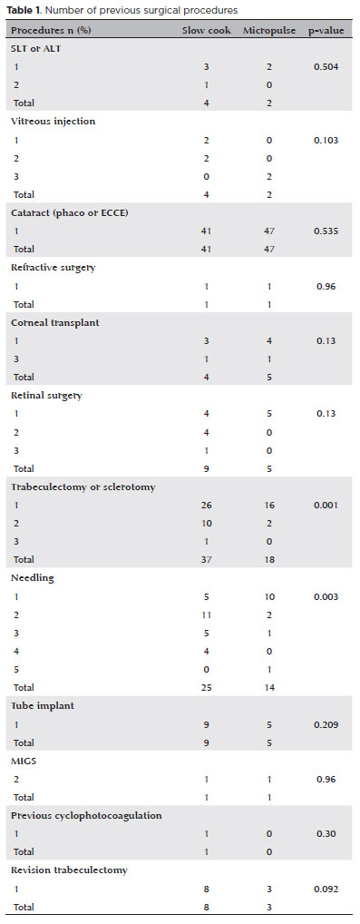
PURPOSE: Glaucoma is one of the leading causes of irreversible blindness worldwide. When topical hypotensive agents or laser trabeculoplasty fail to adequately control the disease, escalation of therapy becomes necessary, with transscleral cyclophotocoagulation being one of the available options. Several variations of transscleral cyclophotocoagulation exist, including traditional continuous wave, MicroPulse, and slow-coagulation techniques. We propose a novel variation – custom slow-coagulation transscleral cyclophotocoagulation – which combines elements of both continuous wave and slow-coagulation approaches. This study aimed to evaluate the outcomes of this technique in patients with refractory glaucoma.
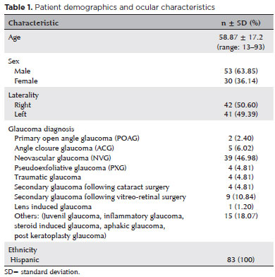
METHODS: This retrospective, interventional study included 104 eyes of 83 patients with refractory glaucoma who underwent custom slow-coagulation transscleral cyclophotocoagulation. Changes in intraocular pressure, visual acuity, the number of glaucoma medications, and postoperative complications were analyzed. A paired t test was used to compare changes in intraocular pressure and visual acuity, while the Wilcoxon signed-rank test was applied to categorical variables. Success rates following custom slow-coagulation transscleral cyclophotocoagulation were estimated using Kaplan–Meier survival analysis.
RESULTS: Mean intraocular pressure decreased significantly from 38.9 ± 15.8 mmHg at baseline to 16.3 ± 9.9 mmHg at Month 12 (p<0.001). The mean number of glaucoma medications also decreased significantly from 3.6 ± 0.6 to 1.8 ± 1.4 (p<0.001). No significant reduction in mean visual acuity was observed during follow-up.
CONCLUSIONS: Custom slow-coagulation transscleral cyclophotocoagulation effectively reduced baseline intraocular pressure and the number of glaucoma medications, with a low rate of complications and no decline in visual acuity over a 12-month follow-up period. This novel technique demonstrated a high safety profile in a Hispanic population and represents a low-cost, minimally invasive procedure with rapid recovery and promising efficacy in intraocular pressure control.
Keywords: Glaucoma/surgery; Sclera; Filtering surgery; Laser coagulation/methods; Lasers, semiconductor/therapeutic use; Intraocular pressure; Blindness/prevention & control; Vision, low/epidemiology; Visual acuity
Arq. Bras. Oftalmol. 2026;89 (3 )
:1-8
| DOI: 10.5935/0004-2749.2025-0043
Abstract
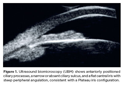
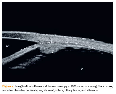
PURPOSE: To evaluate the effect of single-session transscleral diode laser cyclophotocoagulation on intraocular pressure in refractory glaucoma and to determine structural changes using ultrasound biomicroscopy.
METHODS: Forty-three eyes were evaluated. Intraocular pressures at baseline and at the first, third, and sixth months after transscleral diode laser cyclophotocoagulation were compared. Ciliary body thickness, ciliary muscle thickness, ciliary process thickness, iris root thickness, and scleral thickness were assessed at baseline and at the third and sixth months post-treatment.
RESULTS: Reductions in intraocular pressure were significant between baseline and the first month (p=0.018), third month (p<0.001), and sixth month (p<0.001) as well as between the first and third months (p=0.034) and the first and sixth months (p=0.036). Compared with baseline, intraocular pressure reduction rates at the first, third, and sixth months were 34.6%, 56.5%, and 55.3%, respectively, while success rates were 30.2%, 62.8%, and 55.8%, respectively. Decreases in ciliary body thickness, ciliary muscle thickness, and ciliary process thickness were significant between baseline and the third month (p<0.05) and between baseline and the sixth month (p<0.05), whereas changes between the third and sixth months were not significant (p>0.05). Iris root and scleral thicknesses did not change after treatment (p>0.05). At the third and sixth months, significant positive correlations were observed between changes in intraocular pressure and changes in ciliary body thickness and ciliary process thickness (p<0.05).
CONCLUSIONS: To the best of our knowledge, this is one of the few studies comprehensively investigating structural changes after transscleral diode laser cyclophotocoagulation using ultrasound biomicroscopy. Moreover, the relationships between intraocular pressure changes and variations in the ciliary body, ciliary muscle, ciliary process, iris root, and scleral thicknesses were examined in detail. Single-session treatment did not affect iris root or scleral thickness but significantly reduced ciliary body, ciliary muscle, and ciliary process thicknesses. Greater reductions in ciliary body and ciliary process thickness may contribute to more pronounced intraocular pressure reduction.
Keywords: Intraocular pressure; Laser coagulation/methods; Lasers, semiconductor; Microscopy, acoustic; Glaucoma; Ciliary body
Arq. Bras. Oftalmol. 2025;88 (2 )
:1-9
| DOI: 10.5935/0004-2749.2023-0292
Abstract
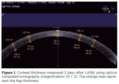
PURPOSE: Myopia, or nearsightedness, is one of the most common eye conditions worldwide. However, a comparison of the effectiveness of different laser-assisted interventions is lacking. Thus, we aimed to compare the efficacy and safety of LASIK and IntraLASIK in addressing myopia.
METHODS: The study was conducted in two ophthalmology clinics in Beijing, China, in 2022. A total of 84 patients (152 eyes) with different degrees of myopia were examined and underwent LASIK (n=46, 80 eyes) or IntraLASIK (n=38, 72 eyes). Keratometry, corneal topography, pachymetry, visual acuity evaluation, and corneal biomechanical analysis were performed before and after the intervention.
RESULTS: IntraLASIK produced more precise flaps than LASIK, with deviations of <8 mm and 0.1 mm from the intended thickness and diameter, respectively. LASIK resulted in nonuniform flaps, with thickness deviations of 5-86 mm. IntraLASIK demonstrated a superior efficacy for patients with severe myopia and thin corneas, with a mean spherical equivalent of 0.9 D at 6 months compared to the 1.4 D for LASIK. Approximately 91% and 83% of the patients with mild to moderate and severe myopia, respectively, achieved results within ± 0.49 D from the refractive target with IntraLASIK.
CONCLUSIONS: Corneal hysteresis and corneal resistance factor decreased with an increase in laser intensity, and they decreased faster with thinner corneas. Thus, IntraLASIK is more useful than LASIK in patients with thin corneas and severe myopia.
Keywords: Myopia; Lasers; Cornea; Keratomileusis; Laser in situ
Arq. Bras. Oftalmol. 2026;89 (4 )
:1-6
| DOI: 10.5935/0004-2749.2025-0373
Abstract
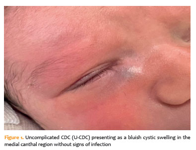
PURPOSE: The purpose of this study was to assess the clinical factors associated with dacryocystitis and the need for surgical intervention in infants with congenital dacryocystocele.
METHODS: This retrospective study included 26 infants diagnosed with congenital dacryocystocele and divided them into two groups: complicated-congenital dacryocystocele (with dacryocystitis or preseptal cellulitis) and uncomplicated-congenital dacryocystocele (without infection). Demographic and perinatal characteristics such as age at diagnosis, gender, birth weight, gestational age, delivery method, and laterality were compared between the groups. For an uncomplicated-congenital dacryocystocele, treatment included conservative management and intravenous antibiotics, followed by probing with intraoperative nasal endoscopy and endonasal marsupialization for a complicated-congenital dacryocystocele.
RESULTS: Of the 26 infants, 14 (53.8%) had complicated-congenital dacryocystocele, while 12 (46.2%) had uncomplicated-congenital dacryocystocele. There were no significant differences between the groups in terms of demographic or perinatal characteristics (p>0.05). Surgical intervention was necessary for all complicated-congenital dacryocystocele cases (100%) and two uncomplicated-congenital dacryocystocele cases (16.7%; p<0.001). During a 6-month median follow-up period, all patients demonstrated complete clinical recovery with no intraoperative complications.
CONCLUSION: In conclusion, approximately half of infants with congenital dacryocystocele developed infection-related complications. While perinatal factors were similar across groups, infectious presentation was linked to the need for surgical intervention. These findings suggest that early detection, prompt conservative management, and close follow-up can help reduce the risk of dacryocystitis and the need for surgery.
Keywords: Dacryocystocele; Dacryocystitis; Lacrimal duct obstruction; Marsupialization; Postoperative complication; Gestational age
Arq. Bras. Oftalmol. 2024;87 (4 )
:1-5
| DOI: 10.5935/0004-2749.2023-0143
Abstract
PURPOSE: The purpose of this study is to assess the long-term outcomes of modified transcanalicular diode laser dacryocys torhinostomy in a large cohort of patients affected by primary acquired nasolacrimal duct obstruction.
METHODS: This study, conducted from January 17 to June 2022, encompassed 141 patients (159 procedures) who underwent modified transcanalicular diode laser dacryocystorhinostomy (MT-DCR). The procedure employed an 810-nm diode laser. Patients were monitored for at least a year after the intervention. Anatomical success was determined by ostium patency upon irrigation, while functional success referred to epiphora resolution. Parameters studied included patient demographics, procedure duration, complications, and both anatomical and functional success. Statistical analysis was performed using the Statistical Package for the Social Sciences software, with results considered significant at a 95% confidence interval (p≤0.05).
RESULTS: A total of 159 lacrimal drainage systems (141 patients: 112 women and 29 men) were included in this study. Among them, 18 underwent bilateral procedures. The average patient age was 58 years (range: 34-91 years), and the average surgical duration was 24 minutes (range: 18-35 minutes). One year after the surgery, MT-DCR exhibited anatomical and functional success rates of 84.9% (135/159) and 83% (132/159), respectively.
CONCLUSION: MT-DCR achieved an anatomical success rate of 84.9%, reflecting an excellent outcome. However, further extensive studies with larger sample sizes and longer follow-up periods are necessary to substantiate these findings.
Keywords: Lacrimal duct obstruction; Nasolacrimal duct/surgery; Dacryocystorhinostomy; Lacrimal apparatus diseases; Laser therapy/methods; Lasers, semiconductor/therapeutic use; Regeneration
 ABO is licensed under a Creative Commons Attribution-NonComercial 4.0 Internacional.
01-fig01.jpg)
11-fig01.jpg)
15-fig01.jpg)
14-fig01.jpg)
09-qua01.jpg)