Arq. Bras. Oftalmol. 2022;85 (2 )
:136-143
| DOI: 10.5935/0004-2749.20220022
Abstract
Objetivo: Estimar a epidemiologia do pterígio; sua correlação com sintomas de olho seco e com potenciais preditores sistêmicos e oculares.
Métodos: Estudo transversal, de base populacional, no qual foram realizadas visitas domiciliares aleatórias a 600 participantes, com 40 anos ou mais de idade, em Ribeirão Preto-SP (n=420) e Cassia dos Coqueiros-SP (n=180), Brasil. Uma entrevista estruturada com um questionário detalhado foi usada para coletar informações sobre demografia e possíveis fatores de risco. Em um segundo momento, participantes aleatórios com pterígio (n=63) ou não (n=110) foram avaliados quanto a alterações na superfície ocular.
Resultados: A frequência de pterígio em Ribeirão Preto foi de 21%; 15.7% entre as mulheres e 32.1% entre os homens (p=0,0002). Em Cássia dos Coqueiros, essa taxa foi de 19.4%; onde 17.3% eram mulheres e 25.5% eram homens (p=0,28). A média de idade naqueles afetados pelo pterígio foi superior à dos participantes sem pterígio, 65,6 ± 10,5 e 61,2 ± 12,0 anos, respectivamente (p=0,02). Houve uma correlação positiva entre o pterígio e história prévia de radioterapia e quimioterapia (p<0,0001 para ambos). Houve maior coloração de fluoresceína na córnea e maior coloração de lissamina verde na conjuntiva em olhos com pterígio (p=0,0003 e 0,0001, respectivamente).
Conclusão: Encontramos uma alta frequência de pterígio em duas populações adultas brasileiras, principalmente em homens e idosos. Danos na superfície ocular e história prévia de radioterapia e/ou quimioterapia foram associados ao pterígio.
Keywords: Pterígio/epidemiologia; Síndrome do olho seco; Prevalência; Fatores de risco
Arq. Bras. Oftalmol. 2025;88 (1 )
:1-7
| DOI: 10.5935/0004-2749.2022-0367
Abstract
PURPOSE: This study aimed to examine the prevalence of myopic eyes over 11 years (2008-2018) in a private clinic and a public assistance service.
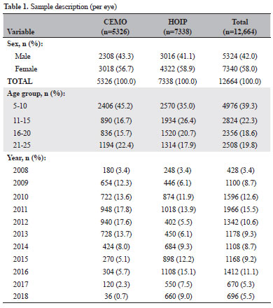
METHODS: We retrospectively evaluated 6332 individuals (12,664 eyes)
between 5 and 25 years old, seen at a private clinic-CEMO (2,663 individuals) and a public service-HOIP (3,669 individuals) from 2008 to 2018. We evaluated the prevalence of myopic eyes (EE ≤-0.50) and high myopic eyes (EE ≤-6.00).
RESULTS: Sex and services did not show statistical differences. The variation in the prevalence of myopic and high myopic eyes showed a random pattern during the study period (this prevalence could not be increased). Prevalences ranged from 20.7% (in 2017) to 32.4% (in 2015) for myopic eyes and from 1.6% (in 2009 and 2016) to 3.3% (in 2015) for eyes with high myopia. The prevalence of myopia showed a statistically significant increase based on the age group.
CONCLUSION: The prevalence of myopic eyes did not increase in our study. The mean prevalence of myopic eyes was similar in the private clinic and public service.
Keywords: Myopia; Refractive errors; Epidemiology; Prevalence
Arq. Bras. Oftalmol. 2021;84 (3 )
:241-248
| DOI: 10.5935/0004-2749.20210032
Abstract
OBJETIVO: Determinar o papel do receptor da vitamina D na patogênese do pterígio. Os níveis de expressão do receptor da vitamina D no tecido do pterígio, os níveis sanguíneos de vitamina D e a frequência de alguns polimorfismos do gene do receptor da vitamina D (BsmI, FokI e TaqI) foram comparados entre pacientes com pterígio e participantes saudáveis.
MÉTODOS: Foram incluídos pacientes com pterígio (n=50) e voluntários saudáveis (n=50). Os níveis séricos de vitamina D foram medidos em ambos os grupos. Foi feita uma coloração imuno-histoquímica para o receptor da vitamina D em cortes obtidos do pterígio e dos tecidos conjuntivais saudáveis adjacentes dos mesmos indivíduos. A existência de polimorfismos do receptor da vitamina D (BsmI, FokI e TaqI) no genoma foi analisada em DNA obtido do sangue venoso dos participantes, usando métodos de Polymerase chain reaction (PCR) e RFLP.
RESULTADOS: Não foi observada nenhuma diferença entre os níveis séricos de vitamina D dos pacientes com pterígio e os dos controles saudáveis. Entretanto, a expressão tissular do receptor da vitamina D foi maior nas células endoteliais dos microvasos do pterígio (p=0,002), nas células estromais sub-epiteliais (p=0,04) e nas células inflamatórias intravasculares (p=0,0001), quando comparada à expressão no tecido conjuntival saudável adjacente. Além disso, embora o haplótipo BBtt tenha sido duas vezes mais frequente, o haplótipo bbTt foi 2,5 vezes menos frequente e o haplótipo BbTT foi 2,25 vezes menos frequente no grupo de controle do que no grupo com pterígio (p<0,001).
CONCLUSÕES: Os níveis séricos de vitamina D não apresentaram diferenças entre o grupo de pessoas saudáveis e o com pterígio. A expressão do receptor da vitamina D mostrou-se maior no grupo com pterígio do que no tecido saudável adjacente. Entretanto, a análise dos polimorfismos do receptor da vitamina D nos pacientes com pterígio não revelou qualquer diferença significativa nos polimorfismos BsmI, FokI ou TaqI em comparação com os voluntários saudáveis.
Keywords: Pterígio; Vitamina D; Polimorfismo genético; Imuno- histoquímica
Arq. Bras. Oftalmol. 2020;83 (4 )
:1-6
| DOI: 10.5935/0004-2749.20200053
Abstract
Objetivo: Determinar a frequência de neoplasia escamosa da superfície ocular associada ao pterígio com apresentação clínica, em um centro de referência em Oftalmologia da região central do México.
Métodos: Revisamos os laudos histopatológicos e as lâminas de biópsia de todos os pacientes que foram submetidos à cirurgia de pterígio de 2014 a 2016 no Instituto Mexicano de Oftalmologia, na cidade de Querétaro.
Resultados: Estudamos 177 amostras de biópsia; 66% eram de pacientes do sexo feminino, sendo a mediana da idade de 52 anos. Encontramos neoplasia escamosa da superfície ocular em 11,29% (n=20). Uma amostra de biópsia mostrou um carcinoma queratinizante infiltrativo pouco diferenciado.
Conclusões: A prevalência da neoplasia escamosa da superfície ocular nessa região parece ser maior do que a indicada por outras pesquisas. Mais estudos de âmbito nacional são necessários para determinar a verdadeira prevalência da neoplasia escamosa da superfície ocular no México e examinar os fatores de risco relacionados.
Keywords: Pterígio; Neoplasias da túnica conjuntiva; Neoplasias oculares; Histopatologia; Carcinoma de células escamosas
Arq. Bras. Oftalmol. 2025;88 (6 )
:1-8
| DOI: 10.5935/0004-2749.2025-0053
Abstract
PURPOSE: This pilot study evaluated the diagnostic accuracy of a deep learning model for detecting pterygium in anterior segment photographs taken using smartphones in the Brazilian Amazon. The model’s performance was benchmarked against assessments made by experienced ophthalmologists, considered the clinical gold standard.
METHODS: In this cross-sectional study, 38 participants (76 eyes) from Barcelos, Brazil, were enrolled. Trained nonmedical health workers captured high-resolution anterior segment images using smartphones. These images were analyzed using a deep learning model based on the MobileNet-V2 convolutional neural network. Diagnostic metrics–including sensitivity, specificity, accuracy, positive predictive value, negative predictive value, and area under the receiver operating characteristic curve–were calculated and compared with the ophthalmologists’ evaluations.
RESULTS: The deep learning model achieved a sensitivity of 91.43%, specificity of 90.24%, positive predictive value of 88.46%, negative predictive value of 92.79%, and an area under the curve of 0.91. Logistic regression revealed no statistically significant association between pterygium and demographic variables such as age or gender.
CONCLUSIONS: The deep learning model demonstrated high diagnostic performance in identifying pterygium in a remote Amazonian population. These preliminary findings support the potential use of artificial intelligence–based tools to facilitate early detection and screening in underserved regions, thereby enhancing access to ophthalmic care.
Keywords: Pterygium/diagnostic imaging; Smartphone; Diagnostic techniques, ophthalmological; Deep learning; Telemedicine; Artificial intelligence; Cross-sectional studies; Brazil/epidemiology
Arq. Bras. Oftalmol. 2025;88 (2 )
:1-7
| DOI: 10.5935/0004-2749.2023-0265
Abstract
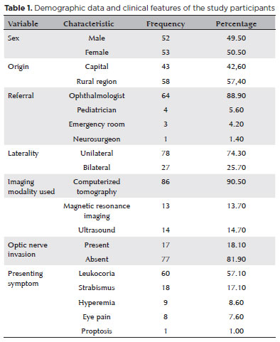
PURPOSE: Although Brazil has a high prevalence of retinoblastoma, there is a lack of epidemiological data on the disease. Thus, in this study, we aimed to evaluate the epidemiological profile of patients diagnosed with retinoblastoma in the ophthalmology department of a pediatric tertiary referral hospital in Ceara, Brazil.
METHODS: A descriptive and cross-sectional study was conducted by retrospectively analyzing the clinical and socioeconomic data from the medical records of pediatric patients followed-up at the hospital between 2007 and 2021. Retinoblastoma was diagnosed on the basis of a fundoscopic or histopathologic examination.
RESULTS: The data of 105 patients were included in the study, and the mean patient age at the time of diagnosis was 1.7 years. Most of the patients were women (50.5%) and hailed from rural areas (57.4%), which was associated with a higher tumor stage. Of the 150 patients, 57.1% initially presented with leukocoria. Ocular hyperemia was associated with more advanced stages of retinoblastoma (p=0.004). Bilateral involvement was observed in 25.7% of the patients and at a significantly younger age (p=0.009). The presence of retinal detachment, vascularized lesions, and vitreous seeds significantly increased the likelihood of requiring enucleation.
DISCUSSION: This study presents an epidemiological description of retinoblastoma in Brazil, which highlights the significance of early detection. Delayed diagnosis is associated with a poorer visual prognosis and higher mortality rate, particularly in patients with unilateral disease. Risk factors for a more severe disease were retinal detachment, vascularized lesions, and vitreous seeds. The correlation between histopathological features and clinical outcomes was limited.
CONCLUSION: Further studies are required to assess the influence of ocular hyperemia, fundoscopic assessment, and histopathologic findings on the prognosis of retinoblastoma. Moreover, it is critical to devise interventions to reduce the time-to-diagnosis in rural areas.
Keywords: Retinoblastoma; Retinal neoplasms; Epidemiology; Prevalence; Risk factors; Delayed diagnosis; Child
Arq. Bras. Oftalmol. 2024;87 (4 )
:1-5
| DOI: 10.5935/0004-2749.2022-0335
Abstract
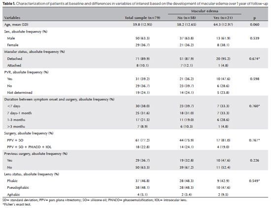
PURPOSE: To clarify the postoperative incidence of macular edema in patients undergoing surgery to repair rhegmatogenous retinal detachment and identify the associated risk factors.
METHODS: In this prospective, observational study, 79 patients who underwent surgery to correct rhegmatogenous retinal detachment using pars plana vitrectomy with silicone oil injection were analyzed. Patients were followed up postoperatively at 7, 30, 90, 180, and 365 days. At each visit, optical coherence tomography was performed to assess the presence or absence of macular edema. were analyzed as possible risk factors for macular edema: age, sex, macular status (attached or detached), presence of vitreoretinal proliferation, history of previous intraocular surgery, reported time of symptoms suggestive of rhegmatogenous retinal detachment up to the date of surgery, and the surgical modality performed.
RESULTS: The 1-year macular edema prevalence rate was 26.6%. In the adjusted analysis, older patients had a higher risk of macular edema, and each 1-year increase in age increased the risk of macular edema by 6% (95% confidence interval = 1.00-1.12). The macular status, vitreoretinal proliferation, the surgical technique used, prior intraocular surgery, and the intraocular lens status were not identified as risk factors. However, the incidence of macular edema increased up to 180 days after surgery, peaking at 10.6%, and then decreased until 365 days after surgery.
CONCLUSION: Macular edema was a common complication after surgery to treat rhegmatogenous retinal detachment, with its incidence peaking between 30 and 180 days after surgery. Age was an important risk factor for macular edema in this cohort.
Keywords: Macular edema; Retinal detachment; Vitrectomy; Tomography, optical coherence; Incidence; Risk factors
Arq. Bras. Oftalmol. 2024;87 (3 )
:1-7
| DOI: 10.5935/0004-2749.2023-0051
Abstract
PURPOSE: To evaluate macular chorioretinal flow changes on optical coherence tomography angiography, in participants who received inactivated and messenger RNA (mRNA) vaccines to prevent coronavirus disease 2019 (COVID-19).
METHODS: In this prospective cohort study, healthy participants who received two doses of an inactivated COVID-19 vaccine (CoronaVac) and then one dose of an mRNA vaccine (BNT162b2) were examined before and after each vaccination. Ophthalmologic examination and imaging with optical coherence tomography angiography were performed during each visit. We evaluated vascular densities in the superficial and deep capillary plexuses in foveal, parafoveal, and perifoveal areas; the foveal avascular zone; and choriocapillaris flows (in 1- and 6-mm-diameter areas).
RESULTS: One eye in each of the 24 participants was assessed. Superficial capillary plexus vascular densities in the parafoveal area were significantly lower after the second dose of the CoronaVac vaccine than after the first dose. In the deep capillary plexus, vascular attenuation was observed only in the parafoveal region after the first CoronaVac dose. However, in all regions, the deep capillary plexus vascular densities and subfoveal choriocapillaris flow were significantly decreased after the second CoronaVac dose. After the BNT162b2 dose, the superficial capillary plexus vascular densities, the deep capillary plexus vascular densities, and subfoveal choriocapillaris flow of most regions were significantly lower than those before vaccinations.
CONCLUSION: Vascular attenuation, observed particularly after the second dose of the CoronaVac vaccine, may explain the pathogenesis of postvaccine ocular ischemic disorders reported in the literature. However, these disorders are extremely rare, and the incidence of thrombotic events caused by COVID-19 itself is higher.
Keywords: Tomography, optical coherence; Angiography; COVID-19 vaccines; COVID-19; Coronavirus infections; SARS-CoV-2; mRNA vaccines; CoronaVac; Incidence
 ABO is licensed under a Creative Commons Attribution-NonComercial 4.0 Internacional.
08-tab01tb.jpg)
09-tab01tb.jpg)
12-fig01.jpg)