Arq. Bras. Oftalmol. 2022;85 (1 )
:13-18
| DOI: 10.5935/0004-2749.20220003
Abstract
Objetivo: Fornecer informações sobre a ocorrência e a eficácia do aconselhamento sobre o uso de tabaco por oftalmologistas a pacientes com doenças oculares associadas à tireoide.
Métodos: Analisamos os prontuários médicos eletrônicos de uma coorte digital de pacientes atendidos por oftalmologistas no Sistema de Saúde da Universidade da Pensilvânia entre o início de 2012 e o final de 2017 com os códigos da Classificação Internacional de Doenças (CID) para a doença de Graves, exoftalmia tireotóxica ou doença ocular associada à tireoide. Os históricos de uso de tabaco foram registrados na primeira e na última visita ao consultório de Oftalmologia, ou na visita mais próxima no tempo. A quantidade de maços/dia (mpd) e todas as anotações feitas nas visitas ao consultório de Oftalmologia foram analisadas para aconselhamento sobre o uso de tabaco.
Resultados: Um total de 435 indivíduos preencheram os critérios de inclusão, dos quais 72 (16,6%) estavam fumando ativamente no momento do primeiro encontro. Apenas 57 (79,2%) desses indivíduos que fumam ativamente registraram queixas relacionadas ao tabagismo, sendo que 34 (59,6%) deles receberam alguma forma de aconselhamento sobre o uso de tabaco. Ao todo, 9 (26,5%) indivíduos dentre os que receberam aconselhamento sobre tabaco e 1 (4,3%) que não teve aconselhamento registrado pararam de fumar (diferença de risco de 22,1%; IC 95%, [1,7%, 39,1%]; p=0,04). Dentre aqueles que receberam aconselhamento, 17 (50,0%) reduziram seus mpd, além de 7 (30,4%) daqueles que não tiveram aconselhamento (diferença de risco de 19,6%; IC 95% [-6,3%, 41,3%]; p=0,18). No geral, 14 (25,5%) dos 55 oftalmologistas que tiveram um paciente fumante ativo registraram evidências de aconselhamento sobre o uso de tabaco.
Conclusões: Os resultados deste estudo revelam tanto as oportunidades perdidas de aconselhamento sobre o uso do tabaco quanto a eficácia do aconselhamento no contexto de doenças oculares associadas à tireoide.
Keywords: Uso de tabaco; Aconselhamento; Doenças da glândula tireóide; Doença de Graves; Oftalmopatias
Arq. Bras. Oftalmol. 2022;85 (6 )
:599-605
| DOI: 10.5935/0004-2749.20220082
Abstract
Objetivo: Avaliar as características clínicas de pacientes pediátricos com blefaroptose adquirida unilateral, transitória e de início agudo.
Métodos: Neste estudo retrospectivo, foram revisados prontuários clínicos entre abril de 2015 e junho de 2020. Os pacientes foram avaliados em termos de características demográficas, manifestações neurológicas e oftalmológicas associadas, duração dos sintomas, etiologia e achados de imagem. Foram excluídos pacientes com blefaroptose congênita e com blefaroptose adquirida de etiologia crônica.
Resultados: Foram incluídos neste estudo 16 pacientes pediátricos (10 masculinos e 6 femininos) com blefaroptose adquirida transitória unilateral de início agudo. A média de idade dos pacientes foi de 6,93 ± 3,16 anos. As causas etiológicas mais comumente identificadas foram trauma em 7 pacientes (43,75%) e infecção (casos parainfecciosos) em 5 pacientes (31,25%). Além disso, a síndrome de Miller-Fisher, a síndrome de Horner secundária a neuroblastoma, a síndrome de Brown adquirida e pseudotumor cerebral foram determinados como causas etiológicas em um paciente cada uma. Achados oculares adicionais estavam associados à blefaroptose em 7 pacientes (58,33%). Foi observada a resolução espontânea da blefaroptose, sem tratamento, em todos os pacientes, exceto nos pacientes com síndrome de Miller-Fisher, neuroblastoma e pseudotumor cerebral. Nenhum paciente precisou de tratamento cirúrgico. Morbidades oculares, como ambliopia, não foram encontradas em nenhum paciente.
Conclusão: Este estudo demonstrou que a blefaroptose transitória unilateral de início agudo, rara na infância, pode regredir sem a necessidade de tratamento cirúrgico na população pediátrica. No entanto, também não deve ser esquecido que patologias graves que requerem tratamento podem se apresentar com blefaroptose.
Keywords: Blefaroptose; Trauma craniocerebral; Síndrome de Miller Fisher; Síndrome de Horner; Criança
Arq. Bras. Oftalmol. 2025;88 (1 )
:1-5
| DOI: 10.5935/0004-2749.2023-0160
Abstract
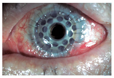
PURPOSE: To determine the clinical outcomes in patients after type 1 Boston keratoprosthesis surgery and the significance of ultrasound biomicroscopy imaging for postoperative follow-up.
METHODS: This retrospective analysis included 20 eyes of 19 patients who underwent corneal transplantation with type 1 Boston keratoprosthesis between April 2014 and December 2021. Data on patient demographics, preoperative diagnosis, visual acuity, and postoperative clinical findings were analyzed.
RESULTS: Type 1 Boston keratoprosthesis implantation resulted in intermediate- and long-term positive outcomes. However, blindness and other serious complications such as glaucoma, retroprosthetic membrane formation, endophthalmitis, or retinal detachment also occurred. The use of ultrasound biomicroscopy imaging allowed for better evaluation of the back of the titanium plate, anterior segment structures, and the relationship of the prosthesis with surrounding tissues, which provided valuable postoperative information.
CONCLUSION: Regular lifetime monitoring and treatment are necessary in patients who undergo Boston type 1 keratoprosthesis implantation for high-risk corneal transplantation. ultrasound biomicroscopy imaging can be a valuable imaging technique for the evaluation of patients with Boston type 1 keratoprosthesis, providing important information on anterior segment anatomy and potential complications. Further studies and consensus on postoperative follow-up protocols are required to optimize the management of patients with Boston type 1 keratoprosthesis.
Keywords: Boston Keratoprosthesis; Corneal transplantation; Ultrasound biomicroscopy; Anterior segment; Prostheses and implants
Arq. Bras. Oftalmol. 2026;89 (4 )
:1-7
| DOI: 10.5935/0004-2749.2024-0409
Abstract
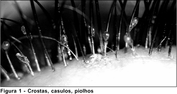
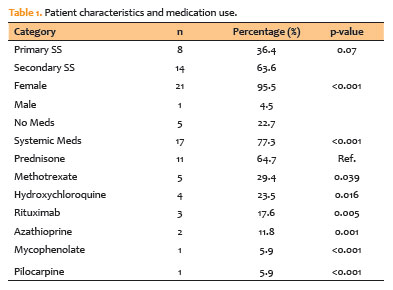
PURPOSE: The purpose of this study is to look into the relationship between tear film osmolarity, tear crystallization, and corneal esthesiometry findings in Sjögren's syndrome patients.
METHODS: This cross-sectional observational study included 43 eyes from patients with a confirmed diagnosis of Sjögren's syndrome. Tear osmolarity was measured with an iPen osmometer, tear crystallization was graded using Roland's classification, and corneal sensitivity was evaluated with a Cochet–Bonnet aesthesiometer. Ocular symptoms were assessed using the Ocular Surface Disease Index questionnaire. Patients who had undergone keratoplasty or worn contact lenses within 4 hours of testing were excluded.
RESULTS: The cohort's mean tear osmolarity was 292.5±15.0 mOsm/L (median: 293 mOsm/L, IQR: 17.5). There was no significant difference between patients with primary Sjögren's syndrome (mean: 289.4 mOsm/L) and those with secondary Sjögren's syndrome (mean: 294.5 mOsm/L; p=0.413). Tear crystallization patterns were more severe in patients with primary Sjögren's syndrome (mean: 3.25, median: 3.5, IQR: 1.25) than in those with secondary Sjögren's syndrome (mean: 3.19, median: 3.0, IQR: 1.0), though the difference was not statistically significant (p=0.87). Corneal sensitivity was reduced by 3.5±1.7 mm (median: 4.0 mm, IQR: 2.13). Tear crystallization has a significant negative correlation with corneal sensitivity (r=−0.313, p=0.041), suggesting that poorer tear quality leads to decreased corneal sensitivity.
CONCLUSION: Tear crystallization patterns and corneal sensitivity were found to be significantly correlated in Sjögren's syndrome patients. The findings also indicate that systemic medication use may affect tear film quality.
Keywords: Sjogren's syndrome, Tear crystallization, Dry eye disease, Cornea, Tears, Osmolarity
Arq. Bras. Oftalmol. 2025;88 (5 )
:1-7
| DOI: 10.5935/0004-2749.2024-0217
Abstract
PURPOSE: This study aimed to evaluate the influence of intrastromal corneal ring segment implants on the intraocular pressure measurements using Goldmann applanation tonometry, rebound tonometry, and noncontact tonometry in keratoconic corneas and analyze the intertonometer agreement.
METHODS: We enrolled 74 eyes in this observational and prospective study. Each participant had a complete eye examination, corneal analysis with Scheimpflug Tomography (Pentacam®), and intraocular pressure evaluation with Goldmann applanation tonometry, rebound tonometry, and noncontact tonometry, before and after intrastromal corneal ring segment implantation (on postoperative days 1, 7, 45, and 90). Intertonometer agreement was assessed using Bland-Altman analysis.
RESULTS: The mean age was 29.9 ± 10.2 years, and 47 (63.5%) eyes had keratoconus grade II. Intraocular pressures were higher for noncontact tonometry preoperatively and on 90 postoperative day (mean ± SD: 12.4 ± and 12.1 ± 2.2 mmHg, respectively), followed by Goldmann applanation tonometry (11.1 ± 3.0 and 11.2 ± 2.7 mmHg, respectively), and were lower for rebound tonometry (9.7 ± and 9.4 ± 3.2 mmHg, respectively). The variation from the Goldmann tonometry on 7 postoperative day to the baseline (p=0.022) and that of noncontact tonometry on 90 postoperative day to the baseline (p=0.021) were statistically significant. The rebound tonometry underestimated intraocular pressure when compared with the Goldmann applanation tonometry by a mean of 1.47 ± 5.19 mmHg. Noncontact tonometry, when compared with Goldmann applanation tonometry, overesti-mated intraocular pressure by a mean of 1.23 ± 4.15 mmHg.
CONCLUSION: Despite statistically significant differences between some postoperative periods, the intraocular pressure measurement differences may not be clinically relevant.
Keywords: Keratoconus; Intraocular pressure; Cornea; Corneal stroma; Postoperative period; Tonometry ocular; Prostheses and implants
Arq. Bras. Oftalmol. 2024;87 (5 )
:1-10
| DOI: 10.5935/0004-2749.2022-0124
Abstract
Objetivo: Avaliar o desempenho de classificação de modelos ou arquiteturas de rede neural convolucional pré-treinadas usando um conjunto de dados de imagem de fundo de olho contendo oito rótulos de doenças diferentes.
Métodos: Neste artigo, o conjunto de dados de reconhecimento inteligente de doenças oculares publicamente disponível foi usado para o diagnóstico de oito rótulos de doenças diferentes. O banco de dados de reconhecimento inteligente de doenças oculares tem um total de 10.000 imagens de fundo de olho de ambos os olhos de 5.000 pacientes para oito categorias que contêm rótulos saudáveis, retinopatia diabética, glaucoma, catarata, degeneração macular relacionada à idade, hipertensão, miopia, outros. Investigamos o desempenho da classificação de doenças oculares construindo três arquiteturas de rede neural convolucional pré-treinadas diferentes, incluindo os modelos VGG16, Inceptionv3 e ResNet50 com otimizador de Momento Adaptativo. Esses modelos foram implementados no Google Colab o que facilitou a tarefa sem gastar horas instalando o ambiente e suportando bibliotecas. Para avaliar a eficácia dos modelos, o conjunto de dados é dividido em 70% para treinamento, 10% para validação e os 20% restantes utilizados para teste. As imagens de treinamento foram expandidas para 10.000 imagens de fundo de olho para cada tal.
Resultados: Observou-se que o modelo ResNet50 alcançou acurácia de 97,1%, sensibilidade de 78,5%, especificidade de 98,5% e precisão de 79,7% e teve a melhor área sob a curva e pontuação final para classificar a categoria da catarata (área sob a curva=0,964, final=0,903). Em contraste, o modelo VGG16 alcançou uma precisão de 96,2%, sensibilidade de 56,9%, especificidade de 99,2% e precisão de 84,1%, área sob a curva 0,949 e pontuação final de 0,857.
Conclusão: Esses resultados demonstram a capacidade das arquiteturas de rede neural convolucional pré-treinadas em identificar doenças oftalmológicas a partir de imagens de fundo de olho. ResNet50 pode ser uma boa solução para resolver problemas na detecção e classificação de doenças como glaucoma, catarata, hipertensão e miopia; Inceptionv3 para degeneração macular relacionada à idade e outras doenças; e VGG16 para retinopatia normal e diabética.
Keywords: Redes neurais de computação; Aprendizado profundo; Processamento de imagem assistida por computador; VGG16; Inceptionv3; ResNet50; Fundo de olho; Oftalmopatias.
Arq. Bras. Oftalmol. 2024;87 (6 )
:1-11
| DOI: 10.5935/0004-2749.2022-0256
Abstract
OBJETIVOS: À medida que a utilização de equipamentos digitais no emprego aumenta, a avaliação do seu efeito na saúde visual necessita de ferramentas válidas e robustas. Este estudo teve como objetivo traduzir, adaptar culturalmente e validar para português o Questionário da Síndrome Visual do Computador (CVS-Q©).
MÉTODOS: O procedimento foi realizado em 5 fases: tradução direta, síntese da tradução, tradução inversa, consolidação por um painel de especialistas, e pré-teste. Para fazer o pré-teste foi realizado um estudo piloto transversal aplicado a uma amostra de 26 participantes que completaram a versão pré-final da versão portuguesa do CVS-Q©, questionando por dificuldades, compreensão e sugestões de melhoria do questionário. Para avaliar a confiança e validade da versão portuguesa do CVS-Q©foi realizado um estudo transversal de validação em uma amostra diferente (280 funcionários).
RESULTADOS: No pré-teste, 96.2% dos participantes não apresentaram dificuldades no preenchimento do questionário, enquanto 84.0% indicaram que era claro e compreensível. Obteve-se, então, o CVS-Q© em português (Questionário da Síndrome Visual do Computador, CVS-Q PT©). A sua validação revelou uma boa consistência interna
da sua escala (Cronbach's alpha=0.793), boa estabilidade temporal (coeficiente de correlação interclasse=0.847; 95% CI 0.764-0.902, kappa=0.839), sensibilidades e especificidades adequadas (78.5% e 70.7%, respetivamente), boa capacidade de discriminação (área abaixo da curva=0.832; 95% CI 0.784-0.879), e uma adequada validade da convergência com o índice de doença da superfície ocular (ocular surface disease index - OSDI; coeficiente de correlação de Spearman=0.728, p<0.001). A análise fatorial revelou um único fator responsável por explicar a variância comum em 37.7%. Um funcionário com uma pontuação ≥7 pontos sofria de síndrome visual do computador.
CONCLUSÃO: O CVS-Q PT© pode ser considerada uma ferramenta intuitiva, de fácil interpretação e com boas propriedades psicométricas para avaliar a síndrome visual do computador em funcionários portugueses expostos a ecrãs digitais. Este questionário facilitará as decisões sobre medidas preventivas, intervenções e tratamento, e a comparação entre as populações expostas em diferentes países de língua portuguesa.
Keywords: Síndrome visual do computador; Dispositivos digitais; Saúde ocular; Estudo de validação; Propriedades psicométricas; Inquéritos e questionários
Arq. Bras. Oftalmol. 2025;88 (2 )
:1-7
| DOI: 10.5935/0004-2749.2024-0029
Abstract
PURPOSE: To evaluate the effect of upper eyelid ptosis repair with Muller muscle-conjunctival resection on meibomian gland function and ocular surface parameters.
METHODS: Thirty-eight patients who underwent ptosis repair with Muller muscle-conjunctival resection were retrospectively reviewed. Meibomian gland loss, Ocular Surface Disease Index OXFORD score, meiboscore, and noninvasive keratograph break-up time were measured preoperatively and at 1st, 3rd, and 6th months postoperatively.
RESULTS: Noninvasive keratograph break-up time values decreased significantly at 1st and 3rd months postoperatively compared to the preoperative level, but were similar to the preoperative level at 6th months postoperatively (p<0.001 and p=0.628, respectively). Ocular surface disease index, OXFORD score, meibomian gland loss, and meiboscore values increased significantly in the 1st and 3rd postoperative months compared to the preoperative period, but these values decreased to preoperative levels in the 6th postoperative month (p<0.001 and p>0.05, respectively).
CONCLUSION: There is a transient deterioration in meibography findings and OSDI score in the early postoperative period after Muller muscle-conjunctival resection. Patients undergoing Muller muscle-conjunctival resection may require topical lubricants, especially in the first 3 postoperative months.
Keywords: Meibomian glands; Blepharoptosis; Preoperative period; Conjunctiva; Muscles; Eyelid diseases; Diagnostic techniques, ophthalmological
Arq. Bras. Oftalmol. 2025;88 (2 )
:1-5
| DOI: 10.5935/0004-2749.2024-0113
Abstract
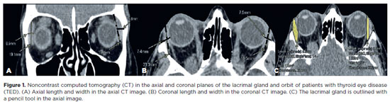
This study aimed to evaluate the morphometric and volumetric dimensions of the lacrimal gland in patients with inactive thyroid eye disease and compare them with the values reported in the literature. This case series evaluated consecutive patients with inactive thyroid eye disease treated at a tertiary eye hospital from 2015 to 2020. The patients' baseline demographics and clinical characteristics were obtained. The axial and coronal length, width, and volume of the lacrimal gland were measured on computed tomography scan images, and the results were statistically analyzed. A total of 21 patients (42 orbits) with inactive thyroid eye disease were evaluated. Their mean age was 49.0 ± 14.6 years, and 12 (57.1%) of them were men. The main complaint was dryness, and the majority of the patients had good vision and mild proptosis. The mean axial length and width of the lacrimal gland were 19.3 ± 3.9 mm and 7.5 ± 2.1 mm, respectively; coronal length and width, 20.4 ± 4.5 mm and 7.5 ± 2.1 mm, respectively; and lacrimal gland volume, 0.825 ± 0.326 mm3. Age, sex, or laterality were not found to be determinants of lacrimal gland enlargement. Patients with thyroid eye disease have enlarged lacrimal gland even in the nonactive phase of the disease multifactorial aspects influence the lacrimal gland in thyroid eye disease, making it difficult to establish a clear correlation with predisposing factors. Further studies are warranted to better understand the association between thyroid eye disease and the lacrimal gland.
Keywords: Graves' ophthalmology; Graves' disease; Lacrimal apparatus; Lacrimal apparatus diseases; X-ray computed tomography
Arq. Bras. Oftalmol. 2026;89 (3 )
:1-6
| DOI: 10.5935/0004-2749.2025-0332
Abstract
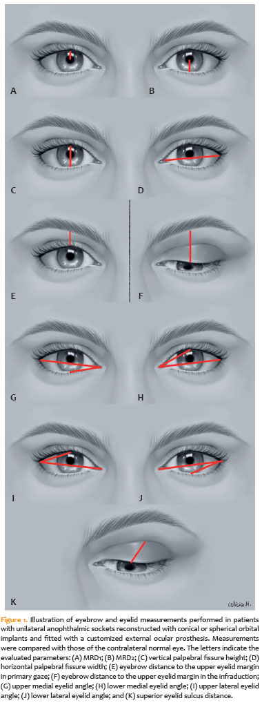
PURPOSE: To quantitatively compare eyebrow and eyelid positions in anophthalmic sockets reconstructed with conical or spherical orbital implants combined with customized external ocular prostheses.
METHODS: This cross-sectional observational study included 38 patients with unilateral anophthalmic sockets, of whom 21 received conical implants, and 17 received spherical implants. Eyelid and eyebrow parameters—including margin reflex distance 1 and 2, vertical and horizontal palpebral fissure dimensions, eyebrow-to-upper-eyelid margin distance in primary gaze and infraduction, medial and lateral eyelid angles in primary gaze, and superior eyelid sulcus depth —were quantitatively assessed using standardized digital photographs analyzed with Image J software. The contralateral healthy eye served as the control. Statistical analyses were performed to compare measurements between groups.
RESULTS: In the primary gaze position, conical and spherical implants showed comparable margin-reflex distance1, margin-reflex distance2, vertical palpebral fissure height, eyelid margin position, and medial and lateral eyelid angles. During infraduction, the upper eyelid margin was significantly lower in sockets reconstructed with conical implants. Compared with contralateral normal eyes, anophthalmic sockets exhibited a reduced horizontal palpebral fissure and a deeper superior eyelid sulcus, irrespective of implant shape.
CONCLUSION: Anophthalmic sockets reconstructed with conical or spherical implants demonstrate similar eyebrow and eyelid positioning in primary gaze. However, conical implants are associated with a lower eyelid margin during infraduction. Independent of implant format, anophthalmic sockets show a narrower horizontal palpebral fissure and increased superior sulcus depth compared with normal eyes.
Keywords: Anophthalmos; Prosthesis implantation; Anophthalmic socket; Conical implants; Spherical implants; Orbital implants; Eyelid measurements
Arq. Bras. Oftalmol. 2024;87 (3 )
:1-7
| DOI: 10.5935/0004-2749.2023-0028
Abstract
PURPOSE: Evaluation of lid contour and marginal peak point changes to compare outcomes of external levator advancement and Müller’s muscle conjunctival resection surgery in unilateral ptosis.
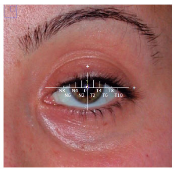
METHODS: We reviewed the charts of unilateral ptosis patients who underwent external levator advancement or Müller’s muscle conjunctival resection. Eyelid contour analysis was conducted on preoperative and 6-month postoperative digital images. This was performed with the multiple margin reflex distances technique, measuring the vertical distance from a line intersecting the center of the pupil to the eyelid margin at 10 positions at 2 mm intervals. The marginal peak point changes were analyzed digitally using the coordinates of the peak point according to the pupil center. Each position’s mean distance was compared preoperatively, postoperatively, and with the fellow eyelid.
RESULTS: Sixteen patients underwent external levator advancement and 16 patients had Müller’s muscle conjunctival resection. The mean margin reflex distance was improved by both techniques (1.46 vs. 2.43 mm and 1.12 vs. 2.25 mm, p=0.008 and p=0.0001 respectively) and approached that of the fellow eyelid (2.43 vs. 2.88 and 2.25 vs. 2.58 mm, p=0.23 and p=0.19, respectively). However, statistically significant lid margin elevation was limited to between the N6 and T6 points in the external levator advancement group. Whereas, significant elevation was achieved along the whole lid margin in the Müller’s muscle conjunctival resection group. The marginal peak point was shifted slightly laterally in the external levator advancement group (p=0.11).
CONCLUSIONS: Both techniques provide effective lid elevation, however, the external levator advancement’s effect lessens toward the canthi while Müller’s muscle conjunctival resection provides more uniform elevation across the lid margin. The margin reflex distance alone is not sufficient to reflect contour changes.
Keywords: Blepharoptosis; Eyelids; Conjunctiva; Oculomotor muscles; Image processing, computer-assisted; Treatment outcome
Arq. Bras. Oftalmol. 2024;87 (3 )
:1-5
| DOI: 10.5935/0004-2749.2023-0038
Abstract
PURPOSE: To assess the effect of the coronavirus disease 2019 (COVID-19) pandemic on cataract surgery by residents who had mandatory surgical simulator training during residency.
METHODS: In this retrospective, observational analytical study, the total number of cataract surgeries and surgical complications by all senior residents of 2019 (2019 class; prepandemic) and 2020 (2020 class; affected by the reduced number of elective surgeries due to the COVID-19 pandemic) were collected and compared. All residents had routine mandatory cataract surgery training on a virtual surgical simulator during residency. The total score obtained by these residents on cataract challenges of the surgical simulator was also evaluated.
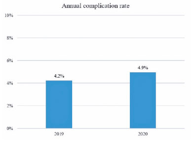
RESULTS: The 2020 and 2019 classes performed 1275 and 2561 cataract surgeries, respectively. This revealed a reduction of 50.2% in the total number of procedures performed by the 2020 class because of the pandemic. The incidence of surgical complications was not statistically different between the two groups (4.2% in the 2019 class and 4.9% in the 2020 class; p=0.314). Both groups also did not differ in their mean scores on the simulator’s cataract challenges (p<0.696).
CONCLUSION: Despite the reduction of 50.2% in the total number of cataract surgeries performed by senior residents of 2020 during the COVID-19 pandemic, the incidence of surgical complications did not increase. This suggests that surgical simulator training during residency mitigated the negative effects of the reduced surgical volume during the pandemic.
Keywords: COVID-19; Pandemics; Cataract extraction/education; Internship and residency/methods; Simulation training/methods; Phacoemulsification/education; Surgery, computer-assisted; Computer simulation; Clinical competence; Ophthalmology/education
 ABO is licensed under a Creative Commons Attribution-NonComercial 4.0 Internacional.
06-tab01tb.jpg)
07-tab01tb.jpg)

10-fig01tb.jpg)
14-tab01tb.jpg)