Arq. Bras. Oftalmol. 2022;85 (1 )
:13-18
| DOI: 10.5935/0004-2749.20220003
Abstract
Objetivo: Fornecer informações sobre a ocorrência e a eficácia do aconselhamento sobre o uso de tabaco por oftalmologistas a pacientes com doenças oculares associadas à tireoide.
Métodos: Analisamos os prontuários médicos eletrônicos de uma coorte digital de pacientes atendidos por oftalmologistas no Sistema de Saúde da Universidade da Pensilvânia entre o início de 2012 e o final de 2017 com os códigos da Classificação Internacional de Doenças (CID) para a doença de Graves, exoftalmia tireotóxica ou doença ocular associada à tireoide. Os históricos de uso de tabaco foram registrados na primeira e na última visita ao consultório de Oftalmologia, ou na visita mais próxima no tempo. A quantidade de maços/dia (mpd) e todas as anotações feitas nas visitas ao consultório de Oftalmologia foram analisadas para aconselhamento sobre o uso de tabaco.
Resultados: Um total de 435 indivíduos preencheram os critérios de inclusão, dos quais 72 (16,6%) estavam fumando ativamente no momento do primeiro encontro. Apenas 57 (79,2%) desses indivíduos que fumam ativamente registraram queixas relacionadas ao tabagismo, sendo que 34 (59,6%) deles receberam alguma forma de aconselhamento sobre o uso de tabaco. Ao todo, 9 (26,5%) indivíduos dentre os que receberam aconselhamento sobre tabaco e 1 (4,3%) que não teve aconselhamento registrado pararam de fumar (diferença de risco de 22,1%; IC 95%, [1,7%, 39,1%]; p=0,04). Dentre aqueles que receberam aconselhamento, 17 (50,0%) reduziram seus mpd, além de 7 (30,4%) daqueles que não tiveram aconselhamento (diferença de risco de 19,6%; IC 95% [-6,3%, 41,3%]; p=0,18). No geral, 14 (25,5%) dos 55 oftalmologistas que tiveram um paciente fumante ativo registraram evidências de aconselhamento sobre o uso de tabaco.
Conclusões: Os resultados deste estudo revelam tanto as oportunidades perdidas de aconselhamento sobre o uso do tabaco quanto a eficácia do aconselhamento no contexto de doenças oculares associadas à tireoide.
Keywords: Uso de tabaco; Aconselhamento; Doenças da glândula tireóide; Doença de Graves; Oftalmopatias
Arq. Bras. Oftalmol. 2022;85 (6 )
:590-598
| DOI: 10.5935/0004-2749.20220081
Abstract
Objetivo: Identificar tendências no campo de pesquisa da orbitopatia de Graves nas últimas duas décadas e analisar os ramos de maior concentração de pesquisas nessa área.
Métodos: O banco de dados Web of Science foi usado para extrair artigos com “orbitopatia de Graves” ou seus sinônimos no título. Dados completos e referências foram exportados para o programa VOSviewer para serem analisados. Mapas e gráficos de visualização foram construídos a partir desses dados.
Resultados: Foram obtidos 1067 artigos sobre a orbitopatia de Graves a partir do banco de dados Web of Science. Os EUA ficaram em primeiro lugar em termos de número de publicações, seguidos pela Itália e pela República Popular da China. Dentre os autores, os artigos de Wiersinga WM tiveram o maior número de citações. Quanto às instituições, os artigos da Universidade de Amsterdã tiveram o maior número de citações, mas a Universidade de Pisa publicou o maior número de artigos. Dentre os periódicos, a revista Thyroid publicou o maior número de artigos. A análise de coautoria mostrou quatro agrupamentos de colaboração entre países. O primeiro agrupamento engloba países europeus; o segundo engloba os EUA, Brasil, Canadá, Coreia do Sul e Taiwan. A República Popular da China compreende um agrupamento por si só. O quarto agrupamento inclui Japão, Austrália e Polônia. A análise das palavras-chave revelou cinco agrupamentos de tópicos de palavras-chave: patogênese, gerenciamento, associação, qualidade de vida e cirurgia. A análise das referências citadas em conjunto revelou cinco agrupamentos: patogênese, manejo, fatores de risco, avaliação clínica e manejo cirúrgico.
Conclusão: A pesquisa no campo da orbitopatia de Graves cresceu nos últimos vinte anos. Os tópicos com a maior concentração de pesquisas são: patogênese, gerenciamento, fatores de risco, qualidade de vida e complicações. As tendências de pesquisa mudaram nas últimas duas décadas. Ficou evidente um aumento do interesse em explorar os mecanismos e associações da orbitopatia de Graves. Observou-se uma cooperação entre países europeus neste campo de pesquisa. Os EUA estabeleceram uma cooperação internacional mais ampla que outros países. Acreditamos que mais colaboração internacional envolvendo países em desenvolvimento seria recomendável.
Keywords: Oftalmopatia de Graves; Bibliometria; Oftalmopatia de Graves; Pesquisa
Arq. Bras. Oftalmol. 2025;88 (6 )
:1-5
| DOI: 10.5935/0004-2749.2024-0321
Abstract
PURPOSE: To report the ophthalmological signs, symptoms, and clinical management observed during an unprecedented outbreak of chemical ocular injuries related to cosmetic hair ointments in Brazil.
METHODS: This descriptive, cross-sectional study reviewed medical records of patients treated at the emergency center of Fundação Altino Ventura for chemical ocular trauma associated with cosmetic hair ointment use between February 2022 and February 2023. Records with incomplete medical information were excluded.
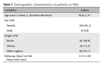
RESULTS: The study included 168 patients (95.2% [n=160] female), with a mean age of 30.8 ± 9.7 years. The most frequently reported symptoms at presentation were pain (167/168, 99.4%) and photophobia (92/168, 54.8%). Severe pain was reported by 137 patients (80%). Keratitis was present in 280 of 336 eyes (83.3%), conjunctival hyperemia in 256 eyes (76.4%), and corneal abrasions in 174 eyes (51.8%). A decrease in visual acuity (worse than 20/25) was documented in 18.5% (31/168) of cases. Lubricants, antibiotics, and re-epithelialization
ointments were prescribed to 64.8% (109/168) of the patients. Topical corticosteroids and oral vitamin C were administered to 34% (57/168) and 1.2% (2/168) of patients, respectively. Followup visits were required in 19% (33/168) of cases.
CONCLUSION: The outbreak of chemical ocular injuries linked to cosmetic ointments used for braiding and hair modeling in Brazil was marked by intense ocular pain, conjunctival hyperemia, keratitis, and corneal abrasions. Most patients were treated with lubricants, antibiotics, and re-epithelialization ointments, although approximately one-fifth required followup care, and one-third received additional treatment with either topical corticosteroids and/or oral vitamin C.
Keywords: Cosmetics; Hair preparations; Eye injuries; Burns, chemical; Eye burns; Keratitis; Cornea; Corneal diseases; Visual low.
Arq. Bras. Oftalmol. 2025;88 (4 )
:1-6
| DOI: 10.5935/0004-2749.2024-0278
Abstract
PURPOSE: This study aimed to evaluate the prevalence of orbital conditions in a tertiary ophthalmic outpatient hospital in Sao Paulo, Brazil, with a focus on the main diagnoses and their distribution.
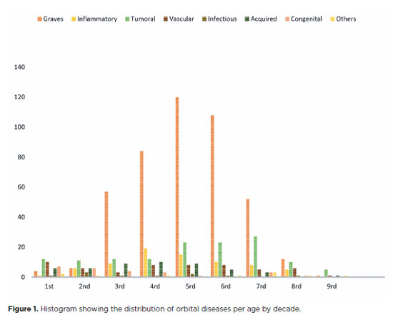
METHODS: A retrospective chart review was conducted involving patients registered and admitted to the orbital disease unit at the Department of Ophthalmology, University of São Paulo Medical School, from January 2004 to March 2018. A total of 838 medical charts were analyzed, of which 37 were excluded due to incomplete data. The remaining charts were categorized into eight diagnostic groups: Graves’ orbitopathy , inflammatory disorders, tumors, vascular lesions, acquired structural abnormalities, congenital structural abnormalities, infectious diseases, and others.
RESULTS: Of the 837,300 ophthalmological appointments, 3,372 (0.4%) were related to orbital diseases. The study included 801 patients, of whom 63.45% were women. The patients’ mean age was 42.86 years. Graves’ orbitopathy was the most common (55%), followed by tumor (17%), inflammatory disorders (9%), vascular lesions (7%), acquired structural abnormalities (5%), congenital structural abnormalities (4%), others (2%), and infectious diseases (1%). The study found significant differences in the incidence and types of orbital diseases, indicating the specialized nature of tertiary care and referral biases.
CONCLUSION: Published data on epidemiological orbital diseases is scarce. Therefore, this study focused on the diverse nature of orbital diseases and their low incidence among ophthalmology appointments. The major trends align with other epidemiological studies, demonstrating a preponderance of Graves’ orbitopathy in middle-aged adults and a bimodal distribution of tumors. These findings are essential in shaping resident training programs and healthcare policies, particularly in tertiary settings. Understanding the epidemiology of orbital diseases can improve diagnostic accuracy, treatment approaches, and patient outcomes as well as support future systemic prospective studies.
Keywords: Orbital diseases; Orbital tumors; Neoplasms; Inflammation; Graves’ ophthalmopathy; Outpatients
Arq. Bras. Oftalmol. 2025;88 (6 )
:1-6
| DOI: 10.5935/0004-2749.2025-0153
Abstract
PURPOSE: This clinical study aimed to assess the effectiveness of microemulsion artificial tears containing povidone and propylene glycol in the management of dry eye disease. Secondary objectives included evaluating improvements in tear-film stability, measured by tear break-up time and corneal staining scores, along with the tolerability and safety of the formulation.
METHODS: This was a prospective, single-arm interventional study involving 30 participants (52 eyes) diagnosed with dry eye disease. Participants self-administered the investigational eye drops twice daily for 28 consecutive days. Primary and secondary outcomes included changes in the Ocular Surface Disease Index, tear break-up time, and corneal staining scores. Adverse events were documented throughout the study period.
RESULTS: Significant improvements in Ocular Surface Disease Index scores were observed, reflecting a reduction in dry eye disease symptoms. Tear break-up time increased notably between baseline and follow-up assessments, with the proportion of eyes exhibiting tear break-up time ≥10 srising from 25.0% to 63.5%. Additionally, the percentage of eyes with a corneal staining score of zero improved from 23.1% to 69.2%. Conjunctival staining also decreased, with the proportion of eyes with scores of 2 and 3 dropping from 11.5% to 3.8% and 5.8% to 0%, respectively.
CONCLUSIONS: The findings suggest that povidone and propylene glycol-based artificial tears significantly enhance tear-film stability and alleviate symptoms in patients with mild to moderate dry eye disease, with minimal adverse effects. This formulation represents a safe and effective short-term treatment option for dry eye disease management.
Keywords: Artificial tears; Dry eye disease; Tear-film stability; Propylene glycol; Povidone; Visual acuity; Surveys and questionnaires
Arq. Bras. Oftalmol. 2025;88 (6 )
:1-8
| DOI: 10.5935/0004-2749.2025-0118
Abstract
PURPOSE: Using advanced imaging techniques, this study aimed to evaluate corneal stability, epithelial remodeling, and tear film changes over a one-year period in first-time soft-contact lens wearers.
METHODS: A retrospective study was conducted on 100 eyes of 50 first-time daily soft-contact lens users aged 21–65 years with no prior rigid gas-permeable lens wear. The Sirius Scheimpflug imaging system was used to assess corneal topography, epithelial thickness, and non-invasive tear break-up time at baseline, 3, 6, and 12 months. Corneal warpage was evaluated using symmetry indices and Baiocchi Calossi Versaci indices. We performed statistical analysis using repeated-measures analyses of variance with Greenhouse-Geisser correction.
RESULTS: The mean baseline central corneal thickness was 537.83 (±7.92) µm, with no significant thinning after one year. The average simulated keratometry values remained stable, indicating no progressive corneal steepening or flattening. There were no significant changes in warpage indices over time, suggesting corneal shape preservation. Higher-order aberrations (coma, trefoil, and spherical aberrations) and non-invasive tear break-up time remained unchanged throughout the study period.
CONCLUSIONS: Modern silicone hydrogel soft-contact lenses do not induce significant corneal warpage, epithelial remodeling, or optical aberrations over a one-year period. We found that corneal morphology and tear film stability were preserved, supporting the safety of soft-contact lens use. These findings provide clinically relevant insights into the long-term impact of contact lens wear. They may facilitate improved lens fitting strategies and preoperative refractive surgery assessments.
Keywords: Contact lenses, hydrophilic; Cornea/surgery; Corneal diseases; Corneal topography; Adaptation, ocular/physiology; Endothelium, corneal/pathology; Refractive errors; Tears/metabolism.
Arq. Bras. Oftalmol. 2026;89 (4 )
:1-7
| DOI: 10.5935/0004-2749.2024-0409
Abstract
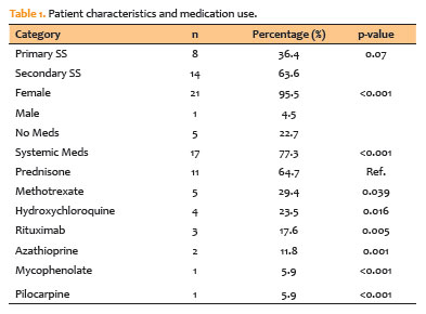
PURPOSE: The purpose of this study is to look into the relationship between tear film osmolarity, tear crystallization, and corneal esthesiometry findings in Sjögren's syndrome patients.
METHODS: This cross-sectional observational study included 43 eyes from patients with a confirmed diagnosis of Sjögren's syndrome. Tear osmolarity was measured with an iPen osmometer, tear crystallization was graded using Roland's classification, and corneal sensitivity was evaluated with a Cochet–Bonnet aesthesiometer. Ocular symptoms were assessed using the Ocular Surface Disease Index questionnaire. Patients who had undergone keratoplasty or worn contact lenses within 4 hours of testing were excluded.
RESULTS: The cohort's mean tear osmolarity was 292.5±15.0 mOsm/L (median: 293 mOsm/L, IQR: 17.5). There was no significant difference between patients with primary Sjögren's syndrome (mean: 289.4 mOsm/L) and those with secondary Sjögren's syndrome (mean: 294.5 mOsm/L; p=0.413). Tear crystallization patterns were more severe in patients with primary Sjögren's syndrome (mean: 3.25, median: 3.5, IQR: 1.25) than in those with secondary Sjögren's syndrome (mean: 3.19, median: 3.0, IQR: 1.0), though the difference was not statistically significant (p=0.87). Corneal sensitivity was reduced by 3.5±1.7 mm (median: 4.0 mm, IQR: 2.13). Tear crystallization has a significant negative correlation with corneal sensitivity (r=−0.313, p=0.041), suggesting that poorer tear quality leads to decreased corneal sensitivity.
CONCLUSION: Tear crystallization patterns and corneal sensitivity were found to be significantly correlated in Sjögren's syndrome patients. The findings also indicate that systemic medication use may affect tear film quality.
Keywords: Sjogren's syndrome, Tear crystallization, Dry eye disease, Cornea, Tears, Osmolarity
Arq. Bras. Oftalmol. 2025;88 (5 )
:1-7
| DOI: 10.5935/0004-2749.2024-0202
Abstract
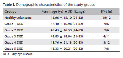
PURPOSE: This study aimed to evaluate the relationship between the objective severity of dry eye disease subjective symptoms, and corneal sensitivity.
METHODS: The study included 62 eyes from 31 healthy volunteers and 150 eyes from 75 patients diagnosed with dry eye disease . Participants underwent the Schirmer I test, tear break-up time assessment, and corneal staining evaluation using the Oxford Scale. Subjective symptoms were assessed through the Ocular Surface Disease Index questionnaire and a modified Ocular Surface Pain Score questionnaire. Corneal sensitivity was measured in five corneal regions using a Cochet-Bonnet esthesiometer. Dry eye disease severity was graded from 1 to 5 based on the Oxford Scale. Comparative analyses were performed.
RESULTS: Schirmer I and tear break-up time values were significantly lower in the DED group, while Ocular Surface Disease Index and Ocular Surface Pain Score were significantly higher (p<0.001 for all). Corneal sensitivity in all quadrants was significantly lower in DED patients (p<0.001 for all). A strong correlation was observed between the Ocular Surface Pain Score and the Ocular Surface Disease Index (r=0.983, p<0.001). Central corneal sensitivity exhibited a moderate positive correlation with Schirmer I and tear break-up time (p<0.001, r=0.583 and 0.657, respectively) and a moderate negative correlation with Ocular Surface Disease Index and Ocular Surface Pain Score (p<0.001, r=0.625 and −0.631, respectively). Disease severity progression was associated with increased Ocular Surface Disease Index and Ocular Surface Pain Score, but no statistically significant difference was found between Grades 3 and 5. Similarly, corneal sensitivity decreased with advancing disease severity, yet no significant difference was observed between Grades 4 and 5.
CONCLUSION: Corneal sensitivity decreases in dry eye disease and is negatively correlated with disease severity. Subjective symptoms increase with disease progression and show a positive correlation with severity. The absence of significant differences between the advanced stages suggests that neuropathic mechanisms and subbasal nerve plexus deterioration play a role in chronic and late-stage dry eye disease.
Keywords: dry eye disease; signs and symptoms; cornea; neuralgia; Cochet-Bonnet esthesiometer; sensory thresholds; surveys and questionnaires
Arq. Bras. Oftalmol. 2025;88 (5 )
:1-7
| DOI: 10.5935/0004-2749.2024-0230
Abstract
PURPOSE: This pilot study was conducted to investigate the presence of various bioactive compounds (copeptin, asprosin, and salusins) in the blood and tears of patients with glaucoma.
METHODS: A total of 83 subjects, including 28 patients with open-angle glaucoma, 28 patients with ocular hypertension, and 27 control volunteers, were enrolled in this study. The levels of salusin-α, salusin-β, copeptin, and asprosin in tears and venous blood samples were measured by enzyme linked immunosorbent assay (ELISA).
RESULTS: Patients with open-angle glaucoma and those with ocular hypertension showed statistically significantly decreased levels of salusin-α and salusin-β in their blood and tears compared with those of control subjects (p<0.05), with the decrease being the most pronounced in patients with ocular hypertension (p<0.05). In contrast, the levels of copeptin and asprosin showed a statistically significant increase in both these patient groups compared with those of control subjects (p<0.05). There was a negative correlation between intraocular pressure and blood and tear salusins.
CONCLUSIONS: Fluids from patients with open-angle glaucoma and ocular hypertension showed lower salusin levels. Patients with ocular hypertension had higher levels of copeptin and asprosin, but not those with open-angle glaucoma (except for asprosin, whose levels showed a slight but remarkable increase in plasma in patients with open-angle glaucoma). The pathogenesis of ocular hypertension and open-angle glaucoma may be significantly impacted by these biomarkers.
Keywords: Glaucoma, open-angle/physiopathology; Intraocular pressure/physiopathology; Retinal ganglion cells/pathology; Biomarkers/blood; Glycopeptides; Fibrillin-1; Tears/chemistry; Intercellular signaling peptides and proteins/blood; Enzyme-linked immunosorbent a
Arq. Bras. Oftalmol. 2025;88 (2 )
:1-5
| DOI: 10.5935/0004-2749.2024-0113
Abstract
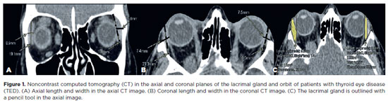
This study aimed to evaluate the morphometric and volumetric dimensions of the lacrimal gland in patients with inactive thyroid eye disease and compare them with the values reported in the literature. This case series evaluated consecutive patients with inactive thyroid eye disease treated at a tertiary eye hospital from 2015 to 2020. The patients' baseline demographics and clinical characteristics were obtained. The axial and coronal length, width, and volume of the lacrimal gland were measured on computed tomography scan images, and the results were statistically analyzed. A total of 21 patients (42 orbits) with inactive thyroid eye disease were evaluated. Their mean age was 49.0 ± 14.6 years, and 12 (57.1%) of them were men. The main complaint was dryness, and the majority of the patients had good vision and mild proptosis. The mean axial length and width of the lacrimal gland were 19.3 ± 3.9 mm and 7.5 ± 2.1 mm, respectively; coronal length and width, 20.4 ± 4.5 mm and 7.5 ± 2.1 mm, respectively; and lacrimal gland volume, 0.825 ± 0.326 mm3. Age, sex, or laterality were not found to be determinants of lacrimal gland enlargement. Patients with thyroid eye disease have enlarged lacrimal gland even in the nonactive phase of the disease multifactorial aspects influence the lacrimal gland in thyroid eye disease, making it difficult to establish a clear correlation with predisposing factors. Further studies are warranted to better understand the association between thyroid eye disease and the lacrimal gland.
Keywords: Graves' ophthalmology; Graves' disease; Lacrimal apparatus; Lacrimal apparatus diseases; X-ray computed tomography
Arq. Bras. Oftalmol. 2024;87 (5 )
:1-7
| DOI: 10.5935/0004-2749.2023-0296
Abstract
PURPOSE: To compare inferomedial wall orbital decompression to balanced medial plus lateral wall orbital decompression in patients with Graves’ orbitopathy in the inactive phase with regard to exophthalmos reduction and the effects on quality of life.
METHODS: Forty-two patients with inactive Graves’ orbitopathy were randomly divided into two groups and submitted to one of two orbital decompression techniques: inferomedial wall orbital decompression or medial plus lateral wall orbital decompression. Preoperative and postoperative assessments included Hertel’s exophthalmometry and a validated Graves’ orbitopathy quality of life questionnaire. The results of the two groups were compared.
RESULTS: Compared to preoperative measurement, exophthalmos reduction was statistically significant in both groups (p<0.001) but more so in patients undergoing medial plus lateral wall orbital decompression (p=0.010). Neither orbital decompression techniques increased the visual functioning subscale score on the Graves’ orbitopathy quality of life questionnaire (inferomedial wall orbital decompression p=0.362 and medial plus lateral wall orbital decompression p=0.727), but a statistically significant difference was observed in the score of the appearance subscale in patients submitted to medial plus lateral wall orbital decompression (p=0.006).
CONCLUSIONS: Inferomedial wall orbital decompression is a good alternative for patients who do not require large exophthalmos reduction. However, medial plus lateral wall orbital decompression offers greater exophthalmos reduction and greater improvement in appearance (higher Graves’ orbitopathy quality of life questionnaire scores), making it a suitable option for esthetic-functional rehabilitation.
Keywords: Graves’ ophthalmopathy; Quality of life; Exophthalmos; Strabismus; Diplopia; Decompression, surgical
 ABO is licensed under a Creative Commons Attribution-NonComercial 4.0 Internacional.
06-tab01tb.jpg)
06-fig01.jpg)


03-fig01.jpg)
02-fig01.jpg)
03-fig01.jpg)