Arq. Bras. Oftalmol. 2026;89 (2 )
:1-8
| DOI: 10.5935/0004-2749.2025-0105
Abstract
PURPOSE: To evaluate structural differences in amniotic membrane fragments subjected to different preservation techniques for potential ophthalmologic applications.
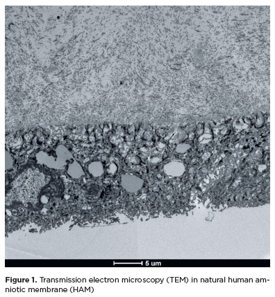
METHODS: Three placentas were collected from healthy donors, and four amniotic membrane fragments were prepared from each placenta. The fragments were divided into four groups with three samples each: cryopreserved, lyophilized, vacuum-dried using a vacuum concentrator, and fresh (control). After processing, the fragments were fixed, sectioned, and analyzed using scanning transmission electron microscopy to assess tissue morphology.
RESULTS: All samples met the established evaluation criteria. No morphological differences were observed among the groups. The structural characteristics of lyophilized and vacuum-dried membranes were comparable with those of cryopreserved and fresh membranes. However, vacuum drying demonstrated the greatest practicality for ophthalmologic use, as it allows membrane availability at any time and storage at room temperature.
CONCLUSION: Vacuum drying using a vacuum concentrator, lyophilization, and cryopreservation preserve the morphological characteristics of the human amniotic membrane similar to those of fresh tissue. A standardized protocol using a vacuum concentrator may be established owing to its advantages in storage convenience and accessibility.
Keywords: Amnion/transplantation; Cryopreservation/methods; Freeze drying; Lyophilization; Ophthalmologic surgical procedures; Regenerative medicine
Arq. Bras. Oftalmol. 2020;83 (4 )
:1-6
| DOI: 10.5935/0004-2749.20200053
Abstract
Objetivo: Determinar a frequência de neoplasia escamosa da superfície ocular associada ao pterígio com apresentação clínica, em um centro de referência em Oftalmologia da região central do México.
Métodos: Revisamos os laudos histopatológicos e as lâminas de biópsia de todos os pacientes que foram submetidos à cirurgia de pterígio de 2014 a 2016 no Instituto Mexicano de Oftalmologia, na cidade de Querétaro.
Resultados: Estudamos 177 amostras de biópsia; 66% eram de pacientes do sexo feminino, sendo a mediana da idade de 52 anos. Encontramos neoplasia escamosa da superfície ocular em 11,29% (n=20). Uma amostra de biópsia mostrou um carcinoma queratinizante infiltrativo pouco diferenciado.
Conclusões: A prevalência da neoplasia escamosa da superfície ocular nessa região parece ser maior do que a indicada por outras pesquisas. Mais estudos de âmbito nacional são necessários para determinar a verdadeira prevalência da neoplasia escamosa da superfície ocular no México e examinar os fatores de risco relacionados.
Keywords: Pterígio; Neoplasias da túnica conjuntiva; Neoplasias oculares; Histopatologia; Carcinoma de células escamosas
Arq. Bras. Oftalmol. 2023;86 (4 )
:365-371
| DOI: 10.5935/0004-2749.20230043
Abstract
Objetivo: Avaliar as alterações da superfície ocular em pacientes com Rosácea, e comparar com grupo controle.
Métodos: Noventa e três indivíduos foram selecionados para este estudo transversal, observacional e não intervencionista, dividido em dois grupos: rosácea (n=40) e controles (n=53). Foram avaliados parâmetros objetivos da superfície ocular (hiperemia conjuntival, estabilidade e volume do filme lacrimal, disfunção da glândula meibomiana, doença do olho seco, coloração da superfície ocular) e comparado indivíduos saudáveis com pacientes com rosácea.
Resultados: 69,23% dos indivíduos com rosácea eram mulheres, com média de idade de 47,34 ± 12,62 anos. Em comparação com controles pareados, não foram evidenciadas diferenças estatisticamente significativas em relação à acuidade visual (p=0,987) e parâmetros do filme lacrimal (altura do menisco lacrimal (p=0,338), tempo de ruptura do filme lacrimal não invasivo (p=0,228), tempo invasivo de ruptura (p=0,471) e teste de Schirmer (p=0,244), bem como hiperemia conjuntival (p=0,106) e coloração com fluoresceína (p=0,489). Associação significativa foi encontrada na avaliação da meibografia (p=0,026), integridade da camada mucosa (p=0,015) e sintomas de superfície ocular (p<0,0001). Pacientes com rosácea também apresentaram alterações importantes na borda palpebral: expressibilidade glandular (p<0,001), padrão de secreção glandular (p<0,001) e telangiectasia (p<0,001).
Conclusão: A disfunção da glândula de Meibômio está frequentemente associada a condições dermatológicas e é caracterizada por achados morfológicos na meibografia, bem como comprometimento da secreção lipídica que leva ao olho seco evaporativo e alterações da superfície ocular e inflamação.
Keywords: Rosácea/complicações; Disfunção da glândula tarsal; Túnica conjuntiva; Síndromes do olho seco; Técnicas de diagnóstico oftalmológico.
Arq. Bras. Oftalmol. 2025;88 (4 )
:1-8
| DOI: 10.5935/0004-2749.2024-0151
Abstract
PURPOSE: To compare the incidence rates of complications following pediatric cataract surgery between the limbal and pars plana approaches.
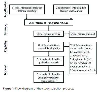
METHODS: PubMed, EMBASE, Web of Science, Scopus, Cochrane Library, and ClinicalTrials.gov were systematically searched for studies comparing the two surgical approaches. We pooled the incidence rates of postoperative complications using a random-effects model.
RESULTS: Seven studies comprising 375 eyes from 260 patients were included. No significant differences in complication rates were observed between the limbal and pars plana approaches. The pooled incidence rates (95% confidence Interval) of postoperative visual axis opacity (VAO), VAO treated with laser or surgery, secondary glaucoma, wound leakage, corneal edema, anterior chamber reaction, posterior iris synechiae, capsular phimosis, intraocular lens dislocation, posterior capsular rupture, and intravitreal lens fragmentation were 4.7% (0.8%10.8%), 3.9% (1.0%-8.1%) , 2.8% (0%-11.4%), 0 (0%-1.3%), 2.9% (0%-11.8%), 5.6% (0.1%-16.5%), 2.4% (0%-8.5%), 3.8% (0.6%-8.9%), 2.2% (0%-6.4%), 9.2% (4.1%-15.8%) and 1.3% (0%-6.3%), respectively. Both surgical approaches demonstrated improved visual acuity postoperatively.
CONCLUSIONS: Pediatric cataract surgery, performed via the limbal or pars plana approach, is effective and safe, with a low incidence of complications when conducted by trained surgeons. Neither method demonstrated a significant difference in the visual acuity improvement or complication rates.
Keywords: Pediatric cataract surgery; Postoperative complications; Limbal route; Pars plana routes; Meta-analysis
Arq. Bras. Oftalmol. 2026;89 (1 )
:1-6
| DOI: 10.5935/0004-2749.2025-0052
Abstract
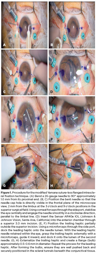
PURPOSE: To evaluate whether two simplified modifications of flanged intrascleral fixation techniques (Yamane and Canabrava) provide comparable refractive outcomes and complication rates while reducing surgical complexity in trocar-assisted vitrectomy.
METHODS: This retrospective observational study included 88 patients who underwent flanged fixation surgery with vitrectomy. In the modified Yamane technique, a single-path sclerotomy with bilateral symmetry was performed instead of an angled sclerotomy. In the modified Canabrava technique, the intraocular lens was inserted first, followed by the creation of a circular polypropylene loop with 2-mm flange spacing. Postoperative refractive parameters, including intraocular lens astigmatism, and complications such as intraocular lens iris capture were analyzed.
RESULTS: Of the 88 patients, 70 underwent the modified Yamane technique, and 18 underwent the modified Canabrava technique. No significant differences were observed between the two techniques regarding refractive outcomes or postoperative complications, except for surgical duration, which was significantly shorter (p<0.001) in one technique. Mean intraocular lens astigmatism was −0.675 D for Yamane and −0.666 D for Canabrava.
CONCLUSION: Optimizing needle engagement for symmetry in the Yamane technique and narrowing flange spacing while ensuring a circular polypropylene configuration in the Canabrava technique may reduce surgical complexity and improve postoperative outcomes.
Keywords: Polypropylenes; Yamane technique; Vitrectomy; Astigmatism; Lenses, intraocular; Postoperative complications; Suture techniques; Iris.
Arq. Bras. Oftalmol. 2025;88 (6 )
:1-5
| DOI: 10.5935/0004-2749.2024-0340
Abstract
PURPOSE: This study aimed to report the surgical outcomes and success predictors of micropulse transscleral cyclophotocoagulation in eyes with refractory glaucoma.
METHODS: This was a noncomparative, interventional case series. Patients with refractory glaucomas, defined as eyes with prior incisional glaucoma surgery failure and uncontrolled intraocular pressure, who underwent micropulse transscleral cyclophotocoagulation between March 2017 and June 2021 were enrolled. A minimum follow-up period of 6 months was required. Preoperative and postoperative intraocular pressure, number of hypotensive medications, surgical complications, and any subsequent related events were recorded. Success criteria were as follows: 1) intraocular pressure reduction ≥20% and intraocular pressure ≤18 mmHg; 2) intraocular pressure reduction ≥30% and intraocular pressure ≤15 mmHg. The need for topical hypotensive medications was not considered a failure.
RESULTS: Seventy-nine (79) eyes (79 patients; mean age, 57.5 ± 20.6 years) were included. Overall, the median follow-up duration was 12.0 (interquartile interval, 6–24) months, and the mean intraocular pressure was reduced from 22.8 ± 6.8 mmHg to 15.5 ± 5.6 mmHg at the last follow-up visit (p<0.001). The mean number of medications was reduced from 2.8 ± 0.7 to 2.0 ± 1.0 (p<0.01). At 12 months postoperatively, the success rates for criteria 1 and 2 were 54.9% and 49.7%, respectively. Aside from one case of corneal ulcer, which fully resolved with clinical treatment, and two cases of persistent hypotony (with no visual acuity loss during follow-up), no other vision-threatening complications were observed during the postoperative period. The magnitude of intraocular pressure reduction at 1 month (adjusted to preoperative intraocular pressure; HR=1.01; p=0.002).
CONCLUSION: Our findings suggest that micropulse transscleral cyclophotocoagulation is a relatively effective alternative for managing refractory glaucomas, with minor postoperative complications. In addition, the initial intraocular pressure reduction was a statistically significant predictor of 1-year success in patients undergoing micropulse transscleral cyclophotocoagulation.
Keywords: Intraocular pressure/physiology; Glaucoma, open-angle/surgery; Trabeculectomy; Laser coagulation/methods; Tonometry, ocular/methods; Postoperative complications; Antihypertensive agents/therapeutic use.
Arq. Bras. Oftalmol. 2024;87 (3 )
:1-7
| DOI: 10.5935/0004-2749.2021-0514
Abstract
Objetivo: Comparar os achados oculares em longo prazo de crianças que se submeteram à cirurgia de catarata congênita antes dos dois anos de idade e receberam uma injeção intracameral de triancinolona no intraoperatório ou usaram prednisolona oral no pós-operatório para modular a inflamação ocular.
Métodos: Neste estudo prospectivo de coorte, todos os pacientes que participaram de um ensaio clínico anterior, que analisou os resultados cirúrgicos de 1 ano da cirurgia de catarata congênita usando triancinolona intracameral (Grupo de Estudo) ou prednisolona oral (Grupo Controle), eram elegíveis para participar. Os prontuários médicos dos pacientes foram revisados e as crianças foram submetidas a um exame oftalmológico completo no acompanhamento final. As principais medidas de desfecho foram: achados biomicroscópicos, pressão intraocular, espessura central da córnea, a necessidade de intervenções cirúrgicas adicionais e achados compatíveis com glaucoma.
Resultados: Vinte e seis olhos (26 pacientes) foram incluídos (Grupo de Estudo = 11 olhos; Grupo de Controle = 15 olhos). O seguimento médio foi de 8,2 ± 1,2 anos e 8,1 ± 1,7 anos nos Grupos de Estudo e Controle, respectivamente (p=0,82). Todos os olhos apresentavam lente intraocular centrada. Não houve diferença estatisticamente significativa entre os grupos com relação à presença de sinéquia posterior (p=0,56), pressão intraocular (p=0,49) ou espessura central da córnea (p=0,21). Nenhum dos olhos preencheu os critérios diagnósticos para glaucoma, apresentou opacificação secundária do eixo visual ou foi reoperado.
Conclusão: Os achados oculares em longo prazo de crianças que se submeteram à cirurgia de catarata congênita e receberam uma injeção intracameral de triancinolona no intraoperatório foram semelhantes aos que usaram prednisolona oral no pós-operatório para modular a inflamação ocular, sugerindo que a triancinolona intracameral pode substituir a prednisolona oral na cirurgia de catarata congênita, facilitando o tratamento pós-operatório e a adesão ao mesmo.
Keywords: Catarata congênita; Triancinolona; Prednisolona; Esteroides; Complicações pós-operatórias; Criança
Arq. Bras. Oftalmol. 2026;89 (4 )
:1-6
| DOI: 10.5935/0004-2749.2025-0373
Abstract
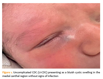
PURPOSE: The purpose of this study was to assess the clinical factors associated with dacryocystitis and the need for surgical intervention in infants with congenital dacryocystocele.
METHODS: This retrospective study included 26 infants diagnosed with congenital dacryocystocele and divided them into two groups: complicated-congenital dacryocystocele (with dacryocystitis or preseptal cellulitis) and uncomplicated-congenital dacryocystocele (without infection). Demographic and perinatal characteristics such as age at diagnosis, gender, birth weight, gestational age, delivery method, and laterality were compared between the groups. For an uncomplicated-congenital dacryocystocele, treatment included conservative management and intravenous antibiotics, followed by probing with intraoperative nasal endoscopy and endonasal marsupialization for a complicated-congenital dacryocystocele.
RESULTS: Of the 26 infants, 14 (53.8%) had complicated-congenital dacryocystocele, while 12 (46.2%) had uncomplicated-congenital dacryocystocele. There were no significant differences between the groups in terms of demographic or perinatal characteristics (p>0.05). Surgical intervention was necessary for all complicated-congenital dacryocystocele cases (100%) and two uncomplicated-congenital dacryocystocele cases (16.7%; p<0.001). During a 6-month median follow-up period, all patients demonstrated complete clinical recovery with no intraoperative complications.
CONCLUSION: In conclusion, approximately half of infants with congenital dacryocystocele developed infection-related complications. While perinatal factors were similar across groups, infectious presentation was linked to the need for surgical intervention. These findings suggest that early detection, prompt conservative management, and close follow-up can help reduce the risk of dacryocystitis and the need for surgery.
Keywords: Dacryocystocele; Dacryocystitis; Lacrimal duct obstruction; Marsupialization; Postoperative complication; Gestational age
Arq. Bras. Oftalmol. 2025;88 (5 )
:1-8
| DOI: 10.5935/0004-2749.2024-0328
Abstract
PURPOSE: Posterior capsule rupture is defined as an intraoperative posterior capsule tear resulting in vitreous loss. This study aimed to analyze the clinical characteristics, preoperative risk factors, intraoperative management strategies, and postoperative complications associated with posterior capsule rupture during phacoemulsification surgery.
METHODS: This was a retrospective observational cohort study of the medical records for 25,224 phacoemulsification surgeries performed at our tertiary eye care center between 2017 and 2022. We collected and collated the demographic characteristics and clinical findings of the patients in our cohort. Intraoperative management strategies and postoperative outcomes over a 1-year followup period were also recorded.
RESULTS: Posterior capsule rupture occurred in 351 eyes (351 patients), giving an overall posterior capsule rupture rate of 1.3%. The mean patient age was 68.6 ± 10.8 years. Pseudoexfoliation syndrome, mature cataracts, brown cataracts, and surgery performed by a resident were identified as risk factors for posterior capsule rupture (p<0.05 for each; the risk ratios were 2.70, 2.15, 2.44, 1.34, respectively). The most common intraoperative complications were dislocated lens fragments in the vitreous (8%) and iris damage (7.1%). The mean best-corrected visual acuity improved from 1.31 ± 0.84 (logMAR) postoperatively to 0.51 ± 0.56 at the end of the 1-year follow-up period (p<0.001). Corneal edema (55.6%) and elevated intraocular pressure (33.3%) were the most common early postoperative complications. Persistently elevated intraocular pressure (11.1%) and cystoid macular edema (5.1%) were the most common late postoperative complications.
CONCLUSION: Posterior capsule rupture is a common complication of phacoemulsification surgery that requires prolonged postoperative follow-up and a multidisciplinary approach. Despite the increased incidence of complications when rupture occurs, appropriate intraoperative and postoperative management can lead to satisfactory visual outcomes.
Keywords: Cataract extraction; Phacoemulsification; Posterior capsule rupture; Corneal edema; Risk factors; Postoperative complications; Intraoperative complications
Arq. Bras. Oftalmol. 2024;87 (2 )
:1-8
| DOI: 10.5935/0004-2749.2022-0328
Abstract
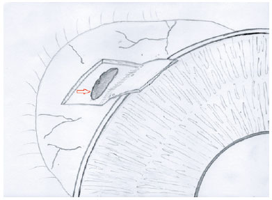
PURPOSE: Wet bio-amniotic membrane plugging combined with transplantation is a novel option that combined amniotic membrane plugging with amniotic membrane transplantation for the treatment of small corneal perforations. This study aimed to evaluate the efficacy of wet bio-amniotic membrane plugging in the treatment of small corneal perforations and compared it with that of the penetrating keratoplasty procedure.
METHODS: Forty patients (41 eyes) with small corneal perforations <3 mm in diameter treated at our hospital between July 2018 and January 2021 were retrospectively included. Among them, 21 eyes were treated with wet bio-amniotic membrane plugging (wet bio-amniotic membrane plugging group), and 20 eyes were treated with penetrating keratoplasty procedure (penetrating keratoplasty procedure group). The best-corrected visual acuity, anterior chamber formation, corneal thickness, primary disease control, postoperative complications, and graft survival rate were assessed.
RESULTS: No significant difference in baseline characteristics was found between the wet bio-amniotic membrane plugging and penetrating keratoplasty procedure groups (p>0.05). The postoperative control rates of primary diseases in the wet bio-amniotic membrane plugging and penetrating keratoplasty procedure groups were 95.2% and 90.0%, respectively (p=0.481). Visual acuity was improved 6 months after the operation in the wet bio-amniotic membrane plugging group and was improved at postoperative 1 month in the penetrating keratoplasty procedure group. The formation time of the anterior chamber in the wet bio-amniotic membrane plugging group was significantly shorter than that in the penetrating keratoplasty procedure group (p=0.023). The corneal thickness of the two groups significantly increased 12 months after the operation; however, the degree of thickening in the penetrating keratoplasty procedure group was higher than that in the wet bio-amniotic membrane plugging group (p<0.001). During the follow-up, postoperative complications were not different between the two groups (p>0.999).
CONCLUSION: The results suggest that wet bio-amniotic membrane plugging is effective and safe in the treatment of small corneal perforations. Thus, it can be used as an emergency treatment alternative to penetrating keratoplasty procedure for small corneal perforations.
Keywords: Amnion; Transplantation; Amniotic membrane; Keratoplasty, penetrating; Corneal perforation; Wet bio-amniotic membrane plugging; Wet bio-amniotic membrane transplantation
Arq. Bras. Oftalmol. 2024;87 (2 )
:1-6
| DOI: 10.5935/0004-2749.2023-2022-0341
Abstract
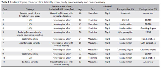
PURPOSE: To evaluate the clinical results of cryopreserved amniotic membrane transplantation as a treatment option for refractory neurotrophic corneal ulcers.
METHODS: This prospective study included 11 eyes of 11 patients who underwent amniotic membrane transplantation for the treatment of refractory neurotrophic corneal ulcers at Hospital de Clínicas da Universidade Federal do Paraná, in the city of Curitiba, from May 2015 to July 2021. Patients underwent different surgical techniques in which the amniotic membrane was applied with the epithelium facing upward to promote corneal re-epithelialization.
RESULTS: The median age of the patients was 60 years (range, 34-82 years), and 64% were men. The predominant etiology of corneal ulcers was herpes zoster (45% of cases). Approximately one-third of the patients (27%) were chronically using hypotensive eye drops, and more than half (54%) had previously undergone penetrating corneal transplantation. At the time of amniotic membrane transplantation, 18% of the eyes had corneal melting, 9% had corneal perforation, and the others had corneal ulceration without other associated complications (73%). The time between clinical diagnosis and surgical treatment ranged from 9 days to 2 years. The corrected visual acuity was worse than 20/400 in 90% of the patients preoperatively, with improvement in 36% after 3 months of the procedure, worsening in 18% and remaining stable in 36%. Of the patients, 81% complained of preoperative pain, and 66% of them reported total symptom relief after the surgical procedure. In one month, 54.6% of the patients presented a closure of epithelial defect, and half of the total group evolved with corneal thinning. The failure rate was 45.5% of the cases.
CONCLUSION: Cryopreserved amniotic membrane transplantation can be considered a good alternative for treating refractory neurotrophic corneal ulcers, as it resulted in significant improvement in pain (66%) and complete epithelial closure (60%) in many patients at 1 month postoperatively. Notably, the high failure rate highlights the need for further studies to identify patient- and ulcer-related factors that may influence the outcomes of this procedure.
Keywords: Amnion/transplantation; Corneal ulcer; Anterior eye segment; Keratitis
 ABO is licensed under a Creative Commons Attribution-NonComercial 4.0 Internacional.
12-fig01.jpg)
12-tab01.jpg)

12-fig01tb.jpg)


10-fig01.jpg)
02-fig01.jpg)
06-fig01.jpg)