Showing of 1 until 15 from 197 result(s)
Search for: Astigmatism; Eye movements; Posture; Rotation; Refractive errors; Reflex, vestibulo-ocular

Abstract
Objetivo: Estudar os custos de correção dos vícios de refração em grupos de pessoas de distinto poder aquisitivo. Métodos: Os autores estudaram cinqüenta pacientes portadores de vícios de refração. Estes foram separados em dois grupos: grupo I com pacientes escolhidos de forma aleatória na primeira consulta ao ambulatório de Oftalmologia do Hospital Evangélico de Curitiba (HUEC), e grupo II com voluntários médicos do HUEC e acadêmicos de medicina da Faculdade Evangélica de Medicina do Paraná (FEMPAR). Foram analisados dados referentes a sexo, faixa etária, profissão, renda, grau de instrução, uso de correção (óculos ou lentes) e seu custo, consultas oftalmológicas. Os pacientes foram submetidos ao exame oftalmológico de rotina. Resultados: Encontramos no grupo I predominância de pacientes de meia idade (48,5 anos), com renda entre 1 a 5 salários mínimos (SM) e hipermétropes; e no grupo II, pacientes jovens (24,4 anos), com renda acima de 20 SM e míopes foram mais freqüentes. Conclusão: O gasto médio anual com óculos fica no mínimo em R$ 46,50 (0,3 SM); com lentes de contato, no mínimo R$ 196,66 (1,4 SM); e com cirurgia refrativa em R$ 800,00 (5,9 SM). O estudo sugere a cirurgia refrativa como boa indicação para ambos os grupos.
Keywords: Vícios de refração; Aspectos socioeconômicos
07-tab01.jpg)
Abstract
Objetivo: Avaliar a qualidade óptica medindo o índice de dispersão objetiva de luz e os parâmetros de qualidade óptica objetiva (Razão de Strehl e Função de Transferência de Modulação) em indivíduos com emetropia e ametropia.
Métodos: Estudo prospectivo, transversal, incluindo 408 olhos. O grupo ametrópico era de olhos com melhor acuidade visual corrigida de 0,0 logMAR ou melhor e apresentando, pelo menos, um erro refrativo de 0,25 D ou mais. Os pacientes foram submetidos a exame com lâmpada de fenda, acuidade visual, refração e qualidade óptica com o HD Analyzer.
Resultados: O índice de dispersão objetiva de luz médio foi de 0,62 ± 0,63, 0,77 ± 0,70, 0,74 ± 0,30, 0,93 ± 0,55, 0,85 ± 0,61 e a média da Razão de Strehl e de Função de Transferência de Modulação foram 38,17 ± 10,4, 37,37 ± 10,06, 29,84 ± 9,71, 33,2 ± 12,11 e 33,13 ± 10,09 em olhos emetrópicos, míopes, hipermétropes, equivalente esférico ≥0 e equivalente esférico <0 respectivamente. Foram encontradas diferenças significativas em todas as variáveis entre olhos emetrópicos e com hipermetropia corrigida, equivalente esférico ≥ 0 e equivalente esférico <0 (p<0,05).
Conclusão: Em condições com lentes corrigidas (com armações de prova), os olhos emetrópicos e com miopia simples apresentaram qualidade óptica significativamente melhor em comparação com os olhos hipermétropes e astigmáticos. O significado clínico destes resultados deve ser estudado posteriormente.
Keywords: Erro de refração; Emetropia; Dispositivo óptico; Técnica de diagnóstico oftalmológico/instrumentação; Refração ocular; Acuidade visual
12-tab01tb.jpg)
Abstract
Objetivo: A refração pós-operatória na cirurgia moderna de catarata por microincisão ganha ainda mais importância em pacientes com cirurgia prévia de ceratomileuse in situ assistida por laser (LASIK). As alterações astigmáticas induzidas cirurgicamente nesses olhos podem diferir não apenas em magnitude, mas também em direção em comparação com córneas virgens. O objetivo deste estudo foi comparar as alterações astigmáticas induzidas cirurgicamente após cirurgia de catarata por microincisão entre córneas pós-LASIK e olhos virgens.
Métodos: Foi revisada uma série de casos de cirurgia de catarata por microincisão em olhos com e sem cirurgia LASIK anterior. Os dados demográficos, o comprimento axial no momento da cirurgia de catarata, a espessura central da córnea, os valores esféricos e cilíndricos, as leituras da ceratometria e o astigmatismo corneano posterior pós-operatório foram avaliados retrospectivamente. O método Alpins modificado foi usado para análise vetorial astigmática e foram avaliados o astigmatismo basal, o astigmatismo induzido cirurgicamente, o vetor de diferença, o efeito de achatamento e o torque.
Resultados: Ao todo, 42 olhos de 24 indivíduos foram avaliados. O Grupo I consistiu em 14 olhos com LASIK prévio; o Grupo II incluiu 28 olhos sem qualquer cirurgia refrativa. A média da espessura corneana central pré-operatória no Grupo I foi significativamente mais fina (p=0,012). Não houve diferença significativa no astigmatismo basal entre os grupos em termos de magnitude e vetores de potência. Após a cirurgia de catarata por microincisão, não houve diferenças significativas nos valores médios esféricos, cilíndricos e leituras médias de ceratometria (todos com p>0,05). No entanto, o astigmatismo induzido cirurgicamente e o vetor de diferença foram significativamente maiores no componente do vetor J45 em olhos pós-LASIK, e o efeito de aumento da inclinação pela cirurgia de catarata por microincisão nas córneas pós-LASIK foi significativo em comparação com olhos virgens (p=0,001, p=0,002 e p=0,018, respectivamente).
Conclusões: A cirurgia de catarata aumentou a inclinação das córneas em ambos os grupos, sendo esse aumento significativamente maior nos olhos pós-LASIK. Certamente, a topografia da córnea antes da cirurgia de catarata é particularmente útil para fornecer interpretações mais precisas do astigmatismo induzido cirurgicamente.
Keywords: Cirurgia de catarata; Ceratomileuse; excimer laser in situ; Cirurgia refrativa; Astigmatismo induzido cirurgicamente; Análise vetorial.
04-tab01tb.jpg)
Abstract
Objetivo: Verificar se pacientes com dislexia do desenvolvimento (DD) apresentam déficits coerentes com uma disfunção magnocelular visual.
Métodos: Participantes com diagnóstico confirmado de dislexia do desenvolvimento (n=62; faixa etária=8 a 25 anos; Média da idade=13.8 anos, desvio padrão=3.9; 77% homens) foram comparados a um grupo controle com desenvolvimento típico, pareado por idade, sexo, dominância ocular, acuidade visual e compreensão de texto. A perimetria Frequency-Doubling Technology avaliou o limiar de sensibilidade ao contraste do campo visual periférico. O rastreador ocular Visagraph-III registrou os movimentos dos olhos durante leitura de texto.
Resultados: O grupo com dislexia do desenvolvimento apresentou piores limiares de sensibilidade no Frequency-Doubling Technology, com tamanho de efeito forte, do que o grupo controle. O grupo com dislexia do desenvolvimento apresentou mais olhos classificados com déficits na sensibilidade à ilusão de frequência duplicada do que o grupo controle. O grupo com dislexia do desenvolvimento apresentou pior habilidade motora ocular e no desempenho de leitura, revelado pela diferença entre os grupos em relação às fixações oculares, regressões, alcance de reconhecimento, taxa de leitura e eficiência relativa. Foi encontrada correlação significativa entre a sensibilidade ao contraste e as habilidades motoras oculares. Os participantes com boa eficiência relativa apresentaram uma sensibilidade ao contraste significativamente melhor do que os participantes com baixa eficiência relativa.
Conclusões: O grupo com dislexia do desenvolvimento apresentou desempenho inferior nas variáveis visuais relacionadas à função visual magnocelular (i.e., perimetria de frequência duplicada e habilidades motoras oculares), quando comparado ao grupo controle pareado. Os profissionais precisam estar cientes da importância de investigar a visão dos pacientes com dislexia do desenvolvimento além da acuidade visual e incluir nos seus procedimentos diagnósticos instrumentos para avaliar o processamento temporal, com limiar de sensibilidade ao contraste.
Keywords: Dislexia; Leitura; Percepção visual; Transtornos da visão; Músculos oculomotores; Movimentos oculares
Abstract
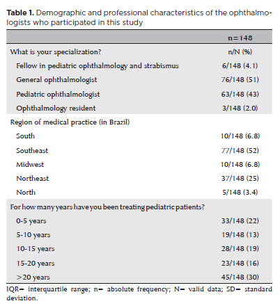
PURPOSE: This study aimed to identify the strategies adopted by Brazilian ophthalmologists to control myopia in clinical practice.
METHODS: This was a prospective cross-sectional study. Data were collected using an online questionnaire.
RESULTS: Responses from 148 participants were collected between March and May 2024. The majority of respondents were general ophthalmologists (51%) and pediatric ophthalmologists (43%). They came from all regions of Brazil, but more than half (52%) were from the Southeast region. Most participants (30%) had over 20 years of clinical practice experience. A significant proportion (89.2%) treated progressive myopia. The most requested complementary exams were optical biometry (83.78%) and corneal topography or tomography (69.59%). Behavioral measures were considered the most effective myopia treatment strategies by 41.2% of the respondents, followed by optical (33.8%) and pharmacological interventions (25%). Most recommended spending more time outdoors (94.59%) and reducing screen time (93.92%). Spectacle lenses for myopia (83.11%) and 0.025% atropine eye drops (54.73%) were the most prescribed treatments after the recommendation of environmental and behavioral changes.
CONCLUSION: This study presents a novel analysis of the clinical strategies for myopia control among Brazilian ophthalmologists. Understanding current clinical practices and identifying possible improvements are essential steps toward developing evidence-based guidelines and professional education aimed at improving patient care.
Keywords: Myopia/epidemiology; Refractive errors; Contact lenses; Myopia/drug therapy; Atropine/therapeutic use; Ophthalmologists; Practice patterns, physicians’; Surveys and questionnaires; Brazil/epidemiology
Abstract
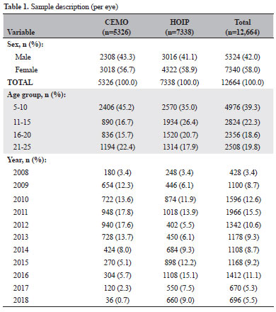
PURPOSE: This study aimed to examine the prevalence of myopic eyes over 11 years (2008-2018) in a private clinic and a public assistance service.
METHODS: We retrospectively evaluated 6332 individuals (12,664 eyes)
between 5 and 25 years old, seen at a private clinic-CEMO (2,663 individuals) and a public service-HOIP (3,669 individuals) from 2008 to 2018. We evaluated the prevalence of myopic eyes (EE ≤-0.50) and high myopic eyes (EE ≤-6.00).
RESULTS: Sex and services did not show statistical differences. The variation in the prevalence of myopic and high myopic eyes showed a random pattern during the study period (this prevalence could not be increased). Prevalences ranged from 20.7% (in 2017) to 32.4% (in 2015) for myopic eyes and from 1.6% (in 2009 and 2016) to 3.3% (in 2015) for eyes with high myopia. The prevalence of myopia showed a statistically significant increase based on the age group.
CONCLUSION: The prevalence of myopic eyes did not increase in our study. The mean prevalence of myopic eyes was similar in the private clinic and public service.
Keywords: Myopia; Refractive errors; Epidemiology; Prevalence
06-tab01.jpg)
Abstract
Objetivo: Avaliar o desempenho clínico do Spot Vision Screener e estabelecer correlações clínicas entre a triagem automatizada e a retinoscopia após indução de cicloplegia em crianças pré-verbais.
Métodos: Neste estudo transversal prospectivo, crianças de 6 a 36 meses foram avaliadas usando o Spot Vision Screener. O exame oftalmológico completo, incluindo refração cicloplégica, foi então realizado, seguido de repetição da triagem automatizada e retinoscopia em todos os casos, a fim de estabelecer correlações quanto à hipermetropia, miopia e astigmatismo após a indução de cicloplegia.
Resultados: O estudo incluiu 185 crianças. A sensibilidade do dispositivo de triagem automática após cicloplegia foi de 100% (IC 95%: 85,18-100%) e a especificidade foi de 87,04% (IC 95%: 80,87-91,79%). Os valores preditivos positivos e negativos foram de 52,27% (42,36 - 62,01%) e 100%, respectivamente. Em comparação com a retinoscopia, o Spot Vision Screener superestimou os valores esféricos em 0,62 D (IC 95%: 0,56 - 0,69) no olho direito e em 0,60 (IC 95%: 0,54 - 0,66) no olho esquerdo e os valores cilíndricos em -0,38 D (IC 95%: -0,42 a -0,33) no olho direito e por -0,39 D (IC 95%: -0,43 a -0,34) no olho esquerdo. A diferença para os valores esféricos e cilíndricos de forma geral foi de 0,61 D (IC 95%: 0,57 - 0,65) e -0,38 D (IC 95%: -0,41 a -0,35), respectivamente.
Conclusão: Foi encontrada correlação substancial entre a retinoscopia e os dados objetivos captados pelo dispositivo. Isso mostra que a tecnologia pode ser usada em conjunto, contribuindo para um diagnóstico mais preciso e identificando os fatores de risco de ambliopia o mais precocemente possível. A técnica automatizada pode fazer a diferença em nível populacional para triagem e intervenção precoce.
Keywords: Erros de refração; Ambliopia; Estrabismo; Refratometria; Retinoscopia
08-fig01.jpg)
Abstract
Objetivo: Determinar o impacto do uso de unidade móvel no acesso à saúde ocular e avaliar o perfil da população que necessita de cuidados oftalmológicos, as doenças oculares mais frequentes e o tratamento. Métodos: Estudo transversal realizado em 14 municípios da região sudoeste do Estado de São Paulo utilizando uma unidade móvel oftalmológica. Os participantes eram usuários do Sistema Único de Saúde que procuraram atendimento oftalmológico, sem restrição quanto a idade, gênero ou condição socioeconômica. Os dados foram transferidos para a tabela Excel para análise estatística. Resultados: Participaram do estudo 6.878 pessoas, com média de idade de 44 anos (variação de 4 meses a 96 anos) e 65,5% eram mulheres. Erros refrativos estavam presentes em 78,6% dos participantes, catarata em 9,6% e pterígio em 8,3%. Para 60% foram prescritos óculos, para 10% foi mantida a correção óptica em uso e para 28% foram necessárias apenas orientações. Exames especializados ou procedimentos cirúrgicos foram indicados para 18,1% dos casos que foram encaminhados para tratamento em serviço terciário. Dentre os pacientes referenciados, 36,4% necessitavam de cirurgia oculoplástica ou para tratar afecções externas do olho e 31,8%, de cirurgia de catarata. Conclusão: A grande maioria dos pacientes que procurou atendimento na unidade móvel necessitava de prescrição de óculos. A unidade móvel oftalmológica possui alto grau de resolutividade para os problemas oculares, com oportunidade de tratar os erros refrativos e referenciar os pacientes que necessitam de atendimento especializado, geralmente relacionado a condições cirúrgicas. Unidades móveis podem ser uma alternativa aos cuidados oftalmológicos básicos, melhorando o acesso, atuando na promoção da saúde ocular e prevenindo a cegueira.
Keywords: Unidades móveis de saúde; Saúde ocular; Transtornos da visão; Erros de refração; Óculos; Cegueira/prevenção & controle
Abstract
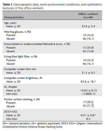
PURPOSE: To examine how ophthalmological features, screen exposure duration, and break habits among office employees affect ocular surface parameters.
METHODS: This single-center cross-sectional study involved two assessments on the same day: one before and one after a visual display terminal task. During the initial assessment, information on screen use was gathered, and refractive error, anterior segment examination, tear breakup time, and Schirmer test measurements were conducted. Participants tracked their screen usage and break durations throughout the day. At the end of the workday, tear breakup time and Schirmer I tests were repeated. Baseline and follow-up results were compared, and regression analysis was performed to identify factors linked to tear breakup time reduction.
RESULTS: The study enrolled 60 female office employees. Their mean screen time was 269.26 ± 70.21 min, with an average break duration of 151.93 ± 46.24 min. Tear breakup time at the second assessment (6.38 ± 2.70) was significantly lower than at baseline (8.62 ± 2.73) (p<0.001), whereas Schirmer test scores showed no significant change (p>0.05). Tear breakup time reduction was noted in 54 participants (90.0%), with a significant association between tear breakup time decrease percentage and screen exposure (p=0.001, r=0.463). Regression analysis showed that uncorrected or undercorrected refractive error was an independent risk factor for a ≥30% tear breakup time reduction, while taking more frequent short breaks (<15 min) acted as a protective factor.
CONCLUSIONS: Taking more frequent short breaks (<15 min) and correcting refractive errors help prevent intra-day tear breakup time decline during visual display terminal use. Structuring breaks to support tear film stability is advisable for occupations that require regular visual display terminal tasks.
Keywords: Tear film; Screen time; Tear breakup time; Office workers; Protective factors; Lacerations; Refractive errors; Risk factors.

Abstract
PURPOSE: Using advanced imaging techniques, this study aimed to evaluate corneal stability, epithelial remodeling, and tear film changes over a one-year period in first-time soft-contact lens wearers.
METHODS: A retrospective study was conducted on 100 eyes of 50 first-time daily soft-contact lens users aged 21–65 years with no prior rigid gas-permeable lens wear. The Sirius Scheimpflug imaging system was used to assess corneal topography, epithelial thickness, and non-invasive tear break-up time at baseline, 3, 6, and 12 months. Corneal warpage was evaluated using symmetry indices and Baiocchi Calossi Versaci indices. We performed statistical analysis using repeated-measures analyses of variance with Greenhouse-Geisser correction.
RESULTS: The mean baseline central corneal thickness was 537.83 (±7.92) µm, with no significant thinning after one year. The average simulated keratometry values remained stable, indicating no progressive corneal steepening or flattening. There were no significant changes in warpage indices over time, suggesting corneal shape preservation. Higher-order aberrations (coma, trefoil, and spherical aberrations) and non-invasive tear break-up time remained unchanged throughout the study period.
CONCLUSIONS: Modern silicone hydrogel soft-contact lenses do not induce significant corneal warpage, epithelial remodeling, or optical aberrations over a one-year period. We found that corneal morphology and tear film stability were preserved, supporting the safety of soft-contact lens use. These findings provide clinically relevant insights into the long-term impact of contact lens wear. They may facilitate improved lens fitting strategies and preoperative refractive surgery assessments.
Keywords: Contact lenses, hydrophilic; Cornea/surgery; Corneal diseases; Corneal topography; Adaptation, ocular/physiology; Endothelium, corneal/pathology; Refractive errors; Tears/metabolism.

Abstract
PURPOSE: To compare the refractive prediction error of Hill-radial basis function 3.0 with those of 3 conventional formulas and 11 combination methods in eyes with short axial lengths.
METHODS: The refractive prediction error was calculated using 4 formulas (Hoffer Q, SRK-T, Haigis, and Hill-RBF) and 11 combination methods (average of two or more methods). The absolute error was determined, and the proportion of eyes within 0.25-diopter (D) increments of absolute error was analyzed. Furthermore, the intraclass correlation coefficients of each method were computed to evaluate the agreement between target refractive error and postoperative spherical equivalent.
RESULTS: This study included 87 eyes. Based on the refractive prediction error findings, Hoffer Q formula exhibited the highest myopic errors, followed by SRK-T, Hill-RBF, and Haigis. Among all the methods, the Haigis and Hill-RBF combination yielded a mean refractive prediction error closest to zero. The SRK-T and Hill-RBF combination showed the lowest mean absolute error, whereas the Hoffer Q, SRK-T, and Haigis combination had the lowest median absolute error. Hill-radial basis function exhibited the highest intraclass correlation coefficient, whereas SRK-T showed the lowest. Haigis and Hill-RBF, as well as the combination of both, demonstrated the lowest proportion of refractive surprises (absolute error >1.00 D). Among the individual formulas, Hill-RBF had the highest success rate (absolute error ≤0.50 D). Moreover, among all the methods, the SRK-T and Hill-RBF combination exhibited the highest success rate.
CONCLUSIONS: Hill-radial basis function showed accuracy comparable to or surpassing that of conventional formulas in eyes with short axial lengths. The use and integration of various formulas in cataract surgery for eyes with short axial lengths may help reduce the incidence of refractive surprises.
Keywords: Cataract; Lenses, intraocular; Axial length, eye; Refractive errors; Artificial intelligence
Abstract
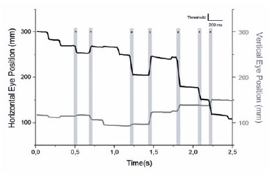
PURPOSE: To evaluate the saccadic movements of patients with visual field loss due to primary open-angle glaucoma.
METHODS: Thirteen patients with good visual acuity (0.2 logMAR or better) (seven patients with primary open-angle glaucoma 65 ± 13 years) and six controls (51 ± 6 years) yielded a comprehensive ophthalmological examination, including Humphrey Visual Field tests (SITA-Standard 24-2), and performed a monocular, exploratory digital visual search task that quantifies the duration for finding the number “4” on a random array of digits distributed on the screen. After individual adjustments of the angle and distance positioning, the screen was spatially matched with the 24-2 visual field, and divided into five areas for analysis. During the task, saccades were simultaneously recorded in the same eye with a video-based eye tracker.
RESULTS: The patients with primary open-angle glaucoma showed a significantly higher number of saccades/screen (median ± interquartile range, 59.00 ± 29.00 vs. 32.50 ± 19.75 saccades (p=0.027) and visual search time per screen (38.50 ± 60.14 vs. 23.75 ± 8.90 seconds (p=0.035) than the controls did. Although the univariate analysis indicated a significant correlation with visual field mean deviation (coefficient=26.19 (p=0.02), only the visual search time/screen was significantly associated with the number of saccades/screen in the multivariate regression model (coefficient=0.55 (p<0.001). Overall, no significant correlation was observed between the sectorial number of saccades and the sensitivity of the five visual field areas.
CONCLUSIONS: The patients with primary open-angle glaucoma show impaired search performance and showed a higher number of saccades needed to find stimuli when performing the exploratory visual task.
Keywords: Glaucoma, open angle; Saccades; Eye movements; Visual fields; Vision disorders
15-tab01tb.jpg)
Abstract
OBJETIVO: Este estudo visou avaliar os mecanismos da lesão e os tipos de fraturas orbitárias e sua relação com commotio retinae simultânea.
MÉTODOS: Este estudo retrospectivo avaliou registros de pacientes com fraturas orbitárias cujos diagnósticos foram confirmados por tomografia computadorizada entre julho de 2017 e setembro de 2019. Foram registrados os dados demográficos, circunstâncias da lesão, os resultados do exame oftalmológico e achados radiológicos. A análise estatística dos dados usou os testes de t-Student bicaudal, qui-quadrado e cálculos de odds ratio. O significado estatístico foi fixada em p<0,05.
RESULTADOS: Dos 204 pacientes com fraturas orbitárias incluídos neste estudo, 154 (75,5%) eram sexo masculino (75,5%). A média de idade foi de 42,1 anos. As fraturas orbitárias envolvendo uma parede orbital (58,8%) foram mais comuns do que as que acometeram várias paredes (41,2%). A maioria das fraturas acometeu a parede inferior (60,3%), sendo as paredes mediais as próximas mais frequentemente afetadas (19,6%). A causda mais comum de lesão foi agressão (59,3%), e a segunda mais comum foi queda (24%). A commotio retinae foi observada em 20,1% dos casos de fratura orbital e foi mais associada a lesões causadas por agressão (OR=5,22, p<0,001) e menos associada com aquelas causadas por quedas (OR=0,06, p<0,001). As restrições de movimentos oculares eram mais comuns na comoção central do que na periférica (OR=3,79, p=0,015) e com fraturas da parede medial do que com fraturas de outras paredes orbitais (OR=7,16, p<0,001). As chances de comoção não foram maiores em pacientes com fraturas orbitais de paredes múltiplas do que naqueles com fraturas de parede simples (p=0,967).
CONCLUSÕES: Na população do estudo, a agressão foi a causa mais comum de fraturas orbitais e resultou em commotio retinae mais grave do que qualquer outra causa. Os oftalmologistas devem estar cientes da probabilidade de commotio retinae em pacientes com fraturas orbitais resultantes de agressão, independentemente da extensão das lesões do paciente.
Keywords: Fraturas orbitárias; Movimentos oculares; Retina; Ferimentos e lesões
Abstract
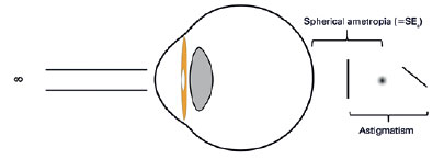
A abordagem de qualquer condição refrativa do olho com astigmatismo regular é mais complicada do que a da miopia ou hipermetropia isoladamente. Requer familiaridade com as imagens complexas coletivamente identificadas como o conóide de Sturm. Felizmente, apenas três deles desempenham um papel crítico na interpretação da ametropia com astigmatismo. Este manuscrito mostra como uma estratégia de prescrição para as ametropias associadas ao astigmatismo regular pode evoluir a partir dessas três imagens principais.
Keywords: Astigmatismo; Erros de refração
Abstract
The paper starts discussing the teleological concept that eye motions - rotations and translations - serve to vision (which supports the notion that torsions are not voluntarily driven, since they do not contribute to expand the visual exploration of space). It proposes that the primary position of the eye (not "of gaze") , the standard condition to measure them, must be defined as the coincidence of the orbital (fixed) and the ocular (movable) system of coordinates. However this becomes only a theoretic concept, since practical operations to obtain it are almost unfeasible. Besides, even a "simple" horizontal or vertical ocular rotation, though always occurring around a (presumably) fixed point (the center of ocular rotation) may be defined by different trajectories and magnitudes, depending on the two systems of measurement of eye positions and motions. Hence, in a graphical (plane) representation of such spherical coordinates, the so-called "tangent screen", an ocular "tertiary" position - a combination of a horizontal and a vertical rotations - may be described by four different points. Or, conversely, a specific eye position may be defined by four sets of angular coordinates. The mathematical representation of variation of three special coordinates in a specific rotation is best made by a matrix disposition, so that, multiplication (not commutative) of three matrices (one for each specific plane) generates six different systems (permutations) of measurements. So, though , actually, there are multiple trajectories possible between two points in space, the order in which rotations are considered influences the final result. With different systems of coordinates for each rotation and different possible orders by which they may be considered, one reaches 48 alternative systems for their measurements. Unfortunately, up to now, there I is no an established convention to express ocular rotations. So, usually, people consider that a vertical prism superimposed to a frontally placed horizontal prism, or vice-versa, correspond to equivalent processes. The paper finishes discussing inconveniences of the clinically used unity to measure eye rotations (the prism-diopter) and proposes other unities as alternative solutions.
Keywords: Angular measurement unity; eye movements; eye position measurement; eye position measurement accuracy; Fick's system; Helmholtz's system; ocular rotation; primary position of gaze; prism-diopter referential systems; superimposition of prisms
 ABO is licensed under a Creative Commons Attribution-NonComercial 4.0 Internacional.
About
Issues
Editorial Board
Submission
Arquivos Brasileiros de Oftalmologia
Official publication of Brazilian Council of Ophthalmology - Conselho Brasileiro de Oftalmologia (CBO)
Rua Casa do Ator, 1.117 - 2nd floor - Zip Code: 04546-004
São Paulo - SP, Brazil
TEL: +55 11 3266-4000
E-mail: [email protected]