Arq. Bras. Oftalmol. 200770
| DOI: 10.1590/S0004-27492007000300002
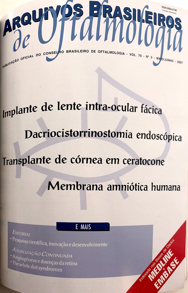
PURPOSE: To observe the preliminary results based on the efficacy, stability and safety of Artisan anterior chamber intra-ocular lens for the correction of myopia in the first 6 months.
METHODS: A retrospective study of 18 patients (34 eyes) that received the implantation of Artisan anterior chamber intraocular lens for the correction of myopia was conducted. Best visual preoperative acuity, best visual postoperative acuity, dynamic preoperative and postoperative refraction, loss and improvement of lines of vision and complications were evaluated.
RESULTS: Eighteen patients (34 eyes) were included in this study. Fourteen patients were females (77.8%) and 4 were males. The mean age was 30 years (SD ± 7.3), range from 21 to 46 years. They were followed up for an average of 8.5 months (SD ± 3.6). The mean endothelial cell loss was 4.75% at 6 months follow-up. The average preoperative uncorrected visual acuity was 0.02 (20/800) range from 0.01 to 0.13. The average preoperative spherical equivalent was -13.25 D, range from -5.75 D to -19,75 D. On the last follow-up, uncorrected visual acuity was 0.64 (20/32), range from 0.33 to 1.00; the average spherical equivalent was -1.18 D (DP ± 0.92) range from +0.25 to -3.0. Complications were observed in 1 case (3.4%) of our study, in one patient who had a dislocation of the lens after an ocular trauma.
CONCLUSION: The use of Artisan anterior chamber intraocular lens in phakic eyes for the correction of myopia was safe, effective and predictable. However, a larger prospective study with a higher number of cases and longer follow-up is necessary to determine long-term safety of the lens.
Keywords: Lens implantation, intraocular; Lenses; intraocular; Myopia
Arq. Bras. Oftalmol. 200770
| DOI: 10.1590/S0004-27492007000300003
PURPOSE: To assess the incidence of operative difficulties in endonasal endoscopic dacryocystorhinostomies, as well as to suggest solutions.
METHODS: This retrospective study searched for reports of complications and intraoperative difficulties in our records of 268 endoscopic endonasal dacryocystorhinostomies, in 241 patients, performed from October 1999 to August 2005. The patients' age ranged from 2 to 92 years (mean: 49.1 years). Uncinectomy (partial excision of the uncinate process) was employed as the entry to the lacrimal fossa, no drills being needed for the osteotomy.
RESULTS: There were operative difficulties in 37 (13.8%) procedures. The most frequent sources of difficulty were hemorrhage (n = 12) and nasal anatomy abnormalities (n = 14). Less common sources were lacrimal sac mucocele, anterior ethmoid cells and resistant uncinate process. Of the nasal alterations disturbing surgery, the most common was septal deviation, followed by polyps, hypertrophied middle turbinate and fracture of the lateral wall. Complications were observed in 5 (1.9%) cases, including postoperative epistaxis (n = 3), subcutaneous emphysema and displacement of the silicone tube.
CONCLUSION: Endonasal endoscopic dacryocystorhinostomy was shown to be a safe procedure, with a minimal incidence of postoperative complications. Its indication, however, is limited. Not all patients can undergo this technique without correction of nasal anatomy alterations. Though most surgeries with this technique present no difficulties, this procedure may be technically difficult, especially in the presence of hemorrhage or reduced nasal space.
Keywords: Dacryocystorhinostomy; Lacrimal apparatus diseases; Lacrimal duct obstruction; Endoscopy; Retrospective studies
Arq. Bras. Oftalmol. 200770
| DOI: 10.1590/S0004-27492007000300004
PURPOSE: Refractive and visual analysis in corneal transplantations performed for keratoconus by two groups of surgeons.
METHODS: Seventy eyes of seventy patients, which had been submitted to penetrating keratoplasty (PK) for keratoconus, were examined and their charts were reviewed retrospectively for long-term results after all sutures had been removed. The patients were divided into two groups, Group I had surgery performed by skillful surgeons and Group II by surgeons in training. The results of best correct visual acuity, refractive error, postoperative complications and computerized videokeratoscopy were compared.
RESULTS: The mean follow-up was 7.9 years with a range of 1.6 - 20 years. Each group consisted of 35 eyes. There were 35 females and 35 males. The donor age was different between the two groups. The surgical technique was the same in both groups, but the number of sutures was different. Preoperative mean logMAR visual acuity was low in the two groups, but statistically different and no relationship was found between the results. All the refractive, topographic and visual acuity results were similar between both groups. Endothelial rejection was more frequent in Group II. Only three grafts lost graft clarity. These patients belonged to Group II.
CONCLUSION: In a corneal referral practice, the visual results after PK were similar to those related to the PK performed by either expert surgeons or in training.
Keywords: Keratoconus; Corneal keratoplasty; Postoperative complications; Refractive error
Arq. Bras. Oftalmol. 200770
| DOI: 10.1590/S0004-27492007000300005
PURPOSE: To evaluate the morphological features of the amniotic membrane denuded by different techniques.
METHODS: Human amniotic membrane was collected at the time of delivery, fixed in increasing concentrations of glycerol (0-50% in DMEM) and preserved at -80ºC until the time of use. The study consisted of 4 groups: intact epithelium (control) and denuded by trypsin (2 mg/mL at 1:250), dispase (1.2 U/mL in Mg2+ and Ca2+ free Hank's balanced salt solution) or ethylenediaminetetraacetic acid (EDTA), 0.02%. Specimens were submitted to electron (scanning and transmission) microscopy analysis.
RESULTS: Scanning electron microscopy disclosed intact epithelium in the control group and its absence in the amniotic membranes denuded by trypsin and dispase. In those denuded by ethylenediaminetetraacetic acid there were areas with and without epithelium. When assessed by transmission electron microscopy, the epithelium was intact and firmly adhered to the basement membrane by hemidesmossomes in controls and in parts of ethylenediaminetetraacetic acid group. There were only collagen fibers in the dispase- and trypsin-treated groups. CONCLUSIONS: Trypsin and dispase treatment of the amniotic membrane may cause complete denuding of the epithelium and basement membrane whereas ethylenediaminetetraacetic acid may leave some intact epithelium-areas and partially destroy the basement membrane in others.
Keywords: Amnion; Edetic acid; Corneal diseases; Trypsin; Endopeptidases; Microscopy; electron
Arq. Bras. Oftalmol. 200770
| DOI: 10.1590/S0004-27492007000300006
PURPOSE: Although it has been proposed that retinal vasculature is fractal, no method of standardization has been performed for vascular segmentation or for dimension calculation, thus resulting in great variability among values of fractal dimensions. The present study was designed to determine if estimation of retinal vessel fractal dimensions is dependent on vascular segmentation and dimensional calculation methods.
METHODS: Ten eye fundus images were segmented to extract their vascular trees by four computational methods ("multithreshold", "scale-space", "pixel classification" and "ridge based detection"). Their information, mass-radius and box counting fractal dimensions were calculated and compared with those of the same vascular trees manually segmented (gold standard).
RESULTS: The mean vascular tree dimension varied among the groups of different segmentation methods, from 1.39 to 1.47 for box counting, from 1.47 to 1.52 for information and from 1.48 to 1.57 for mass-radius dimensions. The utilization of different vascular segmentation methods and different dimension calculation methods introduced significant difference among fractal dimension of vessels.
CONCLUSION: Estimation of retinal vessel fractal dimensions was dependent on both vascular segmentation and dimension calculation methods.
Keywords: Fractals; Retina; Diagnostic techniques and procedures; Biometry
Arq. Bras. Oftalmol. 200770
| DOI: 10.1590/S0004-27492007000300007
PURPOSE: To describe the causes of congenital epiphora in patients with Down syndrome.
METHODS: Retrospective study of 695 patients with congenital epiphora, of the Lacrimal Sector of the Department of Ophthalmology, São Paulo "Santa Casa", Brazil, between October 1998 and July 2005. This study analyzed: the main symptom of continuous epiphora or mucous discharge, which affected one or both eyes, since birth. Subjects were separated in to two groups: group A, with 30 patients with Down syndrome and group B, with 665 control patients. The lacrimal evaluation was performed by the throw irrigation test after general anesthesia.
RESULTS: Both groups were statistically similar regarding age (p=0.07), sex (p=0.63) and race (p=0.68). Bilateral symptoms were more frequent in group A (p=0.0008). Anatomic obstruction of the lacrimal canal was present in 32.73% of group A and in 85.51% of group B (p<0.0001). CONCLUSIONS: The most frequent cause of congenital epiphora in Down syndrome is due to functional block of the lacrimal pump. Eyelid muscle hypotony and eyelid changes, common in Down syndrome, is the main hypothesis of lacrimal dysfunction, but further studies will be necessary for confirmation.
Keywords: Lacrimal apparatus diseases; Lacrimal duct obstruction; Down syndrome; Eye diseases
Arq. Bras. Oftalmol. 200770
| DOI: 10.1590/S0004-27492007000300008
PURPOSE: To compare the results between recession of the lateral recti and monocular recess-resect procedure for the correction of the basic type of intermittent exotropia.
METHODS: 115 patients with intermittent exotropia were submitted to surgery. The patients were divided into 4 groups, according to the magnitude of preoperative deviation and the surgical procedure was subsequently performed. Well compensated orthophoria or exo-or esophoria were considered surgical success, with minimum of 1 year follow-up after the operation.
RESULTS: Success was obtained in 69% of the patients submitted to recession of the lateral recti, and in 77% submitted to monocular recess-resect. In the groups with deviations between 12 PD and 25 PD, surgical success was observed in 74% of the patients submitted to recession of the lateral recti and in 78% of the patients submitted to monocular recess-resect. (p=0.564). In the group with deviations between 26 PD and 35 PD, surgical success was observed in 65% out of the patients submitted to recession of the lateral recti and in 75% of the patients submitted to monocular recess-resect. (p=0.266).
CONCLUSION: recession of lateral recti and monocular recess-resect were equally effective in correcting basic type intermittent exotropia according to its preoperative deviation in primary position.
Keywords: Exotropia; Oculomotor muscles; Ophthalmologic surgical procedures; Visual acuity; Comparative study
Arq. Bras. Oftalmol. 200770
| DOI: 10.1590/S0004-27492007000300009
PURPOSE: To evaluate the diabetic alterations and the impact of short and long-term medical treatment on them.
METHODS: Thirty Wistar rats were divided into 3 groups: control (GC), diabetic (DG), and treated diabetic (TG) and the observations were made 1 month (M1) and 12 months (M2) after diabetes induction. Diabetes was induced by intravenous alloxan (42 mg/kg). The treated group received acarbose orally and insulin by subcutaneous injection. Eyes were prepared for transmission electron microscopy, specifically for ultrastructure of the Bruch membrane and choroidal vessels.
RESULTS: Ultrastructural examination of the diabetic rat coroid showed deposits in the Bruch membrane and accumulation of vesicles, glycogen and dense bodies in endothelial cell cytoplasm. The most affected group was that of the diabetics on month 12 (GDM2). The treated diabetics showed the least alterations on month 12 (GTM2).
CONCLUSION: Diabetic rats develop degenerative alterations in the Bruch membrane and choroidal vessels. These alterations are more evident in animals submitted to chronic disease, but they are also present in acute disease. Degenerative processes were not avoided with short-term treatment. Long-term treatment inhibited the progress of these processes.
Keywords: Choroid; Bruch membrane; Diabetes mellitus, experimental; Rats, Wistar; Alloxan; Insulin; Acarbose
Arq. Bras. Oftalmol. 200770
| DOI: 10.1590/S0004-27492007000300010
PURPOSE: To evaluate the RPS Adenodetector®, a rapid immunochromatographic test, in the diagnosis of patients with clinical overt adenoviral conjunctivitis.
METHODS: Consecutive case series. Patients underwent conjunctiva scraping for RPS Adenodetector® test and culture to identify adenovirus.
RESULTS: A total of 11 patients were studied, and 10 had unilateral disease. Five (45.5%) had symptoms for 2 days, 5 for three days, and 1 for 7 days. Adenovirus culture was positive in 8 patients (73%) and RPS Adenodetector® was positive in 9 (82%) patients. Eight patients had adenovirus identification by both methods. In one patient the RPS Adenodetector® was positive in contrast to a negative culture. The two patients revealing negative RPS Adenodetector® results also had negative cultures. The sensitivity was 100% and the specificity was 67%.
CONCLUSION: The RPS Adenodetector® is a useful tool in the rapid diagnosis of adenovirus conjunctivitis and may contribute to the spread control of this highly contagious disease.
Keywords: Conjunctivitis, viral; Adenoviridae infections; Chromatography; Virus cultivation
Arq. Bras. Oftalmol. 200770
| DOI: 10.1590/S0004-27492007000300011
AIM: To evaluate the incidence of fungi in the ocular conjunctiva of sugar-cane cutting workers as well in a sugar-cane plantation environment. PLACE: Monte Belo - MG, Eye Clinic, Laboratory of Physiology and Biology of Microorganisms of Unifenas, Alfenas - MG.
METHODS: Swabs were made from the ocular conjunctiva of one hundred workers of both sexes and material was collected from leaves, stalk, and the environmental air from the sugar-cane plantation. These were inoculated in specific mold media. After incubation at 25°C for a period of fifteen days, the plates were analyzed and colony forming units (ufc) were identified using conventional mycological techniques.
RESULTS: Of one hundred workers involved in this research, 64 presented one or more genera of fungi, 54 (84.38%) being identified in males and 10 (15.62%) identified in females. The separation of the workers by age range showed that the prevalence of observed fungi by age was not uniform. The highest incidences were found in advanced age ranges, the increase of positivity by age being considered statistically significant (p<0.05). The lowest prevalence (50%) was found in the 11-20-year-old interval, which presents the lowest number of examined persons. The highest positivity was verified in the 61-79 year interval. In 60 workers (93.75%) only one genus was isolated; in 3 (4.69%) two genera, and in only one worker (1.56%) three genera were isolated. The most prevalent isolated fungi were Fusarium sp (43.76%) and Geotrichum sp (23.44%), followed by Cladosporium sp (9.38%), Penicillium sp (7.81%), Mucor sp (9.38%) and Oidium sp (7.81%). The most common genus founded in leaves, stalk and air were Aspergillus, Penicillium, Fusarium, Cladosporium and Rhizopus.
CONCLUSION: The environmental conditions, the socioeconomic status and the general and personal poor hygienic conditions, together with lack of information about prophylactic standards, surely led to the high incidence of ocular conjunctiva fungus isolation (67%). The incidence was much higher in males (84.38%) as (15.62%) in females. The anemophilious fungi and those isolated from stalk and leaves were similar to those already described in the literature. The isolated filamentous fungi were much more numerous than yeast, there being some correlation between these and the conjunctiva isolates.
Keywords: Fungi; Conjunctiva; Eye diseases; Saccharum; Agricultural worker's diseases
Arq. Bras. Oftalmol. 200770
| DOI: 10.1590/S0004-27492007000300012
PURPOSE: There is no uniformity in the literature about the core features required to make the diagnosis of Möbius sequence. Originally, the minimum requirements were the bilateral paralysis of the VI and the VII cranial nerves. The bilateral facial nerve paralysis or paresis, often asymmetric, is common to all patients but some facts show that the isolated VI nerve palsy in the Möbius sequence is not the rule. 1) When there is an esotropia in Möbius sequence, it is often too small to be caused by a bilateral isolated VI nerve palsy. There are many cases in which there is no esotropia in the primary position and even some cases, though rare, with exotropia. 2) In most cases of Möbius sequence, the esotropia can be eliminated with a mere recession of the medial rectus muscles. 3) In most patients with Möbius sequence there is, besides the lateral rectus palsy, a variable degree of adduction limitation, which defines a horizontal gaze palsy. The authors present some arguments to show that the isolated lateral rectus muscle palsy cannot be considered as a sine qua non factor for the diagnosis of Möbius sequence.
METHODS: The binocular alignment in primary position and the incidence of abduction and adduction limitations among 28 of the authors' consecutive patients with Möbius sequence and in patients of 5 other randomly selected publications are presented for comments.
RESULTS: The eyes' position in primary position among 135 of those 6 authors' patients (28 belonging to the authors of this study and 107 to the other 5) were recorded; 55 of them (40.74%) had orthotropia and 9 (6.66%) had exotropia. Among 80 patients of 4 authors (22 belonging to the authors of this study and 52 to the other 3), in whom the horizontal versions were analyzed, 79 (98.75%) had limitation of abduction and 53 (66.25%) had limitation of adduction. COMMENTS: The authors emphasize that the recent studies have shown that inside the VI nerve nucleus there are two types of cells: those which axons form the ipsolateral abducens nerve and those (interneurons) whose axons reach the medial longitudinal fasciculus and ascend for innervating the subnucleus of the contralateral III nerve subserving the contralateral medial rectus. Because of this arrangement, a lesion at the region of the VI nerve nucleus generally causes a paralysis of the ipsolateral lateral rectus and the contralateral medial rectus muscles, which characterizes the ipsolateral horizontal gaze palsy.
CONCLUSION: The definition of the Möbius sequence is the paralysis of the facial nerve and the horizontal gaze palsy, instead of a VI nerve palsy, as seen in most published papers.
Keywords: Mobius syndrome; Facial paralysis; Eye diseases
Arq. Bras. Oftalmol. 200770
| DOI: 10.1590/S0004-27492007000300013
PURPOSE: To asses the correlation between ocular components (axial length, anterior segment length, corneal power, vitreous length and equivalent power of the eye) and refractive error in eyes with higher and lower ametropia of subjects with anisometropia.
METHODS: An analytical survey was carried out in 68 patients with anisometropia of two or more diopters, assisted at the Ophthalmological Clinic from "Hospital das Clínicas da Faculdade de Medicina" of São Paulo University. All participants were submitted to ophthalmologic examination which included objective and subjective cycloplegic refratometry, keratometry and ultrasonic biometry.
RESULTS: No significant difference between measures of ocular components of the eyes with higher and lower ametropias was observed. The eyes with lower ametropia presented the same significant correlations observed in emmetropic eyes: of refraction with anterior segment length and axial length, and of the axial length with corneal power and vitreous length. The eyes with higher ametropia presented significant correlation of refraction with the axial length and of the axial length with vitreous length. Furthermore, we observed in both eyes, significant correlation of the power of the crystalline lens with the anterior chamber depth.
CONCLUSION: The eyes with lower ametropia presented correlations more frequently observed in emmetropia. The eyes with higher ametropia did not present the same correlations observed in emmetropic eyes.
Keywords: Anisometropia; Biometry; Eye; Eye; Refractive errors
Arq. Bras. Oftalmol. 200770
| DOI: 10.1590/S0004-27492007000300014
PURPOSE: The purpose of this study was to compare total testosterone blood level among three groups of postmenopausal women: control, mild to moderate dry eye and severe dry eye.
METHODS: Twenty-nine postmenopausal women were selected. The exclusion criteria were: hormone replacement therapy in the last 8 weeks, mechanical palpebral abnormalities, pterygium, lacrimal obstructions, intraocular inflammation or contact lens use. A blood sample was collected for total testosterone level determination, and the patients were submitted to an ophthalmologic examination (emphasizing on dry eye detection) and answered the OSDI (Ocular Surface Disease Index) questionnaire. Five patients were excluded. Postmenopausal women were divided into three groups according to OSDI score and the ophthalmic examination.
RESULTS: Five patients were classified in the no dry eye group (control), fifteen in the mild to moderate dry eye group and four in the severe dry eye group. There were no statistically significant differences regarding mean age (p=0.3915); instruction level (p=0.9333); number of comorbidities (p=0.2551); medication taken (p=0.2844) and total testosterone level among those groups (p=0.1275).
CONCLUSION: Further research with a greater bigger sample is necessary to establish the relation of androgen levels in dry eye patients.
Keywords: Dry eye syndromes; Testosterone; Hormones; Postmenopause; Questionaires
Arq. Bras. Oftalmol. 200770
| DOI: 10.1590/S0004-27492007000300015
PURPOSE: To evaluate and correlate the peaks and the fluctuation of intraocular pressure seen in the association of the ambulatory intraocular pressure curve with the water-drinking test versus the peaks and the fluctuation seen in the daily intraocular pressure curve.
METHODS: The sample was as follows: 77 eyes belonging to 77 patients who were divided into three groups composed of 31 eyes belonging to 31 patients with primary open-angle glaucoma, 26 eyes belonging to 26 patients with normal tension glaucoma and 20 normal eyes belonging to 20 individuals.
RESULTS: A significant correlation could be observed between the pressure peaks collected from the daily intraocular pressure curve and the pressure peaks seen in the ambulatory intraocular pressure curve, on the water-drinking test, as well as in the association of the ambulatory intraocular pressure curve with the water-drinking test. The procedure which showed the highest rate of correlation between the pressure peaks and the peaks of the daily intraocular pressure curve was the ambulatory intraocular pressure curve (r²= 0.81). However, no statistically significant difference was found between the pressure peaks and the correlation coefficients observed in the other methods. The correlation between the fluctuation of intraocular pressure obtained in the association of the ambulatory intraocular pressure curve with the water-drinking test and the fluctuation of the pressure seen in the daily intraocular pressure curve showed a slight association (r²= 0.21). CONCLUSIONS: The association between ambulatory intraocular pressure curve and water-drinking test was not efficient to estimate peaks and the fluctuation of daily intraocular pressure curve. The ambulatory intraocular pressure curve and the water-drinking test must be analyzed separately. When predicting the peak and the fluctuation of the daily intraocular pressure curve, the ambulatory intraocular pressure curve was the most efficient procedure.
Keywords: Glaucoma, open-angle; Intraocular pressure; Diagnostic techniques, ophthalmological; Tonometry, ocular; Comparative study
Arq. Bras. Oftalmol. 200770
| DOI: 10.1590/S0004-27492007000300016
PURPOSE: To evaluate spontaneous blink activity in the primary eye position in normal preschool children.
METHODS: Two hundred normal children aged 4 to 6 years were prospectively evaluated. They were digitally videotaped in a standard setting, taking the images on frontal and lateral plans, during the vigil state, in the primary eye position. The object of observation was located at the same level of the pupil. A Sony Lithium camera was used. The images were recorded on 8 mm tapes and transferred to a personal computer (MacIntosh G4) and processed with the iMovie software. Opening, closure and total blink times were measured using frame scale. The eyelid movement rhythm per minute was also evaluated, during three minutes. The data were submitted to statistical analysis.
RESULTS: The complete eye blink was more frequently observed than the incomplete eye blink. The blink rate increased with age. The incomplete blink rate was the same for all the evaluated ages. The eyelid opening and closing times and the complete blink time were similar for both sexes, during all evaluation moments. There were no differences between genders regarding eye blink. The closing eyelid time was slower than the opening eyelid time. CONCLUSIONS: The complete blink rate increases with age. The closing and the opening eyelid times and the complete blink were similar for both sexes in all evaluated ages and the opening time is faster than the closing time.
Keywords: Blinking; Saccades; Eyelids; Videotape recording; Child, preschool
Arq. Bras. Oftalmol. 200770
| DOI: 10.1590/S0004-27492007000300017
PURPOSE: To assess ocular biometric parameters and refractive error in Brazilian adults and their relationship with height, age, gender and years of formal education.
METHODS: Cross-sectional study that assessed 173 subjects by keratometry, echobiometry, refraction and measurement of body height. The statistical analysis was performed using Pearson's coefficient and a regression model was constructed.
RESULTS: The correlations found were: each 10 cm - increase in height was related to a 0.32 mm longer axial length, 0.07 mm deeper anterior chamber, 0.26 mm deeper vitreous chamber and 0.50 D flatter keratometry. Each 10-year increase in age, related to a 0.15 mm smaller axial length, 0.25 mm thicker lens, 0.21 mm shallower vitreous chamber and 0.23 D more positive spherical equivalent. Each 10-year increase in education related to a 0.74 D more negative spherical equivalent. Gender did not influence the analyzed biometric parameters. Equations referring to biometric parameters and spherical equivalent were formulated. CONCLUSIONS: Positive correlations were found between: axial length and height; anterior chamber and vitreous cavity depth with height; lens thickness with age; keratometry with height; and spherical equivalent with age. Negative correlations were found between: anterior chamber and vitreous cavity depth with age; spherical equivalent and years of formal education.
Keywords: Eye; Biometry; Refractive errors; Body height; Age factors; Sex factors; Educational status
Arq. Bras. Oftalmol. 200770
| DOI: 10.1590/S0004-27492007000300018
PURPOSE: To report pars plana vitrectomy results of intravitreous use of liquid perfluorocarbon as a short-term postoperative tamponade in retinal detachment due to giant tears in a series of patients.
METHODS: Ten of those patients, all of them complicated by proliferative vitreoretinopathy grade B or worse, with tear extension varying from 90º to 210º were studied. Perfluorocarbon liquid was injected via pars plana until the posterior tear limit, remaining in the postoperative period during five days, with the patients in supine position. After this period, the patients underwent a second surgical procedure to exchange the liquid perfluorocarbon for gas or silicone oil.
RESULTS: The retinas of eight patients were attached (80%) after a mean follow-up of 16.2 ± 12.4 months (from 2 to 43 months). It was necessary to repeat this technique in one (10%) case, and there was no attachment of the retina in two (20%) cases due to advanced proliferative vitreoretinopathy. Visual acuity improved in five (50%) cases.
CONCLUSION: Good results (80%) and improvement of the visual acuity (50%) were observed with the use of intravitreous liquid perfluorocarbon as short-term tamponade in the postoperative period in patients with retinal detachments due to giant tears.
Keywords: Retinal detachment; Retinal diseases; Tears; Vitreoretinopathy, proliferative; Fluorocarbons; Vitrectomy
Arq. Bras. Oftalmol. 200770
| DOI: 10.1590/S0004-27492007000300019
PURPOSE: To report the experience with eyelid pilomatrixoma during 30 years in a university hospital.
METHODS: A review of the Ophthalmic Pathology Laboratory at the "Hospital Universitario Prof. Edgard Santos", was conducted to identify eyelid tumor cases from 1974 to 2004. After identification of pilomatrixoma cases, a chart review retrieved data regarding gender, age, duration of symptoms, gross aspect, anatomic localization and clinical diagnosis. The hematoxilin and eosin stained sections were reviewed.
RESULTS: Of a total of 621 eyelid tumors, four (0.64%) were pilomatrixomas. Three patients were male. The mean age at diagnosis was 25 years (from 8 to 54). Mean duration of symptoms was 5 months (from 2 to 12). In three cases the lesion was located in the inferior lid. Only one patient had a correct clinical diagnosis. Two cases exhibited a pseudo-capsule on histology. The case with 12 months of history was composed mainly of shadow cells. Foreign body reaction was seen in all cases, calcification in only one.
CONCLUSION: This is a very rare benign tumor of young patients. Due to its rarity and variable clinical presentation, clinical misdiagnosis is common.
Keywords: Pilomatrixoma; Pilomatrixoma; Eyelid neoplasms; Diagnosis, differential
Arq. Bras. Oftalmol. 200770
| DOI: 10.1590/S0004-27492007000300020
PURPOSE: To determine the main causes of penetrating keratoplasty indications at "Hospital das Clínicas-UNICAMP" (January, 1999 to December, 2003).
METHODS: A non-comparative, retrospective series of case studies. The authors reviewed the files of 857 patients who underwent penetrating keratoplasty at "Hospital das Clínicas-UNICAMP" between 1999-2003 and classified them into different categories according to diagnostic indication for surgery.
RESULTS: The age range was between 0-88 years (average 44 years ±1.2). The main causes of penetrating keratoplasty were: keratoconus in 427 cases (49.82%); 152 cases (17.74%) of corneal ulceration (perforated or not); corneal graft failure in 87 cases (10.15%); bullous keratopathy, 72 cases (8.40%); Fuchs dystrophy in 59 cases (6.88%); trachoma complications in 28 cases (3.27%); other causes, 32 (3.74%). In children under 10 years of age, the main cause of penetrating keratoplasty indications was infectious ulcer (77.78%) and between 11-50 years of age, keratoconus was the main cause (71.65%).
CONCLUSION: This study was composed of a young population, and the main causes of penetrating keratoplasty were keratoconus and therapeutic keratoplasty.
Keywords: Corneal diseases; Corneal transplantation; Keratoplasty, penetrating; Keratitis
Arq. Bras. Oftalmol. 200770
| DOI: 10.1590/S0004-27492007000300021
PURPOSE: To test the agreement and correlation between the frequency doubling technology and the short wavelength perimetry in glaucoma.
METHODS: Glaucoma patients were selected from the Glaucoma Sector of CEROF - Universidade Federal de Goiás, and then underwent frequency doubling technology and short wavelength perimetry examinations in both eyes (if eligible) on the same day in a random sequence. Pearson's correlation between the global indices (mean deviation - MD e pattern standard deviation - PSD) and the agreement between the examinations (Kappa) were obtained.
RESULTS: Forty-three eyes from 26 patients were included in the study. Comparing the MD (-4.5±4.5 dB for the frequency doubling technology and -8.0±6.8 dB for the short wavelength perimetry, p<0.001) and PSD (6.4±2.8 dB for the FDT and 5.8±2.4 dB for SWAP, p=0.1), only MD was statistically different between the groups. The global indices MD e PSD were highly correlated between the frequency doubling technology and short wavelength perimetry (r=0.644, p<0.001 and r=0.586, p<0.001, respectively). There was a high agreement between the examinations (Kappa=0.319, p<0.001).
CONCLUSION: In the present study, a high correlation between the global indices (MD and PSD) by the frequency doubling technology and short wavelength perimetry was found, as well as a high agreement between the examinations. These observations indicate either that both cell populations are similarly affected by glaucomatous damage or that both methods measure activity in the same cell populations.
Keywords: Glaucoma; Perimetry; Ocular hypertension; Comparative studies
Arq. Bras. Oftalmol. 200770
| DOI: 10.1590/S0004-27492007000300022
Description of a case of acute acquired ocular toxoplasmosis following hantavirus pulmonary syndrome. A 41-year-old man presenting hantavirus pulmonary syndrome, confirmed in the laboratory by detection of IgM antibodies to the virus, was submitted to high doses of intravenous corticosteroids for two months. After clinical improvement of hantavirus pulmonary syndrome the patient presented visual loss in both eyes that was secondary to a toxoplasmosis retinitis. The retinitis resolved with anti-toxoplasma therapy. Acquired toxoplasmic retinochoroiditis can occur following steroid therapy for hantavirus pulmonary syndrome.
Keywords: Hantavirus pulmonary syndrome; Chorioretinitis; Retinitis; Adrenal cortex hormones
Arq. Bras. Oftalmol. 200770
| DOI: 10.1590/S0004-27492007000300023
Sulfamethoxazole-trimethoprim (cotrimoxazole) is an antibiotic combination widely used for infections treatment and prophylaxis. These and others sulfonamides have been implicated in a rare syndrome of choroidal effusion with transient myopia and angle-closure glaucoma. Previous cases reported in literature evolved to complete resolution after drug withdrawal. In contrast, we describe a rare case in which a patient developed the syndrome while taking cotrimoxazole, but did not recover visual acuity. A 49-year-old man started Pneumocystis carini prophylaxis with cotrimoxazole; four days later, the patient presented severe ocular pain, hyperemia and chemosis. Intraocular pressure reached more than 50 mmHg in both eyes a 360° choroidal effusion occurred. Medication was removed soon after the diagnosis was suspected and intraocular pressure decreased in four days. Even so total cataract and phthisis bulbi occurred in both eyes two months later. This would be the first case in the literature in which the outcome was unfavorable despite early diagnosis and withdrawal of the drug.
Keywords: Glaucoma; Glaucoma, angle-closure; Trimethoprim-sulfamethoxazole combination; Case reports
Arq. Bras. Oftalmol. 200770
| DOI: 10.1590/S0004-27492007000300024
Ocular masquerade syndrome was diagnosted in two patients with chronic uveitis. The patients presented non-Hodgkin's lymphoma as the final diagnosis two forms of intraocular retinal pigment epithelium involvement was seen. One case was flecks of the retinal pigment epithelium and another case was a solid retinal pigment epithelium detachment. These unusual presentations of non-Hodgkin's lymphoma is an alert to all involved in lymphoma care.
Keywords: Lymphoma, non-Hodgkin; Uveitis; Syndrome; Central nervous system; Fluorescein angiography; Pigment epithelium of eye; Case reports
Arq. Bras. Oftalmol. 200770
| DOI: 10.1590/S0004-27492007000300025
PURPOSE: To describe a case of vasoproliferative tumor associated with presumed ocular tuberculosis treated with cryotherapy and intravitreal triamcinolone injection. STUDY DESIGN/PATIENT AND
METHODS: Interventional case report. A 42-year-old female patient reported inflammation of the right eye 1 year ago, treated with oral prednisone for 30 days. She referred blurred vision in the right eye since childhood. Ophthalmologic examination showed 20/400 visual acuity in the right eye. Biomicroscopy and intraocular pressure were normal. Fundus examination showed vascularized elevated lesion, associated with serous retinal detachment and hard exudates at the inferior periphery. The lesion was surrounded by extensive area of hyperplastic retinal pigment epithelium. The macula showed attenuation of the foveal reflex. Ocular ultrasound showed a 2.25 mm height vascularized lesion. Serologic examination, hemogram and thorax RX were normal. PPD was considered strong reactor and tuberculosis was diagnosed after positive BK research. Considering the clinical aspects, the ocular diagnosis was vasoproliferative tumor associated with presumed ocular tuberculosis. Treatment with rifampicin, isoniazide and pyridoxine was started. We decided to treat the ocular tumor with cryotherapy and intravitreal triamcinolone injection (4 mg/ml). After 30 days, serous detachment was smaller and the tumor showed atrophic areas. Fluorescein angiography showed areas of vascular hyperfluorescence with slight extravasation and areas of blocked fluorescence due to RPE hyperplasia. Secondary vasoproliferative tumors are retinal glial proliferations and are associated with many ocular conditions that affect retina and choroid. Because of the great number of associated complications, with important visual loss, vasoproliferative tumors should be treated at the moment of diagnosis. Treatment of choice is cryotherapy. Intravitreal triamcinolone can be used as adjuvant treatment because of its angiogenic properties. Until the present moment, no ocular tuberculosis and vasoproliferative tumor association were reported in the literature. Treatment of secondary vasoproliferative tumors with cryoterapy and intravitreal triamcinolone appears as a new therapeutic choice. Further studies are necessary to prove the efficacy of this association.
Keywords: Retinal neoplasms; Retinal neovascularization; Tuberculosis, ocular; Cryotherapy; Triamcinolone; Case reports
Arq. Bras. Oftalmol. 200770
| DOI: 10.1590/S0004-27492007000300026
To describe a case of circumscribed choroidal hemangioma with extensive retinal detachment treated with vitrectomy and endolaser photocoagulation. Interventional case report. A 41-year-old female patient was examined with a 7-day history of blurred vision and progression of visual loss in the right eye in the last 2 days. Ophthalmologic examination showed 20/400 visual acuity in the right eye and an extensive retinal detachment with an elevated red lesion on the posterior pole. Ocular ultrasound showed high a reflective membranes in the vitreous cavity (retinal detachment) and an homogeneous hiperecogenic solid lesion suggestive of choroidal hemangioma. Systemic investigation showed no abnormalities. Posterior pars plana vitrectomy with endophotocoagulation and injection of C3F8 gas was performed. After 15 days, an inferior retinal detachment was observed and the patient was submitted to a second vitrectomy with endophotocoagulation and silicon oil implant. After six months, the retina remains attached and the hemangioma shows no signs of exudation. Visual acuity remains 20/400. Posterior vitrectomy appears as an option for the treatment of circumscribed choroidal hemangiomas with extensive retinal detachment.
Keywords: Hemangioma; Choroid neoplasms; Vitrectomy; Retinal detachment; Case reports
Arq. Bras. Oftalmol. 200770
| DOI: 10.1590/S0004-27492007000300027
Presentation of one case of extraocular muscle enlargement caused by cysticercosis, its clinical, diagnostic and treatment aspects, and review of the literature on this theme. A female 38-year-old patient with extraocular muscle enlargement and a small cystic lesion at the superior rectus muscle insertion was treated with oral prednisone for almost one year, with a non-specific inflammation of right orbit diagnosis. There were important ocular motility restriction and pain. Computerized tomography disclosed a superior rectus muscle thickening with a small cystic and apparently empty lesion at the muscle's insertion. Excisional biopsy and histopathological study confirmed the clinical suspicion of cysticercosis. There was partial resolution of the restricted motility. Extraocular muscle cysticercosis is the most common site of this disease when involving the orbit. Oral albendazole and prednisone are efficient, but a long history of disease can lead to important residual ocular motility restriction.
Keywords: Taenia solium; Cysticercosis; Eye infections, parasitic; Tomography, x-ray computed; Ocular motility disorders; Prednisone; Albendazole; Cases reports
Arq. Bras. Oftalmol. 200770
| DOI: 10.1590/S0004-27492007000300028
The authors report a case of a patient with idiopathic orbital inflammation with extension beyond the orbit. Biopsy was performed to confirm the diagnosis of idiopathic orbital inflammation and computed tomography demonstrated the extraorbital extension. Treatment with methotrexate and radiotherapy was used.
Keywords: Granuloma, plasma cell, orbital; Orbital neoplasms; Methotrexate; Radiotherapy adjuvant; Case reports
Arq. Bras. Oftalmol. 200770
| DOI: 10.1590/S0004-27492007000300029
To report a case of nonarteritic anterior ischemic optic neuropathy (NAION) after uneventful cataract extraction and intraocular lens implantation. Prospective, observational case report and literature review. We report the case history of a 74-year-old woman who underwent phacoemulsification and developed sudden loss of vision on the 13th postoperative day. After complete ocular and systemic evaluation the diagnosis of NAION was made. NAION can be associated with cataract extraction, and surgeons should be aware of this potentially blinding complication.
Keywords: Optic neuropathy, ischemic; Cataract extraction; Phacoemulsification; Optic nerve diseases; Case reports
Arq. Bras. Oftalmol. 200770
| DOI: 10.1590/S0004-27492007000300030
Angiogenesis is the process involving the growth of new blood vessels from preexisting vessels which occurs in both physiologic and pathological settings. It is a complex process controlled by a large number of modulating factors, the pro-and antiangiogenic factors. The underlying cause of vision loss in proliferative retinal diseases, such as age-related macular degeneration and proliferative diabetic retinopathy, are increased vascular permeability and choroidal neovascularization, and vascular endothelial growth factor (VEGF) plays a central role in this process. VEGF is produced in the eye by retinal pigment epithelium (RPE) cells and is upregulated by hypoxia. There are four major biologically active human isoforms, of which VEGF165 is the predominant in the human eye and appears to be the responsible for pathological ocular neovascularization. Besides being a potent and specific mitogen for endothelial cells, VEGF increases vascular permeability, inhibits endothelial cells apoptosis, and is a chemoattractant for endothelial cell precursors. VEGF is not the only growth factor involved in ocular neovascularization. Basic fibroblast growth factor (bFGF), angiopoietins, pigment epithelium-derived factor (PEDF), and adhesion molecules also play a role in the pro- and antiangiogenic balance. Advances in the understanding of the bases of pathological ocular angiogenesis and identification of angiogenesis regulators have enabled the development of novel therapeutic agents. Anti-VEGF antibodies have been developed for intravitreal use, and other approaches are currently under investigation. These new drugs may be powerful tools for the treatment of the leading causes of irreversible blindness in people over age 65.
Keywords: Neovasculatization, pathologic; Macular degeneration; Diabetic retinopathy; Angiogenesis inhibitors; Angiogenesis inducing agents
Arq. Bras. Oftalmol. 200770
| DOI: 10.1590/S0004-27492007000300031
Several entities must be considered when a patient presents with a white dot syndrome. In most cases these can be distinguished from one another based on the appearance or distribution of the lesions, the clinical course, or patient variables such as age, sex, laterality, and functional and image examinations. In this paper we review the distinctive and shared features of the white dot syndromes, highlighting the clinical findings, diagnostic test results, proposed etiologies, treatment, and prognosis.
Keywords: Choroid diseases; Pigmentation disorders; Eletroretinography; Retinal diseases; Syndrome; Fluorescein angiography; Glucocorticoids; Macula lutea; Visual acuity


 ABO is licensed under a Creative Commons Attribution-NonComercial 4.0 Internacional.

