Arq. Bras. Oftalmol. 2025; 88 (6): 10.5935/0004-2749.2025-0031
Total: 709
Mariana Calheira Gontijo; Mariana Gouveia Bastos Meirelles; Ricardo Luz Leitão Guerra
DOI: 10.5935/0004-2749.2025-0031
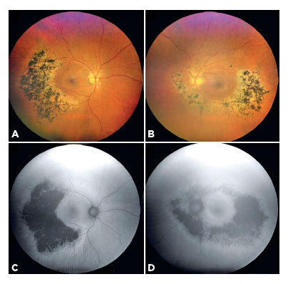
A 58-year-old woman presented with bilateral, asymmetric sector retinitis pigmentosa (RP) (Figure A, B). Fundus autofluorescence imaging revealed hyperautofluorescent rings surrounding hypoautofluorescent regions corresponding to bone spicule pigmentation and retinal degeneration (Figure C, D). The left eye also exhibited nuclear cataract–induced media opacities. Despite the asymmetry, macular integrity was relatively well-preserved in both eyes. Asymmetric and sector RP is a rare variant of the disease characterized by localized retinal involvement, typically affecting specific quadrants and displaying less symmetry compared with the classic diffuse form of RP(1,2).
REFERENCES
1. Lal T, Yu ZX, Guan B, Bender C, Chan CC, Cukras CA, et al. Clinical and histopathologic correlates of asymmetric retinitis pigmentosa. JAMA Ophthalmol. 2021;139(9):1029-32.
2. Coussa RG, Basali D, Maeda A, DeBenedictis M, Traboulsi EI. Sector retinitis pigmentosa: Report of ten cases and a review of the literature. Mol Vis. 2019;25:869-89.
Submitted for publication:
January 30, 2025.
Accepted for publication:
May 9, 2025.
Informed consent was obtained from all patients included in this study.
Data availability statement: All data generated or analyzed during this study are included in this published article.
Funding: This study received no specific financial support.
Disclosure of potential conflicts of interest: The authors declare no potential conflicts of interest.

How to cite this article:
 ABO is licensed under a Creative Commons Attribution-NonComercial 4.0 Internacional.