

Arq. Bras. Oftalmol. 2026; 89 (4): 10.5935/0004-2749.2026-0010
Total: 942
Hugo Diehl de Souza1; José de Mello Rosatelli Neto2; Sofia Cabral Saqueti3; Maria Antônia Vicente Camargo3; Raíssa Gomes Nunes4; Renan Nola Schmoeller5; Jefferson Luiz Brum Marques6
DOI: 10.5935/0004-2749.2026-0010
ABSTRACT
PURPOSE: To evaluate changes in scotopic pupil diameter before and after cataract surgery performed by phacoemulsification with intraocular lens implantation.
METHODS: This prospective longitudinal observational study included patients who underwent cataract surgery. Scotopic pupil diameter was measured preoperatively and 30-40 days postoperatively using an automated keratometer after a standardized dark-adaptation period under controlled ambient illumination. Each eye was considered an independent unit of observation. Because some participants contributed both eyes, intraindividual correlation was accounted for using a linear mixed-effects model with random patient intercepts. Time of assessment (preoperative versus postoperative), age, sex, and eye laterality were included as fixed effects.
RESULTS: A total of 354 eyes from 251 patients were analyzed. The mean patient age was 69.3±7.2 yr. Mean scotopic pupil diameter decreased from 5.3±0.9mm preoperatively to 4.8±0.8mm postoperatively, representing a mean reduction of 0.5mm (9.4%). In the linear mixed-effects model, cataract surgery was associated with a significant reduction in pupil diameter, with an adjusted mean difference of 0.45mm (95% confidence interval [95% CI], 0.39-0.51; p<0.001). Age (p=0.061), sex (p=0.920), and eye laterality (p=0.152) were not significantly associated with the magnitude of pupil diameter change.
CONCLUSION: Phacoemulsification with intraocular lens implantation was associated with a significant reduction in scotopic pupil diameter, independent of age, sex, and eye laterality. This finding should be considered during preoperative planning, particularly when selecting intraocular lenses whose optical performance depends on postoperative pupil size.
Keywords: Cataract; Pupil; Phacoemulsification; Lens implantation, intraocular; Lenses, intraocular; Pseudophakia
INTRODUCTION
Cataract, defined as opacification of the crystalline lens, is most commonly associated with aging and remains the leading reversible cause of blindness worldwide(1). In Brazil, approximately 73.3% of individuals older than 75yr are affected by the condition(2). Cataract-related visual impairment is considered reversible because vision can be restored through phacoemulsification with intraocular lens (IOL) implantation, in which the opacified crystalline lens is replaced with an artificial lens.
Several studies have shown that cataract surgery performed by phacoemulsification with IOL implantation reduces pupil diameter by approximately 10% in the general population under both photopic and mesopic conditions(3-6). This reduction has been attributed primarily to improved ciliary muscle function following cataract extraction. Replacing the crystalline lens, whose anteroposterior thickness frequently exceeds 4mm, with an IOL of approximately 1mm thickness increases the available intraocular space and deepens the anterior chamber(6). In addition, postoperative inflammatory mechanisms affecting the ciliary muscle and anterior chamber have been proposed as contributing factors to the observed reduction in pupil diameter(7).
More recently, increasing attention has been directed toward predicting postoperative pupil diameter because the performance of multifocal and extended depth-of-focus IOLs is highly dependent on pupil size(8). These advanced intraocular implants can provide improved visual performance across a range of focal distances, enabling patients to perform near tasks, such as reading and sewing as well as distance activities, including driving and sports. However, the effectiveness of these technologies is closely linked to pupillary function, which can be assessed before surgery.
The number of cataract surgeries performed within the Brazilian Unified Health System has increased substantially in recent years and has, for the first time, reached a level sufficient to offset the incidence of new cataract cases in the country(9). In addition to highlighting the public health relevance of cataract treatment, this trend creates opportunities for further advances in surgical techniques and clinical knowledge. Therefore, investigating postoperative changes in pupil diameter is important for improving the understanding of ocular physiology and optimizing outcomes following phacoemulsification surgery.
METHODS
This prospective, longitudinal, observational study evaluated scotopic pupil diameter before cataract surgery and approximately 1 month after the procedure. The study was conducted as part of a research project approved by the Human Research Ethics Committee of the Federal University of Santa Catarina (UFSC; Certificate of Ethical Approval No. 81492624.0.0000.0121). Written informed consent was obtained from all participants before enrollment.
Data collection
The study included patients who underwent cataract surgery at Hospital IGAPS, Tijucas, Santa Catarina, Brazil, between October 2024 and January 2025. All procedures were performed by a single experienced surgeon using the Constellation Vision System (Alcon Laboratories, Fort Worth, Texas). A biconvex hydrophilic acrylic intraocular lens (MFR2; Mediphacos, Belo Horizonte, Brazil) was implanted in the capsular bag in all cases. Data collected included age, sex, operated-eye laterality, and preoperative and postoperative pupil diameters.
Because some participants contributed data from both eyes, each eye was treated as an observational unit, and intraindividual correlation was accounted for in the statistical analysis using a linear mixed-effects model. Exclusion criteria included pupillary abnormalities, intraoperative complications, glaucoma, use of topical medications known to affect pupil size (e.g., brimonidine or pilocarpine), white or traumatic cataracts, a history of uveitis, and previous intraocular surgery.
Horizontal pupil diameter under scotopic conditions was measured before phacoemulsification and repeated 30-40 days after surgery. Before each assessment, participants underwent dark adaptation for at least 15 min. Measurements were obtained using the pupillometry function of the PRK-8000 keratometer (Potec, Daejeon, South Korea) by a trained examiner. Ambient illumination was standardized using identical artificial lighting conditions and verified with a lux meter positioned between the participant and the keratometer.
All data were recorded in a digital database under investigator supervision and analyzed in anonymized form.
Statistical analysis
Statistical analyses were performed using Statistical Package for the Social Sciences version 23.0 for Windows (IBM Corp., Armonk, New York). Quantitative variables were expressed as mean±standard deviation (SD) when normally distributed and as median and interquartile range (IQR) when normality assumptions were not met. Categorical variables were presented as absolute and relative frequencies.
To account for intraindividual correlation arising from bilateral surgeries, a linear mixed-effects model with a random intercept for each participant was fitted. Time point (preoperative vs. postoperative), age, sex, and eye laterality were included as fixed effects. Model parameters were estimated using restricted maximum likelihood.
Additional exploratory analyses were performed to characterize the preoperative profile of the study population. The distribution of preoperative pupil diameter was assessed using the Shapiro-Wilk test. As the data were normally distributed, Student’s t-test was used to compare preoperative pupil diameter between sexes. The association between age and preoperative pupil diameter was evaluated using Pearson’s correlation coefficient.
RESULTS
A total of 354 eyes from 251 patients were included in the analysis. The demographic and clinical characteristics of the study population are summarized in table 1. Participants who underwent bilateral surgery contributed two observations, one for each eye, to analyses involving age and sex.
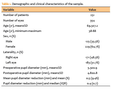
The mean age of the participants was 69.3±7.2 years (range, 38-88 yr). Of the 354 eyes evaluated, 171 (48.3%) were right eyes and 183 (51.7%) were left eyes. Female patients accounted for 229 eyes (64.7%), whereas 125 eyes (35.3%) belonged to male patients.
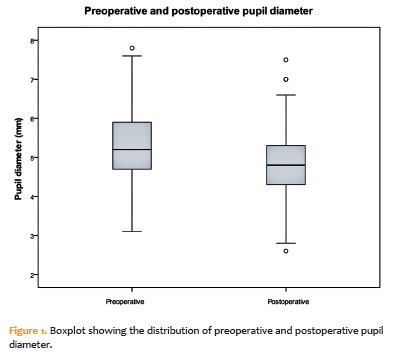
Mean preoperative scotopic pupil diameter was 5.3±0.9mm, decreasing to 4.8±0.8mm postoperatively (Figure 1). This corresponded to a mean reduction of 0.5mm, representing a 9.4% decrease in pupil diameter. The median reduction was 0.4mm (IQR, 0.7mm).
Preoperative pupil diameter did not differ significantly between male and female patients (5.34±0.9mm vs. 5.24±0.9 mm, respectively; p=0.320). Likewise, no significant correlation was observed between preoperative pupil diameter and age (Pearson’s r=−0.089; p=0.096).
The linear mixed-effects model demonstrated a significant reduction in pupil diameter following surgery, with an adjusted mean difference of 0.45 mm between preoperative and postoperative measurements (95% confidence interval [95% CI], 0.39-0.51; p<0.001). In the adjusted analysis, age (p=0.061), sex (p=0.920), and eye laterality (p=0.152) were not significantly associated with the magnitude of pupil diameter change.
Postoperatively, pupil diameter decreased in 271 eyes (76.6%), remained unchanged in 25 eyes (7.1%), and increased in 58 eyes (16.4%).
DISCUSSION
The study sample was well balanced with respect to eye laterality, with a predominance of female patients at an approximate ratio of 2:1. The mean age of 69.3 yr was consistent with expectations for a cataract population, as this disease predominantly affects older individuals. Nevertheless, younger patients were also included (youngest age, 38 yr), which may be explained by factors associated with cataract development, including corticosteroid use, smoking, and diabetes(10,11).
Analysis of preoperative pupil diameter revealed no significant difference between sexes, with a mean scotopic pupil diameter of 5.3mm. A weak negative correlation between age and preoperative pupil diameter was observed, consistent with the age-related miosis reported in the general population(12,13), although this association was not statistically significant.
This study demonstrated a significant and consistent reduction in pupil diameter following cataract surgery, with a mean decrease of approximately 9.4% under scotopic conditions. This finding remained significant after adjustment for intraindividual correlation using a linear mixed-effects model, supporting the robustness of the results and confirming that phacoemulsification has a measurable effect on pupillary dynamics.
The magnitude of the reduction observed is consistent with previous reports describing a decrease in pupil diameter of approximately 10% following phacoemulsification(3,5,6) under both photopic and mesopic conditions. Previous studies have suggested that this phenomenon is partly attributable to the replacement of the relatively thick crystalline lens with a thin IOL(6), resulting in increased anterior chamber depth and more efficient ciliary muscle contraction. In addition, transient inflammatory mechanisms associated with the surgical procedure have been proposed as contributors to postoperative pupillary changes.
From a clinical perspective, the reduction in postoperative pupil diameter is particularly relevant given the increasing use of multifocal and extended depth-of-focus intraocular lenses(14). The optical performance of these lenses is closely dependent on postoperative pupil size, and overestimation of pupil diameter based solely on preoperative measurements may lead to suboptimal visual outcomes. Therefore, the present findings highlight the importance of considering the tendency toward postoperative miosis during surgical planning and IOL selection.
In the adjusted model, pupil diameter variation was not significantly associated with sex, laterality, or age, suggesting that postoperative pupillary reduction is primarily related to the surgical procedure itself rather than to demographic or anatomical characteristics.
The large sample size also enabled characterization of individual pupillary behavior. Although a clear population-level trend toward reduced pupil diameter was observed, a smaller proportion of eyes exhibited stable or increased pupil diameter postoperatively. This finding highlights the persistence of interindividual variability and reinforces the need for individualized clinical assessment.
This study has several limitations. Pupil diameter is influenced by multiple physiological, environmental, and clinical factors. Although efforts were made to standardize these variables, complete control was not possible. Postoperative pupil diameter was assessed at a single time point, precluding evaluation of temporal changes. In addition, measurements were performed exclusively under scotopic conditions, limiting extrapolation of the findings to photopic or mesopic environments. Finally, examiner masking was not feasible because preoperative and postoperative assessments were conducted during separate clinical visits.
Despite these limitations, the present study provides objective and statistically robust data on pupil diameter changes following cataract surgery in a Brazilian population. The findings reinforce the importance of preoperative pupillary assessment and suggest that postoperative miosis following phacoemulsification should be routinely considered during surgical planning, particularly when implanting IOL whose optical performance depends on pupil diameter. Future studies involving different populations, longer follow-up periods, and multiple lighting conditions may further improve understanding of this phenomenon and its clinical implications.
ACKNOWLEDGMENTS
The authors thank the Ophthalmology Academic League of the University of Southern Santa Catarina (LAO-UNISUL) for assistance with data collection
AUTHORS’ CONTRIBUTIONS
Significant contribution to conception and design: Hugo Diehl de Souza, José de Mello Rosatelli Neto, Jefferson Luiz Brum Marques Data Acquisition: Hugo Diehl de Souza, José de Mello Rosatelli Neto, Sofia Cabral Saqueti, Maria Antônia Vicente Camargo, Renan Nola Schmoeller Data Analysis and Interpretation: Hugo Diehl de Souza, Raíssa Gomes Nunes, Jefferson Luiz Brum Marques Manuscript Drafting: Hugo Diehl de Souza, Sofia Cabral Saqueti, Maria Antônia Vicente Camargo, Raíssa Gomes Nunes, Renan Nola Schmoeller Significant intellectual content revision of the manuscript: José de Mello Rosatelli Neto, Jefferson Luiz Brum Marques Have given final approval of the submitted manuscript (mandatory participation for all authors): Hugo Diehl de Souza, José de Mello Rosatelli Neto, Sofia Cabral Saqueti, Maria Antônia Vicente Camargo, Raíssa Gomes Nunes, Renan Nola Schmoeller, Jefferson Luiz Brum Marques Statistical analysis: Hugo Diehl de Souza, Raíssa Gomes Nunes, Jefferson Luiz Brum Marques Obtaining funding: Not applicable Supervision of administrative, technical, or material support: Hugo Diehl de Souza, José de Mello Rosatelli Neto Research group leadership: Jefferson Luiz Brum Marques
REFERENCES
1. Flaxman SR, Bourne RR, Resnikoff S, Ackland P, Braithwaite T, Cicinelli MV; Vision Loss Expert Group of the Global Burden of Disease Study. Global causes of blindness and distance vision impairment 1990-2020: a systematic review and meta-analysis. Lancet Glob Health. 2017;5(12):e1221-34.
2. Ottaiano JA, Ávila MP, Umbelino CC, Taleb AC. As condições da saúde ocular no Brasil. São Paulo: Conselho Brasileiro de Oftalmologia; 2019.
3. Fang X, Xue W, Yu C, Tao J, Wang Y. Correlation between pupillary size and depth of focus after the implantation of extended depth of focus intraocular lenses. Graefes Arch Clin Exp Ophthalmol. 2024;262(12):3897-903.
4. Alió JL, Plaza-Puche AB, Férnandez-Buenaga R, Pikkel J, Maldonado M. Multifocal intraocular lenses: an overview. Surv Ophthalmol. 2017;62(5):611-34.
5. Fernández J, Rodríguez-Vallejo M, Martínez J, Tauste A, Piñero DP. Biometric factors associated with the visual performance of a high addition multifocal intraocular lens. Curr Eye Res. 2018;43(8):998-1005.
6. Kanellopoulos AJ, Asimellis G, Georgiadou S. Digital pupillometry and centroid shift changes after cataract surgery. J Cataract Refract Surg. 2015;41(2):408-14.
7. Totsuka K, Kato S, Shigeeda T, Honbo M, Kataoka Y, Nakahara M, et al. Influence of cataract surgery on pupil size in patients with diabetes mellitus. Acta Ophthalmol. 2012;90(3):e237-9.
8. Fernández J, Rodríguez-Vallejo M, Martínez J, Burguera N, Piñero DP. Pupil dependence assessment with multifocal intraocular lenses through visual acuity and contrast sensitivity defocus curves. Eur J Ophthalmol. 2021;31(6):2989-96.
9. Kara-Junior N, Rossi S. Strategies and challenges for cataract surgery in Brazil. Arq Bras Oftalmol. 2025;88(5):e20241018.
10. Cankurtaran V, Ilhan C, Tekin K, Citirik M, Dirican E, Gurkan E. Use of automated quantitative pupillometric evaluation for monitoring the severity of diabetic retinopathy. Arq Bras Oftalmol. 2021;84(1):37-44.
11. Simsek A, Toptan M. The evaluation of pupil diameter by using Sirius before and after phacoemulsification in healthy, diabetic and hypertension patients. Medicine (Baltimore). 2023;102(16):e33223.
12. Kiel M, Grabitz SD, Hopf S, Koeck T, Wild PS, Schmidtmann I, et al. Distribution of pupil size and associated factors: results from the population-based Gutenberg Health Study. J Ophthalmol. 2022;2022:9520512.
13. Fernández J, Rodríguez-Vallejo M, Martínez J, Burguera N, Piñero DP. Pupil diameter in patients with multifocal intraocular lenses. J Refract Surg. 2020;36(11):750-6.
14. Romano V, Madrid-Costa D, Alfonso JF, Alio J, Allan B, Angunawela R, et al. Recommendation for presbyopia-correcting intraocular lenses: a Delphi consensus statement by the ESASO study group. Am J Ophthalmol. 2023;253:169-80.
Submitted for publication:
January 23, 2026.
Accepted for publication:
May 8, 2026.
Approved by the following research ethics committee: Universidade Federal de Santa Catarina - UFSC (CAAE: 81492624.0.0000.0121).
Data Availability Statement: The datasets generated and/or analyzed during the current study are already available.
Edited by
Editor-in-Chief: Newton Kara-Júnior
Associate Editor: Camila Koch
Funding: This study received no specific financial support.
Disclosure of potential conflicts of interest: The authors declare no potential conflicts of interest.

How to cite this article:
 ABO is licensed under a Creative Commons Attribution-NonComercial 4.0 Internacional.

