
Arq. Bras. Oftalmol. 2026; 89 (6): 10.5935/0004-2749.2025-0383
Total: 16
Fatma Isil Sozen-Delil1; Raziye Dönmez Gün1
DOI: 10.5935/0004-2749.2025-0383
ABSTRACT
PURPOSE: To characterize conjunctiva-scleral thickness over the Ahmed glaucoma valve tube in eyes clinically considered to be at increased risk of tube exposure based on a superficially positioned tube on anterior segment optical coherence tomography, and to explore the association between post-operative follow-up duration and conjunctiva-scleral thickness.
METHODS: This retrospective study included 22 eyes of 21 patients with 23 Ahmed glaucoma valve tubes. One patient contributed both eyes, and another had two Ahmed glaucoma valve tubes in one eye. All eyes had a minimum post-operative follow-up of 6 months and a clinically apparent superficial tube trace on slit-lamp examination. Over-the-tube conjunctiva-scleral thickness and total conjunctiva-scleral thickness were measured at corresponding locations using anterior segment optical coherence tomography. Associations between conjunctiva-scleral thickness measurements and post-operative follow-up duration were evaluated using correlation analysis and generalized estimating equation models to account for clustering.
RESULTS: The mean age was 56 ± 12 years (range, 38–80 years). Best-corrected visual acuity was 1.00 (range, 0.10–3.00) LogMAR, and intraocular pressure was 16 (range, 7–27)mmHg. The median post-operative follow-up duration was 40 months (range, 6–135 months), and the median CST overlying the tube at its most superficial location was 190 μm (range, 54–552 μm). A modest inverse correlation was observed between post-operative follow-up duration and conjunctiva-scleral thickness overlying the tube (ρ=−0.44, p=0.034). generalized estimating equation analysis showed a similar association (p=0.019). However, when eyes were categorized according to post-operative follow-up duration (≤1 year, 1–5 years, and ≥5 years), no statistically significant differences in conjunctiva-scleral thickness measurements were detected were observed among the groups: 273 (79–552), 190 (54–402), and 119 (64–227) μm, respectively (p=0.087).
CONCLUSION: In this preselected high-risk cohort, a modest inverse correlation was observed between post-operative follow-up duration and conjunctiva-scleral thickness overlying the tube; however, this association was not consistently demonstrated across analytical approaches and should therefore be considered exploratory. Anterior segment optical coherence tomography allows objective characterization of tissue coverage over the Ahmed glaucoma valve tube. Further prospective studies with larger cohorts and appropriate control groups are warranted to determine the clinical significance of these findings.
Keywords: Glaucoma; Ahmed glaucoma valve; Intraocular pressure; Anterior segment; Optical coherence tomography; Conjunctiva; Scleral thickness
INTRODUCTION
Glaucoma is the leading cause of irreversible vision loss worldwide. Because the disease is often asymptomatic during its early stages, diagnosis may be delayed(1). Trabeculectomy with mitomycin C and glaucoma drainage devices, including the Ahmed glaucoma valve (AGV), are well-established surgical options for glaucoma management(2).
Tube exposure is a significant complication of AGV implantation and typically occurs within 1–2 years after surgery(3), with a reported incidence of 2.5% to 8.9%(4). Exposure of the tube can cause ocular surface inflammation and increase the risk of late-onset endophthalmitis by providing a pathway for microorganisms to enter the anterior chamber(5).
Anterior segment optical coherence tomography (AS-OCT) has been widely used to assess bleb morphology after AGV implantation(6,7) and to evaluate graft thinning in eyes receiving pericardial or amniotic membrane patch grafts(8). A recent study demonstrated progressive thinning of pericardial patch grafts and stromal tissue covering the AGV tube during a 6-month follow-up period(9). Although AS-OCT has been used to evaluate graft thickness, its role in quantitatively characterizing the conjunctiva-scleral complex over non-grafted, scleral flap-covered tubes remains insufficiently explored. This represents an important gap in the current literature.
Therefore, this study aimed to characterize over-the-tube CST and total CST using AS-OCT in a preselected cohort of eyes considered to be at increased risk of tube exposure and to explore their association with post-operative follow-up duration (PFD).
METHODS
The study was conducted in accordance with the Declaration of Helsinki, and we obtained written informed consent from all participants. The Scientific Research Ethics Committee of our hospital approved the study.
Patients who underwent AGV implantation using the scleral flap technique at the Department of Ophthalmology of our hospital were eligible for inclusion. Among the 156 patients followed in the glaucoma unit between February 2022 and 2024, 21 patients with 22 eyes (23 AGV tubes) and at least 6 months of post-operative follow-up were identified as having a clinically apparent superficial tube trace on slit-lamp biomicroscopy and were referred for AS-OCT evaluation. Superficial tube prominence was defined as a visible tube contour beneath the conjunctiva with minimal overlying tissue coverage on slit-lamp biomicroscopy, suggesting reduced conjunctival protection and a potential risk of tube exposure. One patient contributed both eyes to the study. In another patient, one eye contained two AGV tubes after implantation of a second AGV in the inferonasal quadrant following failure of the initial superotemporal AGV.
Clinical classification was independently performed by two experienced glaucoma specialists, and inclusion was based on consensus. Because of the retrospective study design, the examiners were not masked to the clinical information, and formal interobserver agreement was not assessed. The area of greatest tube prominence was identified clinically, followed by AS-OCT imaging to obtain objective CST measurements. All AS-OCT measurements were performed by an experienced glaucoma specialist. Because only eyes with clinically suspected superficial tube prominence were included, the study was not designed to compare high- and low-risk eyes or establish CST thresholds predictive of tube exposure.
AS-OCT scans
Over-the-tube CST, defined as the combined thickness of the conjunctiva, Tenon’s capsule, and overlying scleral tissue covering the AGV tube, was measured using swept-source AS-OCT (Triton DRI, Topcon, Tokyo, Japan). To localize the area of greatest tube prominence, the most superficial portion of the tube identified on slit-lamp biomicroscopy was marked before imaging (Figure 1). Cross-sectional AS-OCT scans perpendicular to the tube were then obtained (Figure 2). The device provides high-resolution imaging at 100,000 A-scans/s using a 1050-nm wavelength laser and has an imaging depth of 2.6mm(10).
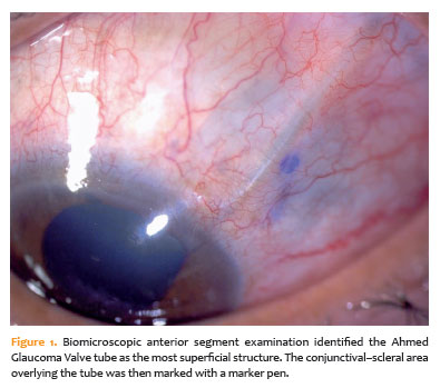
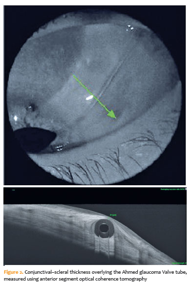
During image acquisition, patients were instructed to look away from the quadrant containing the AGV, while the eyelid was gently elevated to expose the adjacent bulbar conjunctiva. Six-millimeter linear scans were acquired. Minor misalignment between the clinically identified region and the AS-OCT section may have occurred because of eye movement or tissue variability.
Total CST was defined as the combined thickness of the conjunctiva, Tenon’s capsule, and adjacent scleral tissue outside the direct tube elevation. The mean of three manual measurements was used for analysis.
Surgical approach
Sub-Tenon’s anesthesia was administered to all patients before surgery. The FP7 model of the silicone AGV implant (New World Medical Inc., Rancho Cucamonga, California) was used in all cases. One eye underwent implantation of a second AGV in the inferonasal quadrant following failure of the initial superotemporal AGV. In another patient, the AGV was implanted in the inferonasal quadrant because of superior conjunctival scarring.
After an incision was made in the appropriate quadrant for valve placement, a 4 × 5-mm half-thickness, limbus-based scleral flap was created using a fornix-based conjunctival approach. Balanced salt solution was then injected to prime the valve mechanism. The Ahmed valve reservoir was subsequently advanced toward the equator of the globe beneath the conjunctiva and sutured to the sclera with 6-0 Vicryl sutures (Ethicon, New Jersey), 9 mm posterior to the limbus. A 22-gauge needle with a black tip was used to enter the anterior chamber beneath the scleral flap. The tube was angled and trimmed to leave 2–3 mm within the anterior chamber and was then secured in position.
The tube tip was placed in the sulcus in two cases. One of these eyes had extensive 360° synechiae and a history of keratoplasty and graft rejection, whereas the other had peripheral anterior synechiae in the superotemporal region. The scleral flap was sutured with interrupted 10-0 nylon sutures, and a nylon U-suture was used to secure the tube at the scleral flap entry site. Finally, the conjunctiva and Tenon’s capsule were closed with continuous 8-0 Vicryl sutures.
None of the eyes had undergone previous glaucoma surgery, and no antimetabolites, such as mitomycin C, were used during surgery. Postoperatively, patients were prescribed topical antibiotics for 2 weeks and topical corticosteroids for 4 weeks.
Post-operative ophthalmologic examination findings, including best-corrected visual acuity (BCVA) measured in logMAR, anterior and posterior segment findings, intraocular pressure (IOP) measured by Goldmann applanation tonometry, and retinal nerve fiber layer thickness measured using the same OCT device, were recorded. Age, sex, glaucoma diagnosis, number of antiglaucoma medications, AGV tube location, AGV tube tip location, and PFD were also recorded. The association between CST measurements and PFD was evaluated.
Statistical analysis
Statistical analyses were performed using SPSS version 24 (IBM SPSS Statistics, Armonk, New York). Data distribution was assessed using the Shapiro–Wilk test. Normally distributed variables are presented as mean±standard deviation, whereas nonnormally distributed variables are presented as median (range) and interquartile range, as appropriate.
The Kruskal–Wallis test was used to compare independent groups. To account for within-patient correlation arising from clustered observations, including bilateral observations from one patient and two AGV tubes in one eye of another patient, generalized estimating equation (GEE) analysis with an exchangeable correlation structure and robust standard errors was used as the primary analytical approach. Correlations were assessed using Spearman’s rank correlation test, and linear regression analyses were performed when appropriate. Intraobserver repeatability was assessed using intraclass correlation coefficients (ICCs) and coefficients of variation (CVs). A p value <0.05 was considered statistically significant.
RESULTS
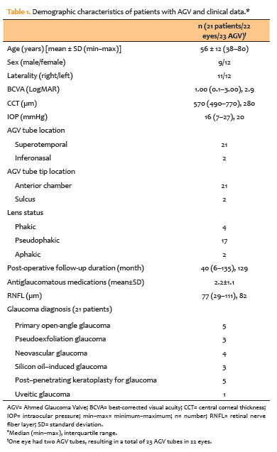
Twenty-one patients (22 eyes; 9 men and 12 women) with a mean age of 56±12 years (range, 38–80 years) were included. One patient contributed both eyes, whereas another had two AGV tubes in one eye, resulting in a total of 23 AGV tubes. The second AGV in the same eye was implanted in the inferonasal quadrant following failure of the initial superotemporal AGV. The median BCVA was 1.00 logMAR (range, 0.10–3.00), the median postoperative IOP was 16mmHg (range, 7–27mmHg), and the median follow-up duration was 40 months (range, 6–135 months). Most eyes were pseudophakic. Indications for AGV implantation included primary open-angle glaucoma, pseudoexfoliative glaucoma (PEXG), neovascular glaucoma (NVG), silicone oil–induced glaucoma, post–penetrating keratoplasty glaucoma, and uveitic glaucoma (Table 1).
When patients were categorized according to PFD (≤1 year, 1–5 years, and >5 years), over-the-tube CST tended to be thinner with longer PFD; however, the differences in over-the-tube CST and total CST were not statistically significant (p=0.087 and p=0.264, respectively; Table 2). Spearman rank correlation analysis demonstrated a modest inverse correlation between PFD (median, 40 months [range, 6–135 months]) and over-the-tube CST (median, 190 μm [range, 54–552 μm]; ρ=−0.44, p=0.034). In contrast, total CST was not significantly correlated with PFD (p=0.275). Simple linear regression did not demonstrate a statistically significant association between PFD and over-the-tube CST (β=−1.25, 95% CI, −2.63 to 0.13; p=0.074).

When eyes were categorized according to IOP (≤15mmHg, 16–21mmHg, and ≥22mmHg), neither over-the-tube CST nor total CST differed significantly among the groups (p=0.490 and p=0.156, respectively; Table 3). In the GEE analysis, PFD was significantly associated with over-the-tube CST. Compared with the reference category, eyes in the longer follow-up group had significantly lower over-the-tube CST values (β=−58.83 μm, 95% CI, −107.91 to −9.75; p=0.019). Categorized IOP levels were not significantly associated with over-the-tube CST (p=0.723). When total CST was considered the dependent variable, neither PFD category (p=0.074) nor IOP category (p=0.339) was significantly associated with CST. Furthermore, IOP was not significantly correlated with either over-the-tube CST or total CST (p=0.366 and p=0.394, respectively).

In this study, AS-OCT showed that over-the-tube CST represented 27.2%±14.5% of the total CST. Intraobserver repeatability was excellent for both over-the-tube CST (ICC=0.995; CV=3.9%) and total CST (ICC=0.993; CV=0.94%), indicating high measurement reliability and low relative variability.
DISCUSSION
In the present study, CST overlying the AGV tube appeared relatively thin in this preselected high-risk cohort, in which superficial tube prominence had already been identified clinically. Although a statistically significant, modest inverse correlation was observed between PFD and over-the-tube CST (ρ=−0.44, p=0.034), this association was not confirmed when the data were analyzed according to clinically relevant post-operative time categories (≤1, 1–5, and ≥5 years), with no significant differences among groups (p=0.087). Furthermore, the association did not reach statistical significance in the linear regression model, and total CST was not associated with the time elapsed since surgery. These findings suggest that any reduction in tissue thickness over the tube may occur gradually rather than as a distinct difference between post-operative intervals. The discrepancy among analytical approaches may reflect the limited sample size and the influence of extreme values. Therefore, the observed association should be interpreted cautiously and cannot be considered evidence of progressive, time-dependent CST thinning. Rather, it should be regarded as exploratory and hypothesis-generating. Clinically, the weak negative correlation may suggest that eyes with longer PFD have thinner CST; however, given the lack of consistency across analyses, this finding cannot currently be used to guide management or predict tube exposure risk.
The AGV is one of the most widely used glaucoma drainage implants worldwide(11). Conjunctival erosion over the tube or plate is a potentially serious complication following AGV implantation. A previous study reported a 5.8% exposure rate for glaucoma drainage devices, with a mean time to exposure of 25.0±21.3 months(12). Risk factors for exposure include younger age, inflammation, uveitic glaucoma, NVG, inferior device placement, and concomitant procedures, particularly cataract surgery or vitrectomy(3,13). Various surgical techniques have been developed to reduce the risk of tube exposure, including the use of different graft materials and scleral flaps, although each approach has limitations(14). Tenon advancement or duplication techniques have demonstrated efficacy in preventing tube exposure(15,16). Techniques involving long scleral tunnels have also shown promise in reducing conjunctival erosion and tube exposure by isolating the tube from the conjunctiva and minimizing trauma caused by tube movement(17–19). More recently, a suture-guided AGV tube implantation technique has been described to improve tube positioning(20).
In this context, our study was designed to descriptively characterize CST overlying the tube in eyes already considered clinically to be at increased risk, rather than to validate AS-OCT as a predictive or risk-stratification tool. Because the cohort consisted exclusively of eyes with clinically apparent superficial tube prominence, the findings cannot be generalized to the broader AGV population or used to establish clinically meaningful CST thresholds associated with tube exposure. Moreover, more than half of the eyes (n=13) had diagnoses associated with increased conjunctival fragility, including NVG (n=4), silicone oil–induced glaucoma (n=3), post–penetrating keratoplasty glaucoma (n=5), and uveitic glaucoma (n=1), which may have contributed to the relatively thin CST measurements.
AS-OCT is a valuable, noninvasive technique for visualizing and quantifying anterior segment structures(21–23). A recent study reported that, in healthy eyes, the mean conjunctival, episcleral, and scleral thicknesses at the superior quadrant were 77±16, 174±54, and 467±52 μm, respectively, yielding an approximate total thickness of 718 μm(24). However, these normative values represent undisturbed tissues and therefore cannot be directly compared with CST overlying an AGV tube. In the surgical setting, the tissue is altered by conjunctival peritomy, scleral flap dissection, and the mechanical presence of the tube, all of which may reduce its native thickness.
In our study population, the median total CST was 766 μm (range, 552–927 μm), which was broadly consistent with published values. In our surgical practice, the AGV tunnel is designed to allow at least half of the total scleral thickness to cover the tube. In this cohort, the median over-the-tube CST was 190 μm (range, 54–552 μm), corresponding to 27.2%±14.5% of the total CST. Although this proportion was relatively small, no immediate tube exposure was observed. The patients were therefore advised to undergo close clinical follow-up, including AS-OCT evaluation.
Few studies have directly evaluated CST overlying an AGV tube using AS-OCT in the context of exposure risk. Previous studies have primarily focused on bleb morphology after AGV implantation(6,7) or XEN stent placement(25). A recent study evaluating pericardial patch grafts demonstrated significant stromal thinning over the central portion of the tube between 1 and 6 months (p=0.015), followed by stabilization(9). Unlike that study, our study did not longitudinally assess the same eyes and therefore cannot establish time-dependent thinning. Longitudinal follow-up might have demonstrated progressive CST reduction within individual eyes. Refractive effects on AS-OCT thickness measurements are generally minimal when repeated measurements are performed in the same eye. In our study, however, most patients were pseudophakic and CST was compared across different PFD categories; therefore, refractive status was not included in the analysis.
The main limitations of this study were its retrospective design and small sample size, which precluded assessment of longitudinal changes in over-the-tube CST within individual patients. Although no control group with deeply positioned AGV tubes was included, over-the-tube CST was compared with each patient’s total CST, providing an internal reference. Consequently, the findings cannot determine whether CST progressively decreases over time, whether specific CST values are associated with an increased risk of tube exposure, or whether AS-OCT measurements can distinguish high-risk from low-risk eyes. The absence of a control group with clinically deep-seated tubes further limits interpretation and generalizability. Nevertheless, this pilot study provides descriptive data from a preselected high-risk cohort and demonstrates that AS-OCT can facilitate detailed visualization and quantification of tissue coverage over the AGV tube. This approach may be useful for structural monitoring of eyes considered clinically at increased risk of tube exposure, including eyes with concomitant surgeries, PEXG, diabetes, or inferior device placement(13,26). The clinical heterogeneity of the cohort may also have influenced conjunctival characteristics, and potential confounders, including glaucoma diagnosis, tube quadrant, lens status, and previous corneal surgery, could not be adequately adjusted for.
In conclusion, this exploratory analysis of clinically selected eyes demonstrated a modest inverse correlation between PFD and over-the-tube CST. However, the absence of consistent findings across analytical approaches and the limited sample size warrant cautious interpretation. No cases of clinical tube exposure occurred during follow-up; therefore, predictive conclusions regarding exposure risk cannot be drawn. AS-OCT enabled objective structural characterization of CST in eyes with clinically suspected reduced tissue coverage over the tube. These findings should not be interpreted as establishing CST thresholds or predictive markers for tube exposure. Further prospective studies with larger cohorts, longitudinal follow-up, and appropriate comparison groups are needed to clarify the clinical significance of CST measurements and their potential relationship with tube exposure risk.
AUTHORS’ CONTRIBUTIONS
Significant contribution to conception and design: Fatma Isil Sozen-Delil and Raziye Dönmez Gün. Data acquisition: Fatma Isil Sozen-Delil, Raziye Dönmez Gün. Data analysis and Interpretation: Fatma Isil Sozen-Delil. Manuscript drafting: Fatma Isil Sozen-Delil. Significant intellectual content revision of the manuscript: Fatma Isil Sozen-Delil, Raziye Dönmez Gün. Final approval of the submitted manuscript: Fatma Isil Sozen-Delil, Raziye Dönmez Gün. Statistical analysis: Fatma Isil Sozen-Delil. Obtaining funding: not applicable. Supervision of administrative, technical, or material support: Fatma Isil Sozen-Delil, Raziye Dönmez Gün. Research group leadership:Fatma Isil Sozen-Delil, Raziye Dönmez Gün.
REFERENCES
1. Weinreb RN, Aung T, Medeiros FA. The pathophysiology and treatment of glaucoma: a review. JAMA. 2014;311(18):1901–11.
2. Lim R. The surgical management of glaucoma: A review. Clin Exp Ophthalmol. 2022;50(2):213–31.
3. Chaku M, Netland PA, Ishida K, Rhee DJ. Risk factors for tube exposure as a late complication of glaucoma drainage implant surgery. Clin Ophthalmol. 2016;10:547–53.
4. Wishart PK, Choudhary A, Wong D. Ahmed glaucoma valves in refractory glaucoma: a 7-year audit. Br J Ophthalmol. 2010;94(9):1174–9.
5. Al-Torbak AA, Al-Shahwan S, Al-Jadaan I, Al-Hommadi A, Edward DP. Endophthalmitis associated with the Ahmed glaucoma valve implant. Br J Ophthalmol. 2005;89(4):454–8.
6. Jung KI, Lim SA, Park HY, Park CK. Visualization of blebs using anterior-segment optical coherence tomography after glaucoma drainage implant surgery. Ophthalmology. 2013;120(5):978–83.
7. Wagdy FM. Bleb Morphology Using anterior-segment optical coherence tomography after ahmed glaucoma valve surgery with tenon capsule resection. J Ophthalmol. 2020;2020(1):8386135.
8. Sheha H, Tello C, Al-Aswad LA, Sayed MS, Lee RK. Outcomes of the shunt tube exposure prevention study: a randomized clinical trial. Ophthalmol Glaucoma. 2019;2(6):392–401.
9. Akbas YB, Alagoz N, Sari C, Altan C, Yasar T. Evaluation of pericardium patch graft thickness in patients with Ahmed glaucoma valve implantation: an anterior segment OCT study. Jpn J Ophthalmol. 2024;68(3):192–9.
10. Li XX, Wu W, Zhou H, Deng JJ, Zhao MY, Qian TW, et al. A quantitative comparison of five optical coherence tomography angiography systems in clinical performance. Int J Ophthalmol. 2018;11(11):1784–95.
11. Arikan G, Gunenc U. Ahmed Glaucoma Valve Implantation to Reduce Intraocular Pressure: updated Perspectives. Clin Ophthalmol. 2023;17:1833–45.
12. Levinson JD, Giangiacomo AL, Beck AD, Pruett PB, Superak HM, Lynn MJ, et al. Glaucoma drainage devices: risk of exposure and infection. Am J Ophthalmol. 2015;160(3):516–521.e2.
13. Trubnik V, Zangalli C, Moster MR, Chia T, Ali M, Martinez P, et al. Evaluation of risk factors for glaucoma drainage device-related erosions: a retrospective case-control study. J Glaucoma. 2015;24(7):498–502.
14. Thakur S, Ichhpujani P, Kumar S. Grafts in glaucoma surgery: a review of the literature. Asia Pac J Ophthalmol (Phila). 2017;6(5):469–76.
15. Tamcelik N, Ozkok A, Sarıcı AM, Atalay E, Yetik H, Gungor K. Tenon advancement and duplication technique to prevent postoperative Ahmed valve tube exposure in patients with refractory glaucoma. Jpn J Ophthalmol. 2013;57(4):359–64.
16. Tamçelik N, Sarici AM, Yetik H, Ozkök A, Ozkiris A. A novel surgical technique to prevent postoperative Ahmed valve tube exposure through conjunctiva: tenon advancement and duplication. Ophthalmic Surg Lasers Imaging. 2010;41(3):370–4.
17. Kugu S, Erdogan G, Sevim MS, Ozerturk Y. Efficacy of long scleral tunnel technique in preventing Ahmed glaucoma valve tube exposure through conjunctiva. Semin Ophthalmol. 2015;30(1):1–5.
18. Kusaka M, Kujime Y, Yamakawa M, Akimoto M. Baerveldt tube shunt implantation through a long scleral tunnel. Eur J Ophthalmol. 2019;29(4):458–63.
19. Eslami Y, Azaripour E, Mohammadi M, Kiarudi MY, Fakhraie G, Zarei R, et al. Single long scleral tunnel technique for prevention of Ahmed valve tube exposure. Eur J Ophthalmol. 2019;29(1):52–6.
20. Şimşek Ş, Karadağ E, Gün RD. New approach to AGV implantation with scleral tunnel method by prolene suture: şimşek Technique. Eur J Ophthalmol. 2024;34(4):1286–92.
21. Gün RD, Yazıcıoğlu T, Oklar M, Gökkaya N. Assessment of Schlemm’s canal with swept-source optical coherence tomography in Graves’ ophthalmopathy. Graefes Arch Clin Exp Ophthalmol. 2024;262(7):2219–26.
22. Gupta N, Varshney A, Ramappa M, Basu S, Romano V, Acharya M, et al. Role of AS-OCT in Managing Corneal Disorders. Diagnostics (Basel). 2022;12(4):918.
23. Desmond T, Tran V, Maharaj M, Carnt N, White A. Diagnostic accuracy of AS-OCT vs gonioscopy for detecting angle closure: a systematic review and meta-analysis. Graefes Arch Clin Exp Ophthalmol. 2022;260(1):1–23.
24. Teeuw GJ, Vergouwen DP, Ramdas WD, Sanchez-Brea L, Andrade De Jesus D, Rothova A, et al. Assessment of conjunctival, episcleral and scleral thickness in healthy individuals using anterior segment optical coherence tomography. Acta Ophthalmol. 2024;102(5):573–80.
25. Teus MA, Paz Moreno-Arrones J, Castaño B, Castejon MA, Bolivar G. Optical coherence tomography analysis of filtering blebs after long-term, functioning trabeculectomy and XEN® stent implant. Graefes Arch Clin Exp Ophthalmol. 2019;257(5):1005–11.
26. Souza C, Tran DH, Loman J, Law SK, Coleman AL, Caprioli J. Long-term outcomes of Ahmed glaucoma valve implantation in refractory glaucomas. Am J Ophthalmol. 2007;144(6):893–900.
Submitted for publication:
December 17, 2025.
Accepted for publication:
August 19, 2026.
Approved by the following research ethics committee: Kartal Dr. Lutfi Kirdar City Hospital (No. 2024/010.99/2/5)
Data Availability Statement: The datasets produced and/or analyzed in this study can be provided to referees upon request.
Edited by
Editor-in-Chief: Newton Kara-Júnior
Associate Editor: Heloísa Russ
Funding: This study received no specific financial support.
p>Disclosure of potential conflicts of interest: The authors declare no potential conflicts of interest.

How to cite this article:
 ABO is licensed under a Creative Commons Attribution-NonComercial 4.0 Internacional.