
Arq. Bras. Oftalmol. 2026; 89 (6): 10.5935/0004-2749.2026-0225
Total: 20
Birumut Gedik1; Berna Dogan2; Muhammet Kazim Erol2; Fulya Duman2; Elcin Suren2; Sibel Yavuz2; Ozdemir Yuksel2; İsmet Selcuk Ertunc2
DOI: 10.5935/0004-2749.2026-0225
ABSTRACT
PURPOSE: This study aimed to compare retinal, choroidal, and optic disc vascular density parameters before infection and at 6 months and 5 years after infection in individuals with a history of coronavirus disease 2019 (COVID-19) using optical coherence tomography angiography.
METHODS: A total of 17 eyes from 17 healthcare workers were included in this study. Retinal, choroidal, and optic disc vascular parameters were evaluated using optical coherence tomography angiography before infection and at 6 months and 5 years after infection. The Friedman test and Wilcoxon signed-rank test with Bonferroni correction were used for statistical analysis.
RESULTS: The foveal avascular zone area was significantly larger at 5 years after infection than at 6 months after infection (p=0.002). Inner retinal parafoveal (p=0.022) and perifoveal (p=0.012) thicknesses were significantly lower at 5 years after infection than before infection. Total optic disc vascular density was significantly lower at 5 years after infection than at 6 months after infection (p=0.005). Furthermore, intraoptic disc vascular density was significantly lower at 5 years after infection than before infection (p=0.009).
CONCLUSION: Microvascular impairment following COVID-19 may persist over the long term. COVID-19 may also have lasting effects on retinal and optic nerve head microcirculation beyond the acute phase of infection. Larger prospective studies with longer follow-up periods are warranted to clarify the clinical significance of these alterations and their potential impact on visual function.
Keywords: COVID-19; Coronavirus disease; Microcirculation; Microvascular density; Optic disk; Tomography, optical coherence; Angiography; Health personnel
INTRODUCTION
In late 2019, coronavirus disease 2019 (COVID-19) emerged and rapidly evolved into a global pandemic. Although initially recognized as a respiratory tract infection, COVID-19 has subsequently been established as a multisystemic disease. It presents with a broad clinical spectrum, ranging from asymptomatic or mild disease to severe illness and death. Clinical manifestations include fever, dry cough, fatigue, myalgia and arthralgia, sore throat, headache, loss of taste and smell, dyspnea, pneumonia, sepsis, septic shock, multiorgan failure, and thromboembolic events(1,2). Ocular manifestations associated with COVID-19 include ocular redness, tearing, burning, foreign body sensation, itching, diplopia, chemosis, retinal microhemorrhages, keratitis, optic neuropathy, and conjunctivitis(3).
Optical coherence tomography angiography (OCTA) is a noninvasive imaging modality that enables visualization of the retinal vascular network by detecting blood flow within retinal vessels. This technique is based on the detection of motion contrast generated by the movement of erythrocytes(4). Previous studies have demonstrated that COVID-19 can affect vascular structures(5,6). In our previous study, we compared retinal and choroidal vascular density parameters before infection and 6 months after infection using OCTA and observed statistically significant differences(7). In the present study, retinal, choroidal, and optic disc vascular density parameters were evaluated and compared in individuals with a history of COVID-19 at three time points: before infection, 6 months after infection, and 5 years after infection.
The aim of this study was to investigate potential long-term alterations associated with COVID-19 in posterior segment structures and vascular density using OCTA.
METHODS
Patient selection
A total of 17 eyes from 17 healthcare workers who had been followed at the Department of Ophthalmology, University of Health Sciences Antalya Training and Research Hospital, were included in this study. For consistency, the right eye was selected for analysis in all participants, and the same eye was evaluated at all three assessment time points. None of the participants had a known systemic or ocular disease. The study cohort comprised individuals who had previously participated as a control group in a separate study conducted before the COVID-19 pandemic and who subsequently developed COVID-19 infection and recovered without requiring intensive care unit admission.
COVID-19 disease severity was classified according to the criteria established by the World Health Organization (WHO). Mild disease was defined as symptomatic COVID-19 without evidence of viral pneumonia or hypoxia, whereas severe disease was defined as severe pneumonia accompanied by a respiratory rate >30 breaths/min, severe respiratory distress, or oxygen saturation <90% on room air(8). All participants in the present study had experienced a mild clinical course according to these criteria.
In a previous study, comparisons between the pre-COVID-19 period and 6 months after COVID-19 infection were performed in 20 patients. In the present study, three patients were excluded because of inadequate follow-up, resulting in the exclusion of data from all three assessment time points (preinfection, 6 months postinfection, and 5 years postinfection). As this study was a retrospective longitudinal analysis of an existing cohort, no a priori sample size calculation was performed. The final sample size was determined by the availability of participants with complete data at all three assessment time points. Comprehensive ophthalmologic examination findings and OCTA images obtained before infection and at 6 months and 5 years after infection were retrospectively retrieved from patient records and compared.
The exclusion criteria were any ocular pathology, particularly glaucoma, uveitis, or diabetic retinopathy; myopia greater than −6.00 diopters; an axial length >26 mm; a history of ocular surgery other than uncomplicated cataract surgery; and any concomitant systemic disease.
Demographic characteristics and ophthalmologic examination findings were obtained from the participants’ medical records. All participants underwent comprehensive slit-lamp biomicroscopic anterior segment and fundus examinations. OCTA images acquired during the pre-COVID-19 period and at 6 months and 5 years after infection were evaluated comparatively.
All OCTA imaging was performed using the AngioVue system (Optovue Inc., Fremont, California), which is based on spectral-domain OCTA technology.
OCTA measurements and parameters
OCTA measurements were obtained using high-resolution 6 × 6 mm retinal angiography and 4.5 × 4.5 mm optic disc angiography protocols. Only images with a signal strength index >8/10 were included in the analysis.
Foveal vascular density was defined as the percentage of vascular density within a circular area 1 mm in diameter centered on the fovea. Parafoveal vascular density (PAFVD) was defined as the vascular density within the annular region between 1 and 3 mm in diameter, whereas perifoveal vascular density (PEFVD) was defined as the vascular density within the 3- to 6-mm annular zone. Total vascular density was defined as the percentage of overall vascular density within the 6-mm fovea-centered scan area. These measurements represented cross-sectional vascular density values of the superficial capillary plexus (SCP) obtained using the device’s automated analysis mode. Vascular density was calculated as the percentage of hyperreflective (white) pixels representing vascular structures relative to the total analyzed area within the OCTA image.
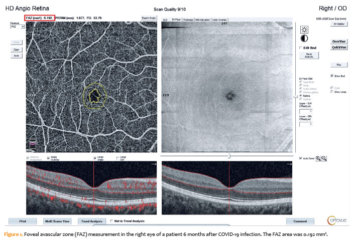
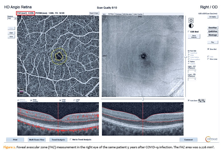
The foveal avascular zone (FAZ) area was determined using the device’s automated segmentation algorithm and recorded in square millimeters (mm²; Figures 1 and 2). Choriocapillaris blood flow (CCBF) was quantified as the area exhibiting flow signals within the choriocapillaris layer, which was automatically identified by the device, in a central region with a 1-mm radius (total area, 3.142 mm²). CCBF was expressed in mm².
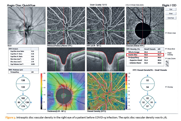
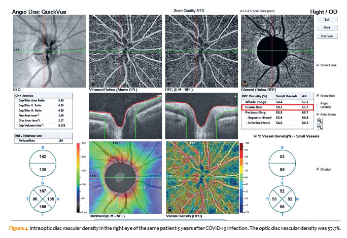
Optic disc vascular density was evaluated as the percentage of vascular density within a circular area 4.5mm in diameter centered on the optic disc (Figures 3 and 4).
Retinal nerve fiber layer (RNFL) thickness was measured using the device’s automated measurement algorithm along a 3.4-mm-diameter peripapillary circular scan centered on the optic disc. Retinal thickness was automatically measured in the foveal, parafoveal, and perifoveal regions as the distance between the internal limiting membrane and the retinal pigment epithelium (RPE).
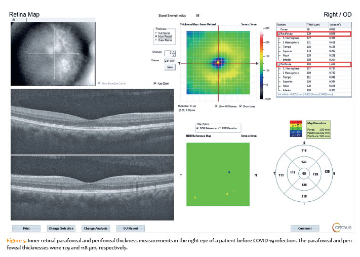
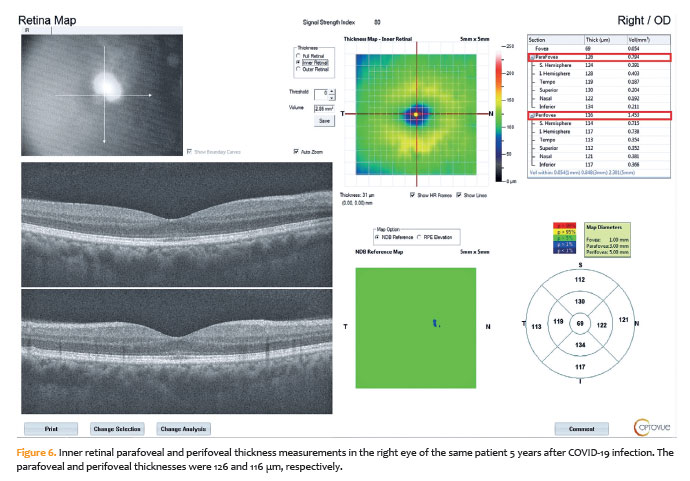
Choroidal thickness was measured manually on “Enhanced HD Line” scans at the subfoveal region by two independent observers. The mean of the two measurements was used for the analysis. Subfoveal choroidal thickness (SCT) was defined as the distance between the RPE and the choroid–scleral interface (Figures 5 and 6).
Statistical analysis
Statistical analyses were performed using SPSS software, version 22.0 (IBM Corp., Armonk, New York). Given the limited sample size, the normality of the data distribution was assessed using analytical methods based on skewness and kurtosis values. Because the data did not conform to a normal distribution, as indicated by skewness and kurtosis values outside the range of −1.5 to +1.5, nonparametric statistical tests were used for subsequent analyses.
Although nonparametric analyses were performed, continuous variables were presented as mean ± standard deviation (SD) to facilitate clinical interpretation. Differences among repeated measurements were evaluated using the Friedman test. When a statistically significant overall difference was identified, post-hoc pairwise comparisons were performed using the Wilcoxon signed-rank test to determine which time points contributed to the observed differences. To control for the type I error associated with multiple comparisons, the Bonferroni correction was applied, and statistical significance for these pairwise comparisons was set at p<0.016 (0.05/3). For all other analyses, p<0.05 was considered statistically significant.
RESULTS
The mean age of the 17 participants was 41.41 ± 1.70 years. Of the participants, 10 were female, and seven were male. Best-corrected visual acuity was 1.0 (20/20) in all 17 eyes at the pre-COVID-19, 6-month, and 5-year follow-up visits, indicating stable visual acuity throughout the follow-up period. None of the participants had pneumonia, and all coagulation parameters, including activated partial thromboplastin time (aPTT), prothrombin time, international normalized ratio (INR), D-dimer, and fibrinogen levels, were within normal limits. Oxygen saturation ranged from 94% to 100% in all participants, and none required supplemental oxygen therapy. All participants received favipiravir and anticoagulant therapy. None required hospitalization or received corticosteroid treatment. No participant had active or persistent COVID-19-related symptoms, including ocular symptoms, at either the 6-month or 5-year follow-up visit. Furthermore, none of the participants experienced a subsequent episode of COVID-19 infection during the 5-year follow-up period.
OCTA measurements obtained before infection and at 6 months and 5 years after infection are presented in Tables 1 and 2. Differences in OCTA measurements across the three time points were initially assessed using the Friedman test. For variables showing statistically significant differences, pairwise comparisons were performed using the Wilcoxon signed-rank test. The Bonferroni correction was applied to control for type I error, and statistical significance for pairwise comparisons was set at p<0.016.
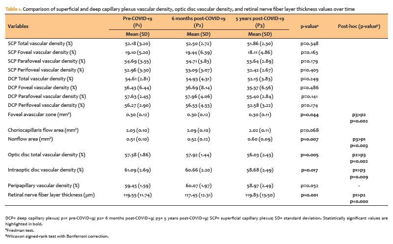
As shown in table 1, SCP and deep capillary plexus (DCP) vascular density, optic disc vascular density, and retinal nerve fiber layer (RNFL) thickness parameters were compared across the three time points. Statistically significant temporal differences were observed in the foveal avascular zone (FAZ) area (p=0.044) and nonflow area (p=0.007). Post-hoc analyses demonstrated a significant increase in the FAZ area at 5 years after infection compared with the 6-month postinfection measurement (p=0.002). In addition, the nonflow area was significantly larger at 5 years after infection than before infection (p=0.003). In the optic disc analysis, total optic disc vascular density was significantly lower at 5 years after infection than at 6 months after infection (p=0.005). Similarly, intraoptic disc vascular density was significantly lower at 5 years after infection than before infection (p=0.009). Regarding RNFL thickness, a statistically significant reduction was observed at 6 months after infection compared with the preinfection measurement (p<0.001). No statistically significant differences were observed in the remaining vascular or structural parameters (p>0.05).
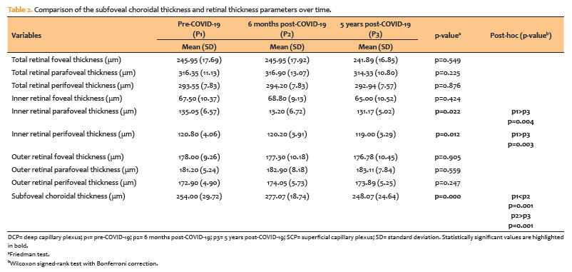
As shown in table 2, retinal thickness parameters and subfoveal choroidal thickness (SCT) were compared across the three time points. Statistically significant differences were observed in inner retinal parafoveal thickness (p=0.022) and inner retinal perifoveal thickness (p=0.012). For both parameters, values at 5 years after infection were significantly lower than the corresponding preinfection measurements (p<0.016). In addition, a statistically significant difference in SCT was observed across the three measurement periods (p<0.001), with SCT increasing significantly at 6 months after infection and subsequently decreasing at 5 years after infection.
DISCUSSION
In this study, long-term alterations in retinal, choroidal, and optic disc vascular structures in individuals with a history of COVID-19 infection were evaluated using OCTA. The principal findings were an increase in the FAZ and nonflow areas, reductions in inner retinal parafoveal and perifoveal thicknesses, an initial increase followed by a long-term decrease in SCT, and reductions in optic disc vascular density parameters. These findings suggest that COVID-19 may have persistent effects on retinal and optic nerve head microvascular structures beyond the acute phase of infection.
Although COVID-19 was initially recognized primarily as a respiratory disease, it has subsequently been established as a multisystemic disorder characterized by endothelial dysfunction, inflammation, activation of coagulation pathways, and microthrombotic processes(1,2,6). Endothelial injury associated with the infection may lead to microcirculatory disturbances, particularly in tissues with dense microvascular networks(2,4,6). The retina and choroid, owing to their high metabolic demands and rich vascular supply, may be particularly susceptible to systemic vascular alterations. Accordingly, OCTA has emerged as an important noninvasive imaging modality for evaluating retinal microvascular changes following COVID-19(7,9–11).
In our study, although no statistically significant differences were observed in superficial and DCP vascular density parameters, significant increases in the FAZ area and nonflow area were observed at 5 years after infection. Enlargement of the FAZ is considered an important indicator of reduced retinal capillary perfusion and microvascular ischemia. Previous OCTA studies conducted after COVID-19 have reported reduced retinal vascular density, FAZ enlargement, and impaired capillary perfusion(12–17). Zapata et al.(12) demonstrated that retinal microvascular abnormalities following COVID-19 may be associated with disease severity. Similarly, a meta-analysis by Kazantzis et al.(17) reported FAZ enlargement in individuals with a history of COVID-19, together with reduced vascular density, particularly in the DCP. The increases in FAZ and nonflow areas observed at 5 years in our study suggest that microvascular alterations following COVID-19 may persist over the long term in some individuals.
Although the precise mechanisms underlying retinal microvascular involvement after COVID-19 remain incompletely understood, endothelial cell injury, inflammatory cytokine responses, complement activation, and microthrombotic processes have been proposed as potential pathogenic mechanisms(2,4,10). The retinal capillary network represents one of the few vascular beds that allows direct in vivo visualization of the systemic microcirculation. Recent studies have reported persistent retinal microvascular changes during the post-COVID-19 and long-COVID periods(13,15,18,19). Our study extends these findings by providing longitudinal data over a 5-year follow-up period.
In our study, inner retinal parafoveal and perifoveal thicknesses were significantly reduced at 5 years after infection, whereas total retinal thickness remained unchanged, suggesting preferential involvement of the inner retinal layers. Previous studies have similarly reported retinal structural alterations following COVID-19, including thinning of the retinal nerve fiber layer, ganglion cell complex, and inner macular structures(15,19–22). These findings may reflect an association between retinal microvascular and neural alterations following COVID-19.
Regarding SCT, our study demonstrated a significant increase at 6 months after COVID-19 infection, followed by a subsequent reduction at the 5-year follow-up. Given its high vascularity, the choroid may be particularly susceptible to systemic inflammation, increased vascular permeability, and hemodynamic alterations. The early postinfection increase in choroidal thickness may be associated with inflammatory activity, vascular congestion, or hyperemia(7,9,15). In contrast, the reduction observed at 5 years suggests that choroidal involvement may be more prominent during the acute and subacute phases, with attenuation of inflammatory effects over the long term. However, whether this late reduction represents a return toward baseline or reflects chronic vascular remodeling remains unclear and warrants further investigation in larger cohorts.
A reduction in optic disc vascular density, particularly at the 5-year follow-up, represents one of the key findings of our study. The microcirculation of the optic nerve head depends heavily on small-vessel perfusion and may therefore be susceptible to systemic endothelial injury. Urfalıoglu et al.(18) reported that OCTA can detect alterations in optic nerve head and retinal microvascular structures even in patients with COVID-19 without pneumonia. Similarly, Batioğlu et al.(19) demonstrated that persistent microvascular changes in the optic disc and macular regions may be present during long-term follow-up after COVID-19 infection. In our study, the significant reduction in total optic disc vascular density at 5 years compared with 6 months after infection, together with the decrease in intraoptic disc vascular density at 5 years compared with preinfection values, suggests that optic nerve head microcirculation may remain altered over the long term following COVID-19.
RNFL thickness decreased at 6 months after infection but returned toward baseline by 5 years, whereas optic disc vascular density remained reduced. This divergent pattern may indicate that vascular alterations persist longer than some neural structural changes and may be more readily detected using OCTA(10,14,19,20).
Compared with our previous findings at 6 months after infection(7), the 5-year results indicate distinct trajectories among the evaluated vascular parameters. The early increase in subfoveal choroidal thickness regressed over time, whereas the FAZ area, nonflow area, and optic disc vascular density demonstrated more persistent alterations.
Physiological aging should also be considered when interpreting the long-term OCTA changes observed in this study. Previous studies have reported age-related reductions in retinal and optic disc vascular density and RNFL thickness(23,24). In our cohort, SCP density showed a small annualized decline of 0.064 percentage points, whereas RNFL thickness did not show a progressive decline over the 5-year period. DCP density showed a numerical decline of 0.492 percentage points per year; however, this change was not statistically significant (p=0.249). Therefore, the contribution of physiological aging cannot be completely excluded, and the observed changes cannot be attributed solely to COVID-19.
Our study has several limitations. First, the relatively small sample size limits the statistical power of the analysis. In addition, because all participants experienced a mild clinical course of COVID-19, the generalizability of the findings to patients with moderate or severe disease may be limited. Nevertheless, a major strength of this study is the availability of pre-COVID-19 OCTA data for all participants, allowing longitudinal comparisons within the same individuals at 6 months and 5 years after infection. To the best of our knowledge, relatively few studies have evaluated retinal and optic disc microvascular structures over such a long follow-up period after COVID-19 infection(19,25). Therefore, our findings provide additional information regarding the potential long-term ocular microvascular effects of COVID-19.
In conclusion, even among individuals who experienced a mild clinical course of COVID-19, long-term alterations in retinal, choroidal, and optic disc microvascular structures may occur. In our study, the increases in FAZ and nonflow areas, together with reduced inner retinal thickness and decreased optic disc vascular density at 5 years, suggest that microvascular alterations following COVID-19 may persist over an extended period. The initial increase in subfoveal choroidal thickness followed by a long-term reduction may indicate that choroidal involvement is more closely associated with transient inflammatory or hemodynamic changes. Although the observed long-term OCTA alterations may be associated with a history of COVID-19, a causal relationship cannot be established. Further prospective studies with larger sample sizes, appropriate control groups, and longer follow-up periods are warranted to clarify the clinical significance of these alterations and their potential impact on visual function.
AUTHORS’ CONTRIBUTIONS:
Significant contribution to conception and design: Birumut Gedik, Berna Dogan, Muhammet Kazim Erol.Data acquisition: Birumut Gedik, İsmet Selcuk Ertunc, Ozdemir Yuksel, Sibel Yavuz. Data Analysis and Interpretation: Birumut Gedik, Berna Dogan, Muhammet Kazim Erol, Fulya Duman, Elcin Suren. Manuscript drafting: Birumut Gedik, Berna Dogan. Significant intellectual content revision of the manuscript: Birumut Gedik, Berna Dogan, Muhammet Kazim Erol, Fulya Duman, Elcin Suren. Final approval of the submitted manuscript: Birumut Gedik, Berna Dogan, Muhammet Kazim Erol, Fulya Duman, Elcin Suren, İsmet Selcuk Ertunc, Ozdemir Yuksel, Sibel Yavuz. Statistical analysis: Birumut Gedik, Berna Dogan. Obtaining funding: not applicable. Supervision of administrative, technical, or material support: Berna Dogan, Muhammet Kazim Erol, Fulya Duman, Elcin Suren. Research group leadership:Birumut Gedik, Berna Dogan, Muhammet Kazim Erol.
REFERENCES
1. Arabi M, Al-Najjar Y, Mhaimeed N, Salameh MA, Paul P, AlAnni J, et al. Severity of the Omicron SARS-CoV-2 variant compared with the previous lineages: A systematic review. J Cell Mol Med. 2023;27(11):1443–64.
2. Xu SW, Ilyas I, Weng JP. Endothelial dysfunction in COVID-19: an overview of evidence, biomarkers, mechanisms and potential therapies. Acta Pharmacol Sin. 2023;44(4):695–709.
3. Al-Dolat W, Abu-Ismail L, Khamees A, Alqudah N, Abukawan MM, Alrawashdeh HM, et al. Is wearing a face mask associated with symptomatic dry eye disease among medical students during the COVID-19 era? An online survey. BMC Ophthalmol. 2022;22(1):159.
4. Jerratsch H, Beuse A, Spitzer MS, Grohmann C. The current status of OCT and OCTA imaging for the diagnosis of long COVID. J ClinTransl Ophthalmol. 2024;2(4):113–30.
5. Vishwakarma N, Goud RB, Tirupattur MP, Katwa LC. The Eye of the storm: investigating the long-term cardiovascular effects of COVID-19 and variants. Cells. 2023;12(17):2154.
6. Tobler DL, Pruzansky AJ, Naderi S, Ambrosy AP, Slade JJ. Long-term cardiovascular effects of covid-19: emerging data relevant to the cardiovascular clinician. Curr Atheroscler Rep. 2022;24(7):563–70.
7. Gedik B, Yuksel O, Kazim Erol M, Duman F, Dogan B, Suren E, et al. Evaluation of the retina, choroid and optic disc vascular structures in individuals with a history of COVID-19. J Fr Ophtalmol. 2024;47(3):104014.
8. World Health Organization (WHO). Clinical management of COVID-19: living guideline, June 2025. Geneva: WHO; 2025.
9. Marzoog BA. Manifestations of COVID-19 in the posterior eye segment - Up-to-date. Oman J Ophthalmol. 2024;17(2):166–72.
10. Teo KY, Invernizzi A, Staurenghi G, Cheung CM. COVID-19-related retinal microvasculopathy: A review of current evidence. Am J Ophthalmol. 2022; 235:98–110.
11. Bajka A, Muth DR, Wiest MR, Said S, Rejdak M, Sidhu S, et al. Analysis of optical coherence tomography and optical coherence tomography angiography parameters in young adults after SARS-CoV-2 infection compared with healthy young controls. Diagnostics (Basel). 2023;13(7):1283.
12. Zapata MA, Banderas García S, Sánchez-Moltalvá A, Falcó A, Otero-Romero S, Arcos G, et al. Retinal microvascular abnormalities in patients after COVID-19 depending on disease severity. Br J Ophthalmol. 2022;106(4):559–63.
13. Zhang C, Cheng S, Chen H, Yang J, Chen Y. New findings on retinal microvascular changes in patients with primary COVID-19 infection: a longitudinal study. Front Immunol. 2024;15:1404785.
14. Noor M, McGrath O, Drira I, Aslam T. Retinal microvasculature image analysis using optical coherence tomography angiography in patients with post-COVID-19 syndrome. J Imaging. 2023;9(11):234.
15. Cosmo E, Frizziero L, Schiavon S, Cattelan AM, Leoni D, Capizzi A, et al. The neurovascular retinal involvement in a large population of patients recovered from COVID-19: an OCT and OCT angiography study. Eye (Lond). 2024;38(9):1674–80.
16. Yılmaz Çebi A, Kılıçarslan O, Uçar D. Evaluation of retinal microvascular impairment after COVID-19 and its clinical correlates using optical coherence tomography angiography. Turk J Ophthalmol. 2022;52(5):324–30.
17. Kazantzis D, Machairoudia G, Theodossiadis G, Theodossiadis P, Chatziralli I. Retinal microvascular changes in patients recovered from COVID-19 compared to healthy controls: A meta-analysis. Photodiagnosis Photodyn Ther. 2023;42:103556.
18. Urfalioğlu S, Akkök B, Özdemir G, Daghan B, Guler M. OCTA evaluation of posterior ocular blood flow in patients after COVID-19 infection without pneumonia. J Fr Ophtalmol. 2023;46(5):468–74.
19. Ozturk M, Kumova Guler D, Oskan EE, Onder F. Long-term effects of COVID-19 on optic disc and retinal microvasculature. Diagnostics (Basel). 2025;15(1):114.
20. Jevnikar K, Meglič A, Lapajne L, Logar M, Vidovič Valentinčič N, Globočnik Petrovič M, et al. The comparison of retinal microvascular findings in acute COVID-19 and 1-year after hospital discharge assessed with multimodal imaging: A prospective longitudinal cohort study. Int J Mol Sci. 2023;24(4):4032.
21. Guçlu ES, Ozer O. Optical coherence tomography analysis of retinal thickness in COVID-19 patients. Kocatepe Tıp Dergisi. 2024;25(2):241–5.
22. Nageeb Louz RE, Salah Eddin MA, Macky TA, Tolba DA. Post COVID-19 retinal evaluation using optical coherence tomography angiography: a case control study. Ocul Immunol Inflamm. 2023;31(6):1175–83.
23. Dastiridou A, Kassos I, Samouilidou M, Koutali D, Mataftsi A, Androudi S, et al. Age and signal strength-related changes in vessel density in the choroid and the retina: an OCT angiography study of the macula and optic disc. Acta Ophthalmol. 2022;100(5):e1095–102.
24. Parikh RS, Parikh SR, Sekhar GC, Prabakaran S, Babu JG, Thomas R. Normal age-related decay of retinal nerve fiber layer thickness. Ophthalmology. 2007;114(5):921–6.
25. Mou K, Gao Y, Zhang Y, Man S, Chen Q, Xu H, et al. Long-term retinal dysfunction following COVID-19 infection: a one-year prospective observational study. BMC Ophthalmol. 2026;26(1):96.
Submitted for publication:
July 31, 2026.
Accepted for publication:
September 9, 2026.
Approved by the following research ethics committee: University of Health Sciences, Antalya Education and Research Hospital (No: 12/20, date: 08/07/2026)
Data Availability Statement: The datasets generated and/or analyzed during the current study are available to referees upon reasonable request.
Edited by
Editor-in-Chief: Newton Kara-Júnior
Associate Editor: Rodrigo P. C. Lira
Funding: This study received no specific financial support.
Disclosure of potential conflicts of interest: The authors declare no potential conflicts of interest.

How to cite this article:
 ABO is licensed under a Creative Commons Attribution-NonComercial 4.0 Internacional.