Showing of 1 until 14 from 173 result(s)
Search for: Eye enucleation; Orbital implants; Eye, artificial; Polyethelene; Image processing, computer-assisted
09-fig01.jpg)
Abstract
Objetivo: Analisar o perfil epidemiológico dos casos de evisceração e enucleação no pronto-socorro oftalmológico de um hospital terciário brasileiro.
Métodos: Análise retrospectiva dos casos tratados no pronto-socorro oftalmológico do Hospital São Paulo (Universidade Federal de São Paulo) entre os anos de 2013 a 2018. Os casos urgentes de evisceração e enucleação foram incluídos e os casos eletivos foram excluídos. A análise dos prontuários médicos foi baseada em: dados demográficos, causas imediatas e associadas ao procedimento, acuidade visual informada, duração dos sintomas antes do atendimento oftalmológico, complicações, distância da residência até o hospital e tempo de hospitalização.
Resultados: 61 enucleações e 121 eviscerações foram incluídas no estudo. Os pacientes tinham uma média de idade de 63,27 ± 18,68 anos; 99 eram do sexo masculino (54,50%) e 83 do sexo feminino (45,60%). As indicações de evisceração e enucleação foram: perfuração corneana com (44,50%) e sem (23,63%) sinais infecciosos, endoftalmite (15,38%), trauma ocular (14,29%), neoplasia (0,55%), queimadura (1,10%) e phthisis bulbi (0,55%). A acuidade visual informada foi de ausência de percepção luminosa (87,36%), percepção luminosa (1.10%), ausência de colaboração (3,30%) e sem dados informados (8,24%). A média de tempo até a busca pelo serviço oftalmológico foi de 18,32 dias. Houve 2 casos de oftalmia simpática após evisceração.
Conclusões: Eviscerações foram predominantemente realizadas em comparação a enucleações em todo o período de estudo. As características demográficas mais comuns foram idade >60 anos e sexo masculino. As principais indicações para procedimentos urgentes de evisceração e enucleação foram perfuração corneana com e sem infecção, endoftalmite e trauma ocular. Este estudo poderia guiar medidas preventivas para evitar procedimentos oculares destrutivos.
Keywords: Evisceração do olho; Enucleação ocular; Úlcera da córnea/epidemiologia; Endoftalmite; Traumatismos oculares; Serviços médicos de emergência; Serviços de saúde ocular.
06-fig01.jpg)
Abstract
Objetivo: Avaliar a resposta tecidual e clínica a um implante orbitário de polimetilmetacrilato, oco e multiperfurado em sua porção posterior em modelo animal após evisceração.
Métodos: Dezesseis coelhos da raça Nova Zelândia foram submetidos à evisceração do globo ocular direito. Todos receberam implante oco de polimetilmetacrilato de 12 mm de diâmetro, multiperfurado em sua semiesfera posterior. O estudo foi dividido em avaliação clínica e histopatológica. A avaliação clínica foi diária até 14 dias pós-evisceração e, a cada sete dias, até completar 180 dias. Os animais foram divididos em grupos de quatro animais e cada um foi submetido à exenteração com 07, 30, 90 e 180 dias e depois à eutanásia. A análise histopatológica teve por fim caracterizar o padrão inflamatório, a estrutura do colágeno e o grau de neovascularização. Para isso, além da tradicional coloração pela hematoxilina-eosina, utilizou-se o corante Picrosirius Red (PSR) e imuno-histoquímica com o marcador CD 34.
Resultados: Não houve sinais de infecção, afinamento conjuntival ou escleral, exposição ou extrusão do implante em nenhum animal durante o estudo. Já no sétimo dia, o tecido neoformado migrou para dentro do implante formando uma rede fibrovascular através dos canais posteriores. A resposta inflamatória diminuiu ao longo do tempo avaliado e não foram encontradas células gigantes multinucleadas.
Conclusão: O implante analisado permite a sua integração aos tecidos orbitários pelo crescimento fibrovascular em seu interior. Os autores acreditam que este modelo de implante orbital pode fazer parte de testes com humanos.
Keywords: Implantes orbitários; Polimetilmetacrilato; Evisceração ocular; Anoftalmia; Procedimentos cirúrgicos oftalmológicos; Coelhos.
Abstract
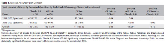
PURPOSE: Natural language models and chatbots, particularly OpenAI’s Generative Pre-Trained Transformer architecture, have transformed human interaction with digital interfaces. The latest versions, including ChatGPT-4o, offer enhanced functionalities compared to their predecessors. This study evaluates the accuracy of ChatGPT-4, ChatGPT-4o, and Claude 3.5 Sonnet in answering questions from the Brazilian Retina and Vitreous Society certification exam.
METHODS: We compiled 200 multiple-choice questions from the Brazilian Retina and Vitreous Society 2018 and 2019 exams. Questions were categorized into three domains: Anatomy and Physiology of the Retina, Retinal Pathology, and Diagnosis and Treatment. Using a standardized prompt developed according to prompt design guidelines, we tested ChatGPT-4, ChatGPT-4o, and Claude 3.5 Sonnet, recording their first responses as final. Three retina specialists performed a qualitative analysis of the answers. Accuracy was determined by comparing responses to the official correct answers. Statistical analysis was conducted using chi-square tests and Cohen’s Kappa.
RESULTS: Claude 3.5 Sonnet achieved the highest overall accuracy (72.5%), followed by ChatGPT-4o (66.0%) and ChatGPT-4 (55.5%). Claude 3.5 Sonnet and ChatGPT-4o significantly outperformed ChatGPT-4 (p<0.01 and p=0.03, respectively), while no significant difference was observed between Claude 3.5 Sonnet and ChatGPT-4o (p=0.16). Model responses agreed 74.5% of the time, with a Cohen’s κ of 0.47. Retinal Pathology was the best-performing domain for all models, whereas Anatomy and Physiology of the Retina and Diagnosis and Treatment were the weakest domains for Claude 3.5 Sonnet and ChatGPT-4, respectively.
CONCLUSIONS: This study is the first to assess Claude 3.5 Sonnet, ChatGPT-4, and ChatGPT-4o in retina specialist certification exams. Claude 3.5 Sonnet and ChatGPT-4o significantly outperformed ChatGPT-4, highlighting their potential as effective tools for studying retina specialist board exams. These findings suggest that the enhanced functionalities of Claude 3.5 Sonnet and ChatGPT-4o offer substantial improvements in medical education contexts.
Keywords: Artificial intelligence; ChatGPT; Retina; Medical education; Ophthalmology, Large language model; Natural language processing
Abstract
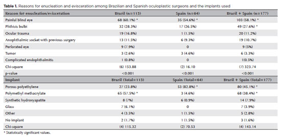
PURPOSE: This study aimed to evaluate the practices employed by oculoplastic surgeons in the assessment and management of anophthalmic sockets and external ocular prostheses.
METHODS: Oculoplastic surgeons from two countries, who specialized in the management of anophthalmic sockets, participated in a web-based survey. Data collected included demographics, types of surgery, implant use, external ocular prostheses management (including fabrication and cleaning), complications encountered, and follow-up times. The frequencies and distributions of the responses were statistically analyzed.
RESULTS: A total of 177 oculoplastic surgeons participated, 113 (63.8%) from Brazil, the remainder from Spain. Evisceration was the preferred surgical procedure of 149 (84.2%) surgeons. The most commonly reported indication for enucleation was a painful blind eye (n=103, 58.1%; both Brazil and Spain, p<0.001). Brazilian surgeons preferred polymethyl methacrylate implants (n=65, 57.5%), while Spanish surgeons favored porous polyethylene implants (n=53, 82.8%; p<0.001). Discharge was the most frequently observed clinical feature during socket evaluation (n=164, 92.6%; p<0.001). Brazilian surgeons recommended daily (n=53, 46.9%) or weekly (n=41, 36.2%) cleaning of external ocular prostheses, while Spanish surgeons more commonly recommended monthly cleaning (n=31, 48.4%; p<0.001). The majority of Brazilian surgeons (n=83, 73.4%) advised patients to remove their external ocular prostheses at night. Only a small number of Spanish surgeons (n=3, 4.6%) suggested this practice (p<0.001). Overall, the follow-up recommendations varied, with 70 (39.5%) surgeons recommending follow-up based on individual case needs, and 59 (33.3%) suggesting annual visits (p<0.001). The primary indications for external ocular prostheses replacement were edge damage (n=75, 42.3%) and loss of volume (n=68, 38.4%). The replacement intervals given typically ranged from 1 to 5 years (n=92, 51.9%; p<0.001).
CONCLUSION: Oculoplastic surgeons in Brazil and Spain demonstrated similar practices in the management of anophthalmic sockets. However, notable differences were observed in the choice of implant materials, cleaning protocols, and recommendations regarding external ocular prostheses removal during sleep.
Keywords: Anophthalmos; Eye, artificial; Polymethyl methacrylate; Polyethylene; Surgeons; Surveys and questionnaires; Brazil; Spain.
Abstract
PURPOSE: This clinical study aimed to assess the effectiveness of microemulsion artificial tears containing povidone and propylene glycol in the management of dry eye disease. Secondary objectives included evaluating improvements in tear-film stability, measured by tear break-up time and corneal staining scores, along with the tolerability and safety of the formulation.
METHODS: This was a prospective, single-arm interventional study involving 30 participants (52 eyes) diagnosed with dry eye disease. Participants self-administered the investigational eye drops twice daily for 28 consecutive days. Primary and secondary outcomes included changes in the Ocular Surface Disease Index, tear break-up time, and corneal staining scores. Adverse events were documented throughout the study period.
RESULTS: Significant improvements in Ocular Surface Disease Index scores were observed, reflecting a reduction in dry eye disease symptoms. Tear break-up time increased notably between baseline and follow-up assessments, with the proportion of eyes exhibiting tear break-up time ≥10 srising from 25.0% to 63.5%. Additionally, the percentage of eyes with a corneal staining score of zero improved from 23.1% to 69.2%. Conjunctival staining also decreased, with the proportion of eyes with scores of 2 and 3 dropping from 11.5% to 3.8% and 5.8% to 0%, respectively.
CONCLUSIONS: The findings suggest that povidone and propylene glycol-based artificial tears significantly enhance tear-film stability and alleviate symptoms in patients with mild to moderate dry eye disease, with minimal adverse effects. This formulation represents a safe and effective short-term treatment option for dry eye disease management.
Keywords: Artificial tears; Dry eye disease; Tear-film stability; Propylene glycol; Povidone; Visual acuity; Surveys and questionnaires

Abstract
PURPOSE: To compare the refractive prediction error of Hill-radial basis function 3.0 with those of 3 conventional formulas and 11 combination methods in eyes with short axial lengths.
METHODS: The refractive prediction error was calculated using 4 formulas (Hoffer Q, SRK-T, Haigis, and Hill-RBF) and 11 combination methods (average of two or more methods). The absolute error was determined, and the proportion of eyes within 0.25-diopter (D) increments of absolute error was analyzed. Furthermore, the intraclass correlation coefficients of each method were computed to evaluate the agreement between target refractive error and postoperative spherical equivalent.
RESULTS: This study included 87 eyes. Based on the refractive prediction error findings, Hoffer Q formula exhibited the highest myopic errors, followed by SRK-T, Hill-RBF, and Haigis. Among all the methods, the Haigis and Hill-RBF combination yielded a mean refractive prediction error closest to zero. The SRK-T and Hill-RBF combination showed the lowest mean absolute error, whereas the Hoffer Q, SRK-T, and Haigis combination had the lowest median absolute error. Hill-radial basis function exhibited the highest intraclass correlation coefficient, whereas SRK-T showed the lowest. Haigis and Hill-RBF, as well as the combination of both, demonstrated the lowest proportion of refractive surprises (absolute error >1.00 D). Among the individual formulas, Hill-RBF had the highest success rate (absolute error ≤0.50 D). Moreover, among all the methods, the SRK-T and Hill-RBF combination exhibited the highest success rate.
CONCLUSIONS: Hill-radial basis function showed accuracy comparable to or surpassing that of conventional formulas in eyes with short axial lengths. The use and integration of various formulas in cataract surgery for eyes with short axial lengths may help reduce the incidence of refractive surprises.
Keywords: Cataract; Lenses, intraocular; Axial length, eye; Refractive errors; Artificial intelligence
10-fig01tb.jpg)
Abstract
Objetivo: Avaliar o desempenho de classificação de modelos ou arquiteturas de rede neural convolucional pré-treinadas usando um conjunto de dados de imagem de fundo de olho contendo oito rótulos de doenças diferentes.
Métodos: Neste artigo, o conjunto de dados de reconhecimento inteligente de doenças oculares publicamente disponível foi usado para o diagnóstico de oito rótulos de doenças diferentes. O banco de dados de reconhecimento inteligente de doenças oculares tem um total de 10.000 imagens de fundo de olho de ambos os olhos de 5.000 pacientes para oito categorias que contêm rótulos saudáveis, retinopatia diabética, glaucoma, catarata, degeneração macular relacionada à idade, hipertensão, miopia, outros. Investigamos o desempenho da classificação de doenças oculares construindo três arquiteturas de rede neural convolucional pré-treinadas diferentes, incluindo os modelos VGG16, Inceptionv3 e ResNet50 com otimizador de Momento Adaptativo. Esses modelos foram implementados no Google Colab o que facilitou a tarefa sem gastar horas instalando o ambiente e suportando bibliotecas. Para avaliar a eficácia dos modelos, o conjunto de dados é dividido em 70% para treinamento, 10% para validação e os 20% restantes utilizados para teste. As imagens de treinamento foram expandidas para 10.000 imagens de fundo de olho para cada tal.
Resultados: Observou-se que o modelo ResNet50 alcançou acurácia de 97,1%, sensibilidade de 78,5%, especificidade de 98,5% e precisão de 79,7% e teve a melhor área sob a curva e pontuação final para classificar a categoria da catarata (área sob a curva=0,964, final=0,903). Em contraste, o modelo VGG16 alcançou uma precisão de 96,2%, sensibilidade de 56,9%, especificidade de 99,2% e precisão de 84,1%, área sob a curva 0,949 e pontuação final de 0,857.
Conclusão: Esses resultados demonstram a capacidade das arquiteturas de rede neural convolucional pré-treinadas em identificar doenças oftalmológicas a partir de imagens de fundo de olho. ResNet50 pode ser uma boa solução para resolver problemas na detecção e classificação de doenças como glaucoma, catarata, hipertensão e miopia; Inceptionv3 para degeneração macular relacionada à idade e outras doenças; e VGG16 para retinopatia normal e diabética.
Keywords: Redes neurais de computação; Aprendizado profundo; Processamento de imagem assistida por computador; VGG16; Inceptionv3; ResNet50; Fundo de olho; Oftalmopatias.

Abstract
PURPOSE: To evaluate the changes in the rates and indications of eye removal procedures during the recent COVID-19 pandemic.
METHODS: The medical records of all patients who underwent eye removal from 2007 to 2022 were retrospectively reviewed. The patient demographic data and indications for surgery were collected. Data from two groups of patients (prepandemic surgery and postpandemic surgery) were compared. Statistical significance was set at p<0.05.
RESULTS: Fifty-nine patients underwent enucleation (69%), evisceration (27%), or exenteration (3%). The mean (SD) age of the patients was 55.9 (19.4) years, and most (69%) of the patients were males. Most (47%) of the study population were Black. The common indications for eye removal were trauma (41%), painful blind eye (34%), and infection/inflammation (24%). The types of trauma were assault (55%), accidental (39%), and self-inflicted (6%). The mean (SD) monthly rates of eye removal increased from 0.25 (0.50) in the prepandemic period to 0.77 (0.91) during the pandemic (p<0.001). These increases were noted in both males (p=0.003) and females (p=0.001) and were the highest among Black patients [0.42 (0.76); p<0.001]. Among the indications of eye removal, painful blind eyes [0.35 (0.75); p<0.001] and ocular trauma [0.31 (0.47); p=0.051] exhibited the greatest increases following the pandemic.
CONCLUSION: The rate of eye removal procedures increased during the recent pandemic. Although delayed care of chronic eye conditions may have contributed to the increased rates of painful blind eyes, the increased trauma-related eye removals may be attributed to the simultaneous spike in violent assaults in New York City.
Keywords: Eye injuries; Eye enucleation; COVID-19; Pandemics; Ethinicity; Inflammation, Trauma centers
14-tab01tb.jpg)
Abstract
OBJETIVOS: À medida que a utilização de equipamentos digitais no emprego aumenta, a avaliação do seu efeito na saúde visual necessita de ferramentas válidas e robustas. Este estudo teve como objetivo traduzir, adaptar culturalmente e validar para português o Questionário da Síndrome Visual do Computador (CVS-Q©).
MÉTODOS: O procedimento foi realizado em 5 fases: tradução direta, síntese da tradução, tradução inversa, consolidação por um painel de especialistas, e pré-teste. Para fazer o pré-teste foi realizado um estudo piloto transversal aplicado a uma amostra de 26 participantes que completaram a versão pré-final da versão portuguesa do CVS-Q©, questionando por dificuldades, compreensão e sugestões de melhoria do questionário. Para avaliar a confiança e validade da versão portuguesa do CVS-Q©foi realizado um estudo transversal de validação em uma amostra diferente (280 funcionários).
RESULTADOS: No pré-teste, 96.2% dos participantes não apresentaram dificuldades no preenchimento do questionário, enquanto 84.0% indicaram que era claro e compreensível. Obteve-se, então, o CVS-Q© em português (Questionário da Síndrome Visual do Computador, CVS-Q PT©). A sua validação revelou uma boa consistência interna
da sua escala (Cronbach's alpha=0.793), boa estabilidade temporal (coeficiente de correlação interclasse=0.847; 95% CI 0.764-0.902, kappa=0.839), sensibilidades e especificidades adequadas (78.5% e 70.7%, respetivamente), boa capacidade de discriminação (área abaixo da curva=0.832; 95% CI 0.784-0.879), e uma adequada validade da convergência com o índice de doença da superfície ocular (ocular surface disease index - OSDI; coeficiente de correlação de Spearman=0.728, p<0.001). A análise fatorial revelou um único fator responsável por explicar a variância comum em 37.7%. Um funcionário com uma pontuação ≥7 pontos sofria de síndrome visual do computador.
CONCLUSÃO: O CVS-Q PT© pode ser considerada uma ferramenta intuitiva, de fácil interpretação e com boas propriedades psicométricas para avaliar a síndrome visual do computador em funcionários portugueses expostos a ecrãs digitais. Este questionário facilitará as decisões sobre medidas preventivas, intervenções e tratamento, e a comparação entre as populações expostas em diferentes países de língua portuguesa.
Keywords: Síndrome visual do computador; Dispositivos digitais; Saúde ocular; Estudo de validação; Propriedades psicométricas; Inquéritos e questionários
Abstract
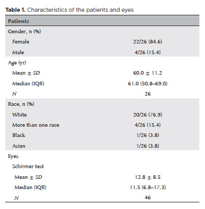
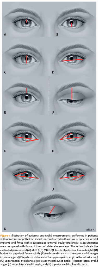
PURPOSE: To quantitatively compare eyebrow and eyelid positions in anophthalmic sockets reconstructed with conical or spherical orbital implants combined with customized external ocular prostheses.
METHODS: This cross-sectional observational study included 38 patients with unilateral anophthalmic sockets, of whom 21 received conical implants, and 17 received spherical implants. Eyelid and eyebrow parameters—including margin reflex distance 1 and 2, vertical and horizontal palpebral fissure dimensions, eyebrow-to-upper-eyelid margin distance in primary gaze and infraduction, medial and lateral eyelid angles in primary gaze, and superior eyelid sulcus depth —were quantitatively assessed using standardized digital photographs analyzed with Image J software. The contralateral healthy eye served as the control. Statistical analyses were performed to compare measurements between groups.
RESULTS: In the primary gaze position, conical and spherical implants showed comparable margin-reflex distance1, margin-reflex distance2, vertical palpebral fissure height, eyelid margin position, and medial and lateral eyelid angles. During infraduction, the upper eyelid margin was significantly lower in sockets reconstructed with conical implants. Compared with contralateral normal eyes, anophthalmic sockets exhibited a reduced horizontal palpebral fissure and a deeper superior eyelid sulcus, irrespective of implant shape.
CONCLUSION: Anophthalmic sockets reconstructed with conical or spherical implants demonstrate similar eyebrow and eyelid positioning in primary gaze. However, conical implants are associated with a lower eyelid margin during infraduction. Independent of implant format, anophthalmic sockets show a narrower horizontal palpebral fissure and increased superior sulcus depth compared with normal eyes.
Keywords: Anophthalmos; Prosthesis implantation; Anophthalmic socket; Conical implants; Spherical implants; Orbital implants; Eyelid measurements
Abstract
PURPOSE: Evaluation of lid contour and marginal peak point changes to compare outcomes of external levator advancement and Müller’s muscle conjunctival resection surgery in unilateral ptosis.
METHODS: We reviewed the charts of unilateral ptosis patients who underwent external levator advancement or Müller’s muscle conjunctival resection. Eyelid contour analysis was conducted on preoperative and 6-month postoperative digital images. This was performed with the multiple margin reflex distances technique, measuring the vertical distance from a line intersecting the center of the pupil to the eyelid margin at 10 positions at 2 mm intervals. The marginal peak point changes were analyzed digitally using the coordinates of the peak point according to the pupil center. Each position’s mean distance was compared preoperatively, postoperatively, and with the fellow eyelid.
RESULTS: Sixteen patients underwent external levator advancement and 16 patients had Müller’s muscle conjunctival resection. The mean margin reflex distance was improved by both techniques (1.46 vs. 2.43 mm and 1.12 vs. 2.25 mm, p=0.008 and p=0.0001 respectively) and approached that of the fellow eyelid (2.43 vs. 2.88 and 2.25 vs. 2.58 mm, p=0.23 and p=0.19, respectively). However, statistically significant lid margin elevation was limited to between the N6 and T6 points in the external levator advancement group. Whereas, significant elevation was achieved along the whole lid margin in the Müller’s muscle conjunctival resection group. The marginal peak point was shifted slightly laterally in the external levator advancement group (p=0.11).
CONCLUSIONS: Both techniques provide effective lid elevation, however, the external levator advancement’s effect lessens toward the canthi while Müller’s muscle conjunctival resection provides more uniform elevation across the lid margin. The margin reflex distance alone is not sufficient to reflect contour changes.
Keywords: Blepharoptosis; Eyelids; Conjunctiva; Oculomotor muscles; Image processing, computer-assisted; Treatment outcome
15-tab01tb.jpg)
Abstract
OBJETIVO: Este estudo visou avaliar os mecanismos da lesão e os tipos de fraturas orbitárias e sua relação com commotio retinae simultânea.
MÉTODOS: Este estudo retrospectivo avaliou registros de pacientes com fraturas orbitárias cujos diagnósticos foram confirmados por tomografia computadorizada entre julho de 2017 e setembro de 2019. Foram registrados os dados demográficos, circunstâncias da lesão, os resultados do exame oftalmológico e achados radiológicos. A análise estatística dos dados usou os testes de t-Student bicaudal, qui-quadrado e cálculos de odds ratio. O significado estatístico foi fixada em p<0,05.
RESULTADOS: Dos 204 pacientes com fraturas orbitárias incluídos neste estudo, 154 (75,5%) eram sexo masculino (75,5%). A média de idade foi de 42,1 anos. As fraturas orbitárias envolvendo uma parede orbital (58,8%) foram mais comuns do que as que acometeram várias paredes (41,2%). A maioria das fraturas acometeu a parede inferior (60,3%), sendo as paredes mediais as próximas mais frequentemente afetadas (19,6%). A causda mais comum de lesão foi agressão (59,3%), e a segunda mais comum foi queda (24%). A commotio retinae foi observada em 20,1% dos casos de fratura orbital e foi mais associada a lesões causadas por agressão (OR=5,22, p<0,001) e menos associada com aquelas causadas por quedas (OR=0,06, p<0,001). As restrições de movimentos oculares eram mais comuns na comoção central do que na periférica (OR=3,79, p=0,015) e com fraturas da parede medial do que com fraturas de outras paredes orbitais (OR=7,16, p<0,001). As chances de comoção não foram maiores em pacientes com fraturas orbitais de paredes múltiplas do que naqueles com fraturas de parede simples (p=0,967).
CONCLUSÕES: Na população do estudo, a agressão foi a causa mais comum de fraturas orbitais e resultou em commotio retinae mais grave do que qualquer outra causa. Os oftalmologistas devem estar cientes da probabilidade de commotio retinae em pacientes com fraturas orbitais resultantes de agressão, independentemente da extensão das lesões do paciente.
Keywords: Fraturas orbitárias; Movimentos oculares; Retina; Ferimentos e lesões
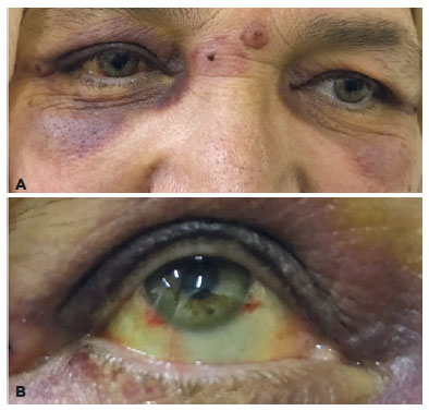
Abstract
Uma paciente de 62 anos procurou nosso ambulatório com queixas de equimose periorbital e hemorragia subconjuntival, visíveis principalmente no olho direito. Descobrimos que suas queixas começaram no dia seguinte a um tratamento para dor de cabeça com sanguessugas na área da glabela. Na glabela, 2 mordidas de sanguessuga foram encontradas próximas ao lado direito. Durante os exames da paciente, foram detectadas equimoses nas pálpebras bilaterais e hemorragia subconjuntival no limbo ínfero lateral e medial do olho direito. Nenhum tratamento foi iniciado, sendo recomendado apenas controle. No acompanhamento, observou-se que as queixas da paciente desapareceram em cerca de um mês.
Keywords: Cefaléia/terapia;Hirudo medicinalis;Aplicação de sanguessugas/efeitos adversos; Doenças orbitárias; Hematoma; Túnica conjuntiva; Hemorragia ocular/etiologia
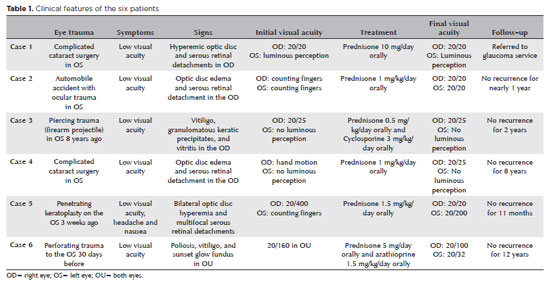
Abstract
A oftalmia simpática consiste em uma panuveíte granulomatosa bilateral rara e potencialmente devastadora, ocorrendo geralmente após trauma ocular cirúrgico ou não cirúrgico. O diagnóstico é baseado em aspectos clínicos e apoiado por exames de imagem, como ultrassonografia ocular e tomografia de coerência óptica. O tratamento consiste em terapia imunossupressora com esteróides e, eventualmente, drogas poupadoras de esteróides, como ciclosporina, azatioprina, ciclofosfamida e micofonato de mofetila. O manejo rápido e eficaz com agentes imunossupressores sistêmicos permite o controle da doença e a obtenção de boa acuidade visual no olho simpatizante. A enucleação, por outro lado, poderia ser considerada apenas em situações em que o olho lesado não tem percepção luminosa ou há trauma grave. Além de uma revisão bibliográfica sobre o tema, foi relatada uma série de 6 casos com diferentes modalidades de tratamento imunossupressor e cirúrgico.
Keywords: Oftalmia simpática; Autoimunidade, Terapia de imunossupressão; Imunossupressores/uso terapêutico; Enucleação ocular; Evisceração do olho; Humanos; Relato de casos
 ABO is licensed under a Creative Commons Attribution-NonComercial 4.0 Internacional.
About
Issues
Editorial Board
Submission
Arquivos Brasileiros de Oftalmologia
Official publication of Brazilian Council of Ophthalmology - Conselho Brasileiro de Oftalmologia (CBO)
Rua Casa do Ator, 1.117 - 2nd floor - Zip Code: 04546-004
São Paulo - SP, Brazil
TEL: +55 11 3266-4000
E-mail: [email protected]