Arq. Bras. Oftalmol. 2023;86 (3 )
:1-5
| DOI: 10.5935/0004-2749.20230031
Abstract
Objetivo: Descrever os resultados clínicos do tratamento do crescimento epitelial através da técnica de remoção manual seguido da utilização de um compressor de ar comprimido aquecido após a cirurgia de laser in situ keratomileusis (LASIK).
Métodos: Vinte olhos de 17 pacientes foram incluídos no estudo. Cada paciente havia sido submetido a cirurgia de LASIK com presença de crescimento epitelial e foi submetido a tratamento cirúrgico para sua retirada. O objetivo primário foi identificar a presença de crescimento epitelial recorrente ao final de 3 meses de seguimento. Os objetivos secundários foram as medidas de acuidade visual sem correção, acuidade visual com correção, e complicações pós-operatórias.
Resultados: Dez pacientes (58,8%) eram homens e 7 mulheres. Oito olhos de sete (41,2%) pacientes apresentavam cirurgia de LASIK primária e 12 olhos de 10 pacientes tinham cirurgia de LASIK com retratamento; dezesseis olhos (80%) utilizaram microcerátomo manual e quatro (20%) laser de femtosegundo. A média de idade no momento da cirurgia de remoção do epitélio era de 37,0 anos ± 9,3 (DP) (variando de 24 a 55 anos). Ocorreu recidiva do crescimento epithelial em dois olhos (10%) após 3 meses de seguimento. A acuidade visual sem correção antes da cirurgia era de 0,07 ± 0,09 logMAR, e após a cirurgia passou para 0,02 ± 0,04 logMAR (p=0,06). A chance (odds ration) de aparecimento do crescimento epithelial após uma reoperação de LASIK é 29,41 vezes maior do que no LASIK primário.
Conclusão: A técnica de remoção epitelial manual seguida da utilização de ar comprimido aquecido é segura e efetiva no tratamento do crescimento epitelial após LASIK. Ao final do último acompanhamento, nenhum olho apresentou perda de linhas de visão.
Keywords: Epitélio/crescimento & desenvolvimento; Endotélio corneano; Doenças da córnea; Ceratomileuse assistida por excimer laser in situ; Ceratectomia fotorrefrativa; Procedimentos cirúrgicos refrativos; Acuidade visual
Arq. Bras. Oftalmol. 2020;83 (4 )
:277-282
| DOI: 10.5935/0004-2749.20200049
Abstract
Objetivo: Este estudo foi realizado para avaliar os resultados do cross-linking corneano acelerado em córneas ceratocônicas com os valores mais baixos de paquimetria <400 μm.
Métodos: O estudo incluiu 28 olhos de 24 pacientes. As acuidades visuais não corrigidas e melhor corrigidas (logMAR), leituras ceratométricas mais planas e íngremes, espessura corneana central no ponto mais fino, aberrações corneanas de mais alta ordem e a sensibilidade ao contraste foram avaliadas antes e em 1, 3, 6, 12 e 24 meses após a realização do do cross-linking.
Resultados: A média da acuidade visual melhor corrigida e a sensibilidade ao contraste aumentaram (p=0,02, p=0,03, respectivamente), enquanto a média da acuidade visual não corrigida não diferiu significativamente (p>0,05) aos 24 meses após o cross-linking, comparada com medidas antes do procedimento. Embora a leitura da média da ceratometria mais plana não tenha apresentado alteração significativa (p=0,58), a leitura ceratométrica mais íngreme diminuiu quando comparada ao seu valor antes do cross-linking (p=0,001). Não foi observada alteração na média da espessura corneana central no ponto mais fino aos 24 meses após o cross-linking em comparação com seu valor antes do procedimento (p=0,12).
Conclusão: O cross-linking corneano acelerado nos olhos ceratocônicos com córneas finas pode interromper a progressão do ceratocone nas córneas mais finas que 400 μm 24 meses após o tratamento.
Keywords: Ceratocone; Paquimetria corneana; Reagentes para ligações cruzadas; Riboblavina/uso terapêutico
Arq. Bras. Oftalmol. 2021;84 (4 )
:324-329
| DOI: 10.5935/0004-2749.20210046
Abstract
OBJETIVO: O ceratocone na população pediátrica apresenta algumas particularidades em relação à população adulta. O maior desafio é devido à doença ser geralmente mais severa e rapidamente progressiva em crianças.
MÉTODOS: Este artigo utiliza uma análise retrospectiva para relatar o uso do crosslinking em jovens menores de 18 anos e sua evolução pelo menos 24 meses após o procedimento. Foram estudados 12 olhos de 10 pacientes, e dados como acuidade visual com e sem correção, ceratometria máxima, espessura corneana, espessura foveal e microscopia endotelial avaliados no pré e pós-operatórios. O crosslinking corneano foi realizado em todos os pacientes pelo mesmo cirurgião.
RESULTADOS: Observou-se uma tendência de redução do valor do Kmax e melhora da acuidade visual corrigida em todos os momentos de pós operatório. Com relação à paquimetria, observou-se afinamento corneano do ponto mais fino, nos primeiros quatro meses de pós-operatório.
CONCLUSÃO: Resultados encorajadores foram obtidos com relação à estabilização da doença, progressão e segurança do procedimento, corroborando com as conclusões de outros autores. A importância do diagnóstico precoce e do acompanhamento a curto prazo do paciente deve ser destacada.
Keywords: Ceratocone/diagnóstico; Ceratocone/tratamento farmacológico; Córnea; Doenças da córnea; Topografia da córnea; Colágeno/metabolismo; Raios ultravioleta; Reagentes para ligações cruzadas/uso terapêutico; Riboflavina/uso terapêutico; Acuidade visual; Adolesc
Arq. Bras. Oftalmol. 2025;88 (4 )
:1-7
| DOI: 10.5935/0004-2749.2023-0356
Abstract
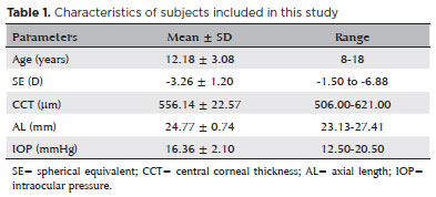
PURPOSE: Although the orthokeratology effects on corneal biomechanics have been proven with clinical trials, reports of stiffness parameter change are scarce. This study investigated the short-term orthokeratology effects in pediatric myopia and compared stiffness parameter changes to those published in recent clinical investigations. This prospective study aimed to investigate corneal biomechanics changes induced by short-term overnight orthokeratology treatment, focusing on stiffness parameter at A1 and stress-strain index
METHODS: Twenty-six children aged 8 to 18 were included in this study using orthokeratology lenses for two different durations: 1 day and 1 week. Corneal biomechanics were assessed using corneal visualization (Corvis) Scheimpflug technology. Measurements were taken at baseline and after each wearing session. Changes in corneal stiffness parameters and corneal curvature were analyzed.
RESULTS: All parameters changed significantly after 1 week of lens wear (p<0.05), except for velocity of corneal apex at the first and second applanation times highest concavity time, radius, stiffness parameter at A1 and stress-strain index. After 1 day, central corneal thickness, first applanation time, second applanation time, deformation amplitude ratio (2 mm), and Corvis biomechanical index (CBI) remained stable (p>0.05). After 1 week, central corneal thickness and first applanation time decreased, whereas second applanation time, deformation amplitude ratio, and Corvis Biomechanical Index significantly increased. With intraocular pressure and central corneal thickness as control variables, no significant correlation was found between stress-strain index and curvature changes (p>0.05). With age as the control variable, no significant correlation was found between stress-strain index and curvature changes (p>0.05).
CONCLUSIONS: Short-term orthokeratology treatment induced notable changes in several corneal biomechanical parameters. Stiffness parameter at A1 and stress-strain index are unaffected by increasing lens wear duration and do not influence the orthokeratology effect.
Keywords: Orthokeratologic procedures; Epithelium, corneal; Corneal topography; Myopia/therapy; Diagnostic techniques, ophthalmological; Biomechanical phenomena; Refraction, ocular; Visual acuity; Humans; Children; Adolescent
Arq. Bras. Oftalmol. 2025;88 (1 )
:1-4
| DOI: 10.5935/0004-2749.2023-0056
Abstract
PURPOSE: This study aimed to analyze variations in intraoperative corneal thickness during corneal cross-linking in patients with keratoconus and to investigate its possible correlation with presurgical maximal keratometry (Kmax) and pachymetry.
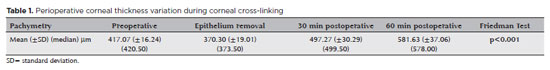
METHODS: This was a prospective case series. We used a method similar to the Dresden protocol, with the application of hydroxypropyl methylcellulose 0.1% hypo-osmolar riboflavin in corneas between 330 and 400 µm after epithelium removal. Corneal thickness was measured using portable calipers before and immediately after epithelium removal, and 30 and 60 min after the procedure.
RESULTS: The 30 patients in this study were followed up for one year. A statistically significant difference was observed in pachymetry values during the intraoperative period (p<0.0001) and an increase of 3.05 µm (95%CI: 0.56–5.54) for each diopter was seen after epithelium removal (p0.019). We found an average Kmax difference of −2.12 D between men and women (p0.013). One year after treatment, there was a statistically significant reduction in pachymetry (p<0.0001) and Kmax (p0.0170) values.
CONCLUSIONS: A significant increase in pachymetry measurements was seen during the procedure, and most patients showed a regression in Kmax and pachymetry values one year after surgery.
Keywords: Corneal pachymetry; corneal topography; cross-linking reagents/therapeutic use; hypromellose derivatives; keratoconus/surgery; riboflavin/therapeutic use
Arq. Bras. Oftalmol. 2025;88 (6 )
:1-8
| DOI: 10.5935/0004-2749.2025-0118
Abstract
PURPOSE: Using advanced imaging techniques, this study aimed to evaluate corneal stability, epithelial remodeling, and tear film changes over a one-year period in first-time soft-contact lens wearers.
METHODS: A retrospective study was conducted on 100 eyes of 50 first-time daily soft-contact lens users aged 21–65 years with no prior rigid gas-permeable lens wear. The Sirius Scheimpflug imaging system was used to assess corneal topography, epithelial thickness, and non-invasive tear break-up time at baseline, 3, 6, and 12 months. Corneal warpage was evaluated using symmetry indices and Baiocchi Calossi Versaci indices. We performed statistical analysis using repeated-measures analyses of variance with Greenhouse-Geisser correction.
RESULTS: The mean baseline central corneal thickness was 537.83 (±7.92) µm, with no significant thinning after one year. The average simulated keratometry values remained stable, indicating no progressive corneal steepening or flattening. There were no significant changes in warpage indices over time, suggesting corneal shape preservation. Higher-order aberrations (coma, trefoil, and spherical aberrations) and non-invasive tear break-up time remained unchanged throughout the study period.
CONCLUSIONS: Modern silicone hydrogel soft-contact lenses do not induce significant corneal warpage, epithelial remodeling, or optical aberrations over a one-year period. We found that corneal morphology and tear film stability were preserved, supporting the safety of soft-contact lens use. These findings provide clinically relevant insights into the long-term impact of contact lens wear. They may facilitate improved lens fitting strategies and preoperative refractive surgery assessments.
Keywords: Contact lenses, hydrophilic; Cornea/surgery; Corneal diseases; Corneal topography; Adaptation, ocular/physiology; Endothelium, corneal/pathology; Refractive errors; Tears/metabolism.
Arq. Bras. Oftalmol. 2024;87 (3 )
:1-8
| DOI: 10.5935/0004-2749.2022-0076
Abstract
MÉTODOS: Córneas humanas de treinamento disponibilizadas foram randomizadas em quatro grupos: Pachy-100 (profundidade de incisão = espessura corneana central - margem de segurança de 100 µm), Pachy-50 (margem de segurança de 50 µm), Pachy-0 (sem margem de segurança) e Pachy+50 (profundidade de incisão = espessura corneana central + 50 µm). Todas as lamelas foram dissecadas através um método padronizado e já publicado (Pachy-DSEK). As espessuras das lamelas (centro, zona de 3,0mm e zona de 6,0mm) foram medidas com tomografia de coerência óptica. A razão de espessura centro-periferia foi calculada aos 3,0 e 6,0 mm de diâmetro.
RESULTADOS: Perfuração endotelial ocorreu apenas no grupo Pachy+50 (n=3, 30%). A espessura central da lamela nos grupos Pachy-100, Pachy-50, Pachy-0 e Pachy+50 foi de 185 ± 42 µm, 122 ± 29 µm, 114 ± 29 µm, e 58 ± 31 µm, respectivamente (p<0,001). As razões C/P aos 3,0 e 6,0 mm foram de 0,97 ± 0,06 e 0,92 ± 0,14, respectivamente. Os parâmetros de características do doador não se correlacionaram com os resultados de espessura de lamela. A profundidade planejada de incisão se correlacionou com a maioria dos parâmetros de espessura de lamela (p<0,001). A espessura de lamela se correlacionou negativamente com a profundidade planejada da incisão (p<0.001, r=-0,580). O melhor resultado foi observado no grupo Pachy-0, em que 75% das lamelas mediram abaixo de 130 µm e não houve perfuração endotelial.
CONCLUSÃO: Através de um método padronizado de dissecção, a maioria das lamelas endoteliais apresentou uma configuração planar. O planejamento de profundidade de incisão igual à espessura corneana central resultou em alta porcentagem de lamelas ultrafinas sem ocorrência de perfuração.
Keywords: Transplante de córnea; Ceratoplastia lamelar; Endotélio corneano; Dissecção; Tomografia de coerência óptica
Arq. Bras. Oftalmol. 2026;89 (4 )
:1-9
| DOI: 10.5935/0004-2749.2025-0034
Abstract
PURPOSE: To evaluate the impact of the COVID-19 pandemic and characterize the serological profile of discarded corneal donations in the coverage area of the Banco de Olhos de Londrina, through reverse transcription-polymerase chain reaction testing for COVID-19 and serological screening of cornea donors excluded because of positive test results.
METHODS: This observational retrospective study included 776 cornea donors who’s serological and reverse transcription-polymerase chain reaction test results were processed at the Hospital of Universidade Estadual de Londrina between May 2020 and 2022. The number of corneal donations and tissue utilization rates throughout the years of operation of the Banco de Olhos de Londrina were also analyzed.
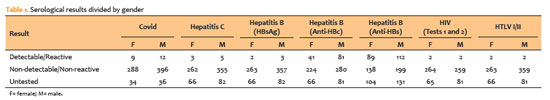
RESULTS: The mean donor age was 53.14 years; 332 donors (43%) were female, and 444 (57%) were male. Positive results were identified in 15.76% of donors for hepatitis B core antibody antibodies, 0.65% for hepatitis B surface antigen, 1.03% for hepatitis C antibodies, and 0.52% for human immunodeficiency virus and human T-lymphotropic vírus. Positive reverse transcription-polymerase chain reaction results for SARS-CoV-2 were observed in 2.7% of cases. Older adults were 2.6 times more likely to test positive for SARS-CoV-2 (95% CI, 1.06-6.34) and 3.0 times more likely to test positive for hepatitis B core antibody (95% CI, 1.95-4.41) than younger individuals. A 75.2% reduction in corneal donations was observed in 2020 compared with 2019, accompanied by a 5% increase in tissue utilization, possibly associated with the effectiveness of donor screening during the pandemic.
CONCLUSION: The COVID-19 pandemic had a profound impact on the number of corneal transplants worldwide, in Brazil, and at the Banco de Olhos de Londrina because of the substantial decline in donations during this period. Hepatitis B was the leading cause of corneal tissue discard due to positive serology in both this study and previous reports, highlighting the importance of prevention programs and improved vaccination coverage. Strict legislation, comprehensive serological screening, and appropriate processing of donated tissue remain essential to eliminate potential sources of infection and ensure transplantation safety.
Keywords: Cornea; Corneal transplantation; COVID-19; Eye banks; Serology
Arq. Bras. Oftalmol. 2025;88 (3 )
:1-7
| DOI: 10.5935/0004-2749.2023-0309
Abstract
PURPOSE: Keratoconus presents certain peculiarities in pediatric patients when compared with adults. The greatest challenge in children is that the disease is more severe and faster in progression. In this retrospective study, we aimed to compare the accelerated and Dresden protocols for corneal crosslinking in patients aged <18 years who were followed-up for at least 12 months.
METHODS: A total of 36 eyes from 27 patients were included in the study. The best corrected and uncorrected visual acuity, maximal keratometry, corneal thickness, foveal thickness, and endothelial microscopy findings were evaluated at baseline and during the postoperative period at one, three, and six months. Thereafter, the patients were evaluated at one, three, six and twelve months postoperative. Corneal crosslinking was performed in all patients via the Dresden protocol (n=21 eyes) or the accelerated protocol (n=15 eyes). Data between the two groups were compared and XY statistical analysis was used.
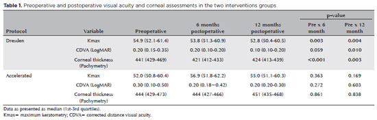
RESULTS: Both protocols were effective in halting keratoconus progression. No patient had progression at the 12-month follow-up. A significant reduction in Kmax and improvement in the corrected distance visual acuity were observed only in the Dresden protocol group. Although the Dresden protocol was superior to the accelerated protocol in reducing Kmax (p=0.002), there was no significant difference in corrected distance visual acuity between the two groups.
CONCLUSION: The accelerated protocol is as efficient as the Dresden protocol in stabilizing keratoconus progression. Although the Dresden protocol was superior to the accelerated protocol in reducing the Kmax, it did not produce better clinical results. Thus, the accelerated protocol is an efficient option. Furthermore, considering the advantages of reduced surgical time, the accelerated protocol is effective in halting keratoconus progression in the pediatric age group.
Keywords: Keratoconus; Corneal diseases; Ultraviolet rays; Cross-linking reagents; Visual acuity
Arq. Bras. Oftalmol. 2024;87 (3 )
:1-8
| DOI: 10.5935/0004-2749.2022-0004
Abstract
OBJETIVO: Examinar os efeitos do tratamento de reticulação unilateral do colágeno corneano na acuidade visual e os achados topográficos em olhos não tratados de pacientes com ceratocone progressivo bilateral.
MÉTODOS: Foram rastreados retrospectivamente pacientes com ceratocone progressivo submetidos a tratamento de reticulação. Foram incluídos no estudo 188 olhos não tratados de 188 pacientes tratado unilateralmente com reticulação padrão ou acelerada e que recusaram o procedimento de reticulação no outro olho. A acuidade visual e os achados topográficos dos olhos não tratados foram obtidos no pré- e pós-operatório no 1o, 3o, 6o, 12o, 24o, 30o e 36o mês.
RESULTADOS: As alterações ao longo do tempo foram semelhantes para as variáveis examinadas nos olhos não tratados de pacientes tratados com métodos de reticulação padrão e acelerado (p>0,05). No 12º mês, 136 olhos não tratados (95,8%) estavam estáveis, de acordo com os critérios de progressão. Apenas quatro olhos (8%) mostraram progressão no 24o mês. Nenhuma progressão foi observada nos 16 pacientes que tiveram um acompanhamento de 36 meses.
CONCLUSÕES: O estudo mostrou que os olhos não tratados de pacientes com ceratocone progressivo bilateral não apresentaram taxas de progressão significativas após o tratamento unilateral com reticulação.
Keywords: Topografia da córnea; Reagentes de ligações cruzadas; Ceratocone; Fármacos fotossensibilizantes; Colágeno/uso terapêutico; Fotoquimioterapia/métodos; Acuidade visual
Arq. Bras. Oftalmol. 2024;87 (3 )
:1-7
| DOI: 10.5935/0004-2749.2023-0049
Abstract
PURPOSE: To investigate the association of pre-photorefractive keratectomy Schirmer-1 test value with post-photorefractive keratectomy central corneal epithelial thickness, ocular surface disease index score, and uncorrected distance visual acuity.
METHODS: Patients were categorized according to preoperative Schirmer-1 value: the normal Schirmer Group (n=54; Schirmer-1 test value, >10 mm) and the low Schirmer Group (n=52; Schirmer-1 test value, between 6 and 10 mm). We analyzed ablation depth, visual acuity, result of Schirmer-1 test (with anesthesia), tear film break-up time, ocular surface disease index score, central corneal epithelial thickness, and spherical equivalent refraction.
RESULTS: We found significant differences between the groups in Schirmer-1 test value, tear film break-up time, and ocular surface disease index score, both preoperatively and postoperatively (p<0.001). The preoperative central corneal epithelial thicknesses of the two groups were similar (p>0.05). After photorefractive keratectomy, the Schirmer-1 test value and spherical equivalent refraction decreased in both groups (p<0.05), and ocular surface disease index scores and central corneal epithelial thickness values increased in the low Schirmer Group (p<0.001) but not in the normal Schirmer Group (p>0.05). The postoperative central corneal epithelial thicknesses of the low Schirmer Group were significantly higher than those of the normal Schirmer Group (p<0.001). Postoperative uncorrected distance visual acuity did not differ significantly between the two groups (p>0.05).
CONCLUSIONS: In patients with low Schirmer-1 test values before photorefractive keratectomy, the corneal epithelium thickened and ocular surface complaints increased during the postoperative period. However, changes in the corneal epithelium did not affect the postoperative uncorrected distance visual acuity. To reduce postoperative problems on the ocular surface in these patients, we recommend that dry eye be treated before photorefractive keratectomy.
Keywords: Epithelium, corneal; Cornea; Photorefractive keratectomy; Schirmer test; Visual acuity
Arq. Bras. Oftalmol. 2025;88 (5 )
:1-7
| DOI: 10.5935/0004-2749.2024-0368
Abstract
PURPOSE: To compare endothelial corneal cell changes following cataract surgery performed by phacoemulsification with intraocular lens implantation, conducted by surgeons with varying levels of experience.
METHODS: Two hundred and eighty-three eyes diagnosed with cataract were included. Lens opacity was classified into three categories (I, II, and III). Surgeons were categorized into four experience levels (1, 2, 3, and 4), based on years of practice and lifetime surgeries performed. Corneal endothelial characteristics were assessed using non-contact specular microscopy, with measurements taken before surgery and 30-60 days post-surgery.
RESULTS: Pre- and postoperative endothelial analysis showed no significant differences between surgeon levels regarding visual acuity achieved, corneal thickness, and endothelial hexagonality. However, the central endothelial cell density index showed a significantly greater reduction among level 1 surgeons (p=0.026). Grade II cataracts exhibited significant variations in the central endothelial cell density (p=0.011) and average cell size, with level 1 surgeons showing the largest increases (p=0.024).
CONCLUSIONS: The analysis revealed significant differences in visual acuity and endothelial indices between surgeon experience levels, with less experienced surgeons showing greater variations and poorer performance. Clinical protocols should consider these data to establish safer training protocols.
Keywords: Cataract extraction; Phacoemulsification; Endothelium; corneal; Lens implantation, intraocular; Visual acuity; Internship and residency; Surgeons
Arq. Bras. Oftalmol. 2025;88 (3 )
:1-6
| DOI: 10.5935/0004-2749.2024-0207
Abstract
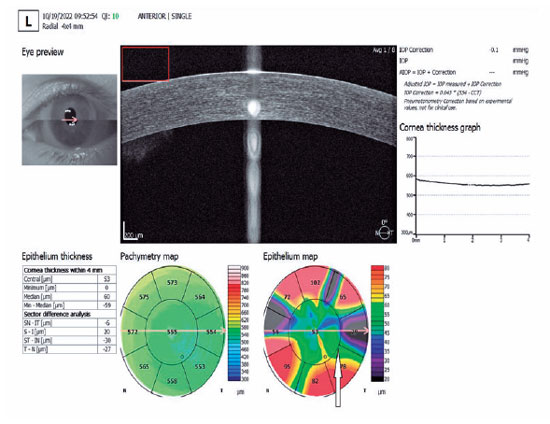
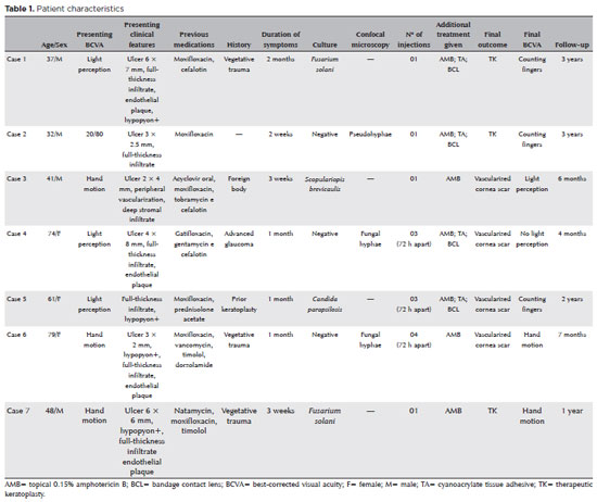
PURPOSE: This study aimed to report the use, efficacy, and safety of intracameral voriconazole as an adjuvant treatment for deep fungal keratitis.
METHODS: This was a prospective case series of seven eyes with fungal keratitis with anterior chamber involvement or a corneal ulcer refractory to conventional topical treatment. In addition to topical treatment with 0.15% amphotericin B eye drops, voriconazole 50 μg/ 0.1 mL
was administered to the anterior chamber of each affected eye up to four times within 72 h. The primary outcome measures were healing (fungal eradication) and the need for therapeutic keratoplasty. Best-corrected visual acuity was a secondary outcome measure.
RESULTS: Three cases were confirmed by confocal microscopy, and four were diagnosed from positive culture tests. At presentation, one patient had a best-corrected visual acuity of 20/80, while all others had hand motion or worse. Four cases received one intracameral injection, two cases received three, and one case received four injections. There were no complications after any of the intracameral voriconazole injections. Four patients had imminent corneal perforations and were treated with cyanoacrylate adhesive and bandage contact lenses. Four patients recovered from the infection, and three underwent therapeutic keratoplasty. The final best-corrected visual acuity was improved in two cases but all patients had a final visual acuity of counting fingers or worse.
CONCLUSION: As an adjuvant treatment for deep fungal keratitis, intracameral voriconazole injection is a feasible option. Although fungal eradication was achieved in all patients, three required therapeutic keratoplasty and all patients had unsatisfactory visual acuity outcomes.
Keywords: Antifungal agents; Fungi; Corneal transplantation; Keratitis; Eye infections, fungal; Voriconazole
 ABO is licensed under a Creative Commons Attribution-NonComercial 4.0 Internacional.
08-fig01.jpg)
02-tab01tb.jpg)
03-tab01.jpg)

06-tab01tb.jpg)
08-tab01.jpg)

09-fig01.jpg)