Arq. Bras. Oftalmol. 2021;84 (3 )
:220-224
| DOI: 10.5935/0004-2749.20210030
Abstract
OBJETIVO: Avaliar a morfologia da córnea e da câmara anterior em olhos fácicos com inflamação intraocular não infecciosa.
MÉTODOS: Esse estudo incluiu 59 olhos com uveíte ativa, 62 olhos com uveíte inativa e 95 olhos saudáveis. A densidade de células endoteliais da córnea, a proporção de células hexagonais, o coeficiente de variação, o volume e a espessura da córnea, a ceratometria máxima e o volume e profundidade da câmara anterior foram medidos com um microscópio especular e uma Pentacam HR.
RESULTADOS: A duração média da uveíte foi de 24,6 ± 40,5 (0-180) meses. O número médio de crises de uveíte foi de 2,8 ± 3,0 (1-20). O coeficiente de variação foi significativamente maior no grupo com uveíte ativa do que no grupo com uveíte inativa (p=0,017, Tukey post-hoc). Não houve diferença significativa nos demais parâmetros do segmento anterior entre os grupos com uveíte ativa, com uveíte inativa e controle (p>0,05). A análise de regressão linear múltipla demonstrou que o coeficiente de variação foi maior na uveíte ativa do que na uveíte inativa, após ajustes para a duração e tipo de uveíte e a presença ou não de doença reumática e de tratamento imunossupressor (p=0,003). A duração da uveíte e o número de crises não demonstraram correlação significativa com os parâmetros oculares (p>0,05, correlação de Spearman). A diferença nos parâmetros não demonstrou correlação significativa com o tipo de uveíte (p>0,05).
CONCLUSÕES: O coeficiente de variação foi maior nos olhos com uveíte ativa do que naqueles com uveíte inativa, ao passo que a densidade de células endoteliais e a morfologia da câmara anterior não mostraram diferenças significativas entre os grupos com uveíte ativa, com uveíte inativa e controle.
Keywords: Câmara anterior; Inflamação; Epitélio posterior; Contagem de células; Uveites
Arq. Bras. Oftalmol. 2020;83 (4 )
:299-304
| DOI: 10.5935/0004-2749.20200051
Abstract
Objetivo: Investigar a utilidade de marcadores inflamatórios sistêmicos (ou seja, contagem de glóbulos brancos e plaquetas, volume médio de plaquetas e suas proporções) como marcadores de diagnóstico da patogênese do edema macular diabético.
Métodos: A coorte do estudo incluiu 80 pacientes com edema macular diabético (40 com retinopatia diabética e 40 sem) e 40 controles saudáveis de acordo com a idade e sexo. As contagens de neutrófilos, linfócitos, monócitos, plaquetas e valores do volume plaquetário médio foram determinados a partir de amostras de sangue periféricdo, e as proporções de monócitos/linfócitos, plaquetas/linfócitos, volume plaquetário médio/linfócitos e neutrófilos/linfócitos foram calculadas e comparadas entre os grupos.
Resultados: A proporção média de neutrófilos/linfócitos dos grupos com edema macular diabético e não-diabético foi maior que a do grupo controle, e o valor do grupo com edema macular diabético foi maior que o do grupo com edema macular não diabético (p<0,001 no com edema macular diabético vs. controle, p=0,04 no com edema macular não diabético vs. controle e p=0,03 no com edema macular diabético vs. o com edema macular não-diabético). Um valor de corte de neutrófilos/linfócitos ≥2,26 foi identificado como um indicador da patogênese do edema macular diabético com sensibilidade de 85% e especificidade de 74%. O volume plaquetário médio do grupo com edema macular diabético foi maior que o dos grupos com edema macular não-diabético e controle, enquanto os do grupo de edema macular não-diabético e controle foram semelhantes (edema macilar diabético vs. Edema macular não-diabético, p=0,08; com edema macular diabético vs. controle, p=0,02; e com edema macular não-diabético vs. controle, p=0,78). Todos os outros parâmetros foram semelhantes entre os grupos (todos p>0,05).
Conclusão: A proporção de neutrófilos/linfócitos e o volume plaquetário médio do grupo com edema macular diabético foram superiores aos do grupo com edema macular não-diabético e controle. Um valor de corte da razão neutrófilos/linfócitos ≥2,26 foi identificado como um indicador da patogênese do edema macular diabético com alta sensibilidade e especificidade. Além disso, a proporção de neutrófilos/linfócitos do grupo com edema macular não-diabético foi superior à do grupo controle.
Keywords: Edema macular; Retinopatia diabética; Volume plaquetário médio; Contagem de linfócitos; Neutrófilos; Inflamação
Arq. Bras. Oftalmol. 2023;86 (4 )
:345-352
| DOI: 10.5935/0004-2749.20230050
Abstract
Objetivo: Investigar a susceptibilidade a antibióticos, o perfil clínico, epidemiológico e microbiológico das ceratites infecciosas.
Métodos: Estudo retrospectivo longitudinal. Registros médicos e laboratoriais de 2015 a 2019 foram revisados retrospectivamente.
Resultados: Trezentos e oitenta patógenos (321 bactérias e 59 fungos) foram isolados das córneas de 352 pacientes. As espécies de Staphylococcus foram os microorganismos mais isolados (45%), seguidos de Pseudomonas (18,4%), fungos (15,5%), Streptococcus (7,9%) e Serratia (3,2%). Não houve resistência das bactérias Gram-positivas à amicacina ou vancomicina, enquanto 14,8% isolados Gram-positivos foram resistentes à ciprofloxacina (p<0,05). Todos os organismos Gram-negativos eram suscetíveis à amicacina. Pacientes do sexo masculino representaram 62,8% de 129 casos com dados clínicos acessíveis. A média de idade foi 53,17 ± 21 anos. O tempo até a apresentação (desde o início dos sintomas) foi de 14,9 ± 19,4 dias (mediana: 7 dias). Úlceras grandes (>5mm em qualquer extensão) representaram 49,6% (64 olhos) dos casos. A duração do tratamento foi de 49 ± 45,9 dias (mediana: 38 dias). Trauma ocular direto foi relatado por 48 (37,2%) pacientes e uso de lentes de contato por 15 (11,6%) pacientes. Foi prescrito tratamento prévio para 72 (55.8%) pacientes. Outras classes de medicamentos foram prescritas para 16 (12.4%). Setenta e nove (61,2%) pacientes tiveram que ser hospitalizados. Como complicações maiores, 53 (41,1%) pacientes apresentaram perfuração corneana, 40 pacientes (31%) foram submetidos à ceratoplastia penetrante tectônica e 28 (21,7%) desenvolveram glaucoma secundário. Oito pacientes (6,2%) evoluíram para endoftalmite. O tratamento empírico da ceratite microbiana foi amplamente empregado, com 94 (72,9%) pacientes em uso de moxifloxacina e 56 (43,4%) em uso de ciprofloxacina antes do resultado da cultura.
Conclusões: Nosso hospital tratou predominantemente de pacientes com úlceras microbianas graves. Embora bactérias Gram-positivas constituíssem a maioria dos isolados, bacilos e fungos Gram-negativos também foram frequentemente identificados nas ceratites microbianas. Os resultados de suscetibilidade sugerem a combinação de vancomicina e amicacina como um regime terapêutico empírico eficaz para essa condição grave com risco de perda visual permanente.
Keywords: Ceratite; Infecções oculares bacterianas; Antibacterianos.
Arq. Bras. Oftalmol. 2025;88 (4 )
:1-6
| DOI: 10.5935/0004-2749.2024-0278
Abstract
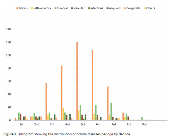
PURPOSE: This study aimed to evaluate the prevalence of orbital conditions in a tertiary ophthalmic outpatient hospital in Sao Paulo, Brazil, with a focus on the main diagnoses and their distribution.
METHODS: A retrospective chart review was conducted involving patients registered and admitted to the orbital disease unit at the Department of Ophthalmology, University of São Paulo Medical School, from January 2004 to March 2018. A total of 838 medical charts were analyzed, of which 37 were excluded due to incomplete data. The remaining charts were categorized into eight diagnostic groups: Graves’ orbitopathy , inflammatory disorders, tumors, vascular lesions, acquired structural abnormalities, congenital structural abnormalities, infectious diseases, and others.
RESULTS: Of the 837,300 ophthalmological appointments, 3,372 (0.4%) were related to orbital diseases. The study included 801 patients, of whom 63.45% were women. The patients’ mean age was 42.86 years. Graves’ orbitopathy was the most common (55%), followed by tumor (17%), inflammatory disorders (9%), vascular lesions (7%), acquired structural abnormalities (5%), congenital structural abnormalities (4%), others (2%), and infectious diseases (1%). The study found significant differences in the incidence and types of orbital diseases, indicating the specialized nature of tertiary care and referral biases.
CONCLUSION: Published data on epidemiological orbital diseases is scarce. Therefore, this study focused on the diverse nature of orbital diseases and their low incidence among ophthalmology appointments. The major trends align with other epidemiological studies, demonstrating a preponderance of Graves’ orbitopathy in middle-aged adults and a bimodal distribution of tumors. These findings are essential in shaping resident training programs and healthcare policies, particularly in tertiary settings. Understanding the epidemiology of orbital diseases can improve diagnostic accuracy, treatment approaches, and patient outcomes as well as support future systemic prospective studies.
Keywords: Orbital diseases; Orbital tumors; Neoplasms; Inflammation; Graves’ ophthalmopathy; Outpatients
Arq. Bras. Oftalmol. 2026;89 (2 )
:1-8
| DOI: 10.5935/0004-2749.2025-0175
Abstract
PURPOSE: Endophthalmitis is one of the most important adverse events after cataract surgery, as it can lead to total vision loss. This study aimed to describe the occurrence of endophthalmitis after phacoemulsification with intraocular lens implantation in patients treated in a community setting in Porto Velho, Rondônia, Brazil.
METHODS: This retrospective cohort study was conducted using a database of 649 medical records of patients who underwent surgery and were followed for three months. Poisson regression analysis was used to estimate relative risks and 95% confidence intervals (95% CIs).
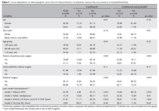
RESULTS: The incidence of confirmed endophthalmitis was 11.94% (95% CI, 9.50-14.76), while the incidence of confirmed and probable cases was 20.50% (95% CI, 17.52-23.73). For confirmed cases, bilateral surgery and the use of lens model 3 were identified as risk factors for endophthalmitis, whereas age over 70 yr and preoperative antibiotic use were protective factors. For confirmed and probable cases, brown and yellow skin color, bilateral surgery, and the use of lens model 3 were also identified as risk factors. Gram-negative bacteria were the predominant etiological agents, and corneal edema was the main clinical manifestation. The mean duration of treatment was eight days, and 27.12% of patients used antibiotics.
CONCLUSION: The incidence observed was substantially higher than that reported in the literature, with a predominance of Gram-negative agents and an association with bilateral surgeries and the Eyeol intraocular lens model. These findings reinforce the need for continuous epidemiological surveillance and the implementation of specific biosafety and infection control protocols during cataract surgery campaigns.
Keywords: Endophthalmitis; Disease outbreaks; Phacoemulsification; Lens implantation, intraocular; Lenses, intraocular; Cataract; Risk factors; Anti-bacterial agents
Arq. Bras. Oftalmol. 2026;89 (5 )
:1-6
| DOI: 10.5935/0004-2749.2025-0154
Abstract
PURPOSE: This study aimed to evaluate the serum levels of thrombospondin-1, growth differentiation factor-15, resolvin D1, and resolvin E1 in patients with keratoconus.
METHODS: Thirty-five patients with keratoconus and 35 control subjects of similar age and sex were evaluated. The keratoconus group was divided into four stages according to the modified Krumeich classification. Commercial kits were used to determine thrombospondin-1, growth differentiation factor-15, resolvin D1, and resolvin E1 levels in serum samples using enzyme-linked immunosorbent assay (ELISA). Serum thrombospondin-1, growth differentiation factor-15, resolvin D1, and resolvin E1 levels were compared between the patient and control groups.
RESULTS: The keratoconus group consisted of 14 females and 21 males, and the control group consisted of 14 females and 21 males, with mean ages of 29.4±8.8 and 30.0±7.8 yr, respectively. There were significant differences in serum thrombospondin-1, growth differentiation factor-15, resolvin D1, and resolvin E1 levels between the serum of the keratoconus and control groups (p=0.006, p<0.001, p<0.001, and p=0.001, respectively). No significant correlations were found between keratoconus stage and serum thrombospondin-1, growth differentiation factor-15, resolvin D1 and resolvin E1 levels (r=−0.025, p=0.892; r=−0.019, p=0.913; r=−0.036, p=0.838; and r=−0.108, p=0.538, respectively).
CONCLUSION: Serum levels of thrombospondin-1, growth differentiation factor-15, resolvin D1, and resolvin E1 were significantly lower in patients with keratoconus. These results suggest that these parameters may be associated with the pathogenesis of keratoconus.
Keywords: Keratoconus/pathology; Thrombospondin 1/blood; Growth differentiation factor 15/blood; Docosahexaenoic acids/blood; Eicosapentaenoic acid/blood; Inflammation mediators
Arq. Bras. Oftalmol. 2025;88 (5 )
:1-8
| DOI: 10.5935/0004-2749.2024-0312
Abstract
PURPOSE: To evaluate the changes in the rates and indications of eye removal procedures during the recent COVID-19 pandemic.
METHODS: The medical records of all patients who underwent eye removal from 2007 to 2022 were retrospectively reviewed. The patient demographic data and indications for surgery were collected. Data from two groups of patients (prepandemic surgery and postpandemic surgery) were compared. Statistical significance was set at p<0.05.
RESULTS: Fifty-nine patients underwent enucleation (69%), evisceration (27%), or exenteration (3%). The mean (SD) age of the patients was 55.9 (19.4) years, and most (69%) of the patients were males. Most (47%) of the study population were Black. The common indications for eye removal were trauma (41%), painful blind eye (34%), and infection/inflammation (24%). The types of trauma were assault (55%), accidental (39%), and self-inflicted (6%). The mean (SD) monthly rates of eye removal increased from 0.25 (0.50) in the prepandemic period to 0.77 (0.91) during the pandemic (p<0.001). These increases were noted in both males (p=0.003) and females (p=0.001) and were the highest among Black patients [0.42 (0.76); p<0.001]. Among the indications of eye removal, painful blind eyes [0.35 (0.75); p<0.001] and ocular trauma [0.31 (0.47); p=0.051] exhibited the greatest increases following the pandemic.
CONCLUSION: The rate of eye removal procedures increased during the recent pandemic. Although delayed care of chronic eye conditions may have contributed to the increased rates of painful blind eyes, the increased trauma-related eye removals may be attributed to the simultaneous spike in violent assaults in New York City.
Keywords: Eye injuries; Eye enucleation; COVID-19; Pandemics; Ethinicity; Inflammation, Trauma centers


 ABO is licensed under a Creative Commons Attribution-NonComercial 4.0 Internacional.


04-tab01.jpg)
06-tab01tb.jpg)
05-tab01.jpg)


02-fig01.jpg)
01-fig01.jpg)
10-fig01.jpg)
10-fig01.jpg)
08-tab01.jpg)