Abstract
Objetivo: O objetivo deste estudo é investigar os efeitos dos antidepressivos tricíclicos, dos inibidores da recaptação da serotonina e dos inibidores da recaptação da serotonina e noradrenalina na superfície ocular.
Métodos: Foram incluídos no estudo 330 olhos de 165 pacientes em uso de antidepressivos e 202 olhos de 101 controles. Foi medido o tempo de ruptura do fluido lacrimal e foram administrados o teste de Schirmer I e o questionário Ocular Surface Disease Index (OSDI). Os Inventários de Depressão e de Ansiedade de Beck foram aplicados ao uso dos medicamentos e foram registrados as dosagens, a duração da doença psiquiátrica e o tempo de remissão.
Resultados: No grupo de estudo, o tempo médio de ruptura do fluido lacrimal foi de 14,29 ± 4,81 segundos (intervalo de 4-26 segundos) e o valor médio do teste de Schirmer I foi de 16,05 ± 5,89 mm (intervalo de 2-28 mm). No grupo controle. o tempo médio de rompimento do fluido lacrimal foi de 18,16 ± 2,12 segundos (intervalo de 15-24 segundos) e o valor do teste de Schirmer I foi de 16,64 ± 2,31 mm (intervalo de 15-24 mm), com p<0,001 e p=0,005, respectivamente. No grupo de estudo, 38,18% (n=63) dos pacientes tinham olho seco, enquanto no grupo controle 17% (n=18) tinham olho seco (p<0,001). O escore médio no OSDI foi de 82,56 ± 16,21 (intervalo 66-100) no grupo dos antidepressivos tricíclicos, 60,02 ± 29,18 (10-100) no grupo dos inibidores da recaptação da serotonina e 22,30 ± 20,87 (0-75) no grupo dos inibidores da recaptação da serotonina e noradrenalina (p<0,001). O tempo médio de rompimento do fluido lacrimal foi de 14,36 ± 3,35 segundos (intervalo de 10-20 segundos) no grupo dos antidepressivos tricíclicos, 13,94 ± 5,81 segundos (intervalo de 4-26 segundos) no grupo dos inibidores da recaptação de serotonina e 14,93 ± 4,20 segundos (intervalo de 6-20 segundos) no grupo dos inibidores da recaptação de serotonina e noradrenalina (p=0,730). O valor médio do teste de Schirmer I foi de 9,90 ± 7,22 mm (intervalo de 2-30 mm) no grupo dos antidepressivos tricíclicos, 15,55 ± 5,15 mm (intervalo de 2-25 mm) no grupo dos inibidores da recaptação da serotonina e 17,71 ± 4,21 mm (intervalo de 10-30 mm) no grupo dos inibidores da recaptação da serotonina e noradrenalina (p<0,001). Não houve diferença estatisticamente significativa no escore OSDI, no tempo de ruptura do fluido lacrimal e nos valores do teste de Schirmer I entre os subgrupos de pacientes em uso de inibidores da recaptação de serotonina e de inibidores da recaptação de serotonina e noradrenalina.
Conclusões: Olho seco é uma queixa comum em usuários de antidepressivos, mas no que diz respeito à superfície ocular, inibidores da recaptação de serotonina e noradrenalina podem ser mais confiáveis que outros antidepressivos. Pacientes em uso de inibidores da recaptação de serotonina e noradrenalina têm escores menores no questionário OSDI. Os inibidores da recaptação da serotonina e noradrenalina, úteis nas síndromes de dor crônica, também podem ter um efeito corretivo nos sintomas de olho seco.
Keywords: Inibidores de recaptação de serotonina; Antidepressivos tricíclicos; Serotonina; Depressão; Dor crônica; Síndromes do olho seco; Norepinefrina; Inquéritos e questionários; Ansiedade; Preparações farmacêuticas.
 ABO is licensed under a Creative Commons Attribution-NonComercial 4.0 Internacional.
10-tab01.jpg)
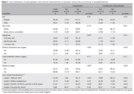
05-tab01.jpg)

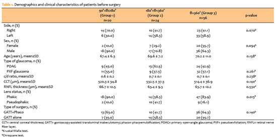
03-tab01.jpg)
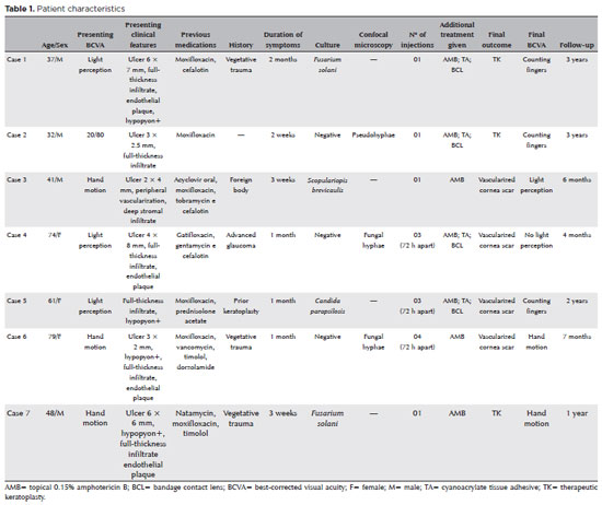
08-tab01.jpg)
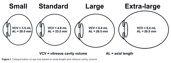
10-fig01.jpg)
07-fig01.jpg)